Biology II
🐶
Biology II
Table of Contents
Part I | Animal Excretory Systems
Osmoregulation balances the uptake and loss of water and solutes An animal’s nitrogenous wastes reflect its phylogeny and habitat Diverse excretory systems are variations on a tubular scheme
The nephron is organized for stepwise processing of blood filtrate Hormonal circuits link kidney function, water balance, and blood pressure
Part II | Nervous System
Neuron structure and organization reflect function in information transfer Ion pumps and ion channels establish the resting potential of a neuron Action potentials are the signals conducted by axons
Part I | Animal Excretory Systems
Osmoregulation balances the uptake and loss of water and solutes
Osmosis
Definition
the process of water entering and leaving cells
occurs when two solutions separated by a membrane differ in total solute concentration
water molecules cross the cell membrane at equal rates in both directions when there is no difference in concentration, thus, there is no net movement of water
water flows from a hypoosmotic solution to a hyperosmotic one
Hyperosmotic — the solution with a higher concentration of solutes
Hypoosmotic — the solution with a lower concentration of solutes
Isoosmotic — when two solutions have the same osmolarity
Osmolarity
the unit of measurement for solute concentration
the number of moles of solute per liter of solution (mOsm/L)
human blood measures 300 mOsm/L, and seawater is 1000 mOsm/L
Osmoregulation in Land Animals
Descriptions
the threat of dehydration is a major regulatory problem for terrestial plants and animals
adaptations such as the body coverings of terrestial plants that reduce water loss are key to survival
waxy layers of insect exoskeletons, shells of land snails, and layers of dead, keratinized skin cells in
vertebrates
maintain water balance in a variety of ways
- drinking and eating moist foods
- producing water metabolically through cell respiration
most terrestrial animals lose water through a variety of routes despite anatomical and behavioral adaptations that try to conserve it
- in urine and feces
- across the skin (sweating)
- epithelial surfaces of gas exchange in organs and airways
Desert Animals
Osmoregulatory Challenges and Mechanisms
Osmoconformers and Osmoregulators
an animal maintains water balance in by such
- Osmoconformer
an animal that is isoosmotic to its surroundings
all are marine animals
due to the animal’s internal osmolarity being the same as its environment, there is no gaining or losing of water
- Osmoregulator
an animal that controls internal osmolarity independent to the external environment
enables animals to live in environments (freshwater and terrestial habitats) that are uninhabitable for osmoconformers
in a hypoosmotic environment, the osmoregulator discharges excess water
in a hyperosmotic environment, the osmoregulator takes in water to offset osmotic loss
many terrestrial animals, especially desert-dwellers, are nocturnal which reduces evaporative water loss from the sun
a number of desert animals are well enough adapted that they can survive for long periods without drinking
camels can tolerate a 7°C rise in body temperature, reducing the amount of water lost in sweat production and can also lose 25% of their body water and still survive
humans losing 12.5% of their body water will result in death from heart failure
Stenohalines and Euryhalines
- Stenohalines — animals that cannot tolerate substantial changes in external osmolarity
- Euryhaline — animals that can survive large fluctuations in external osmolarity
Euryhaline Osmoconformers
— barnacles and mussels
Euryhaline Osmoregulators — striped bass and salmon
Energetics of Osmoregulation
Descriptions
maintaining an osmolarity difference between an animal’s body and its environment carries an energy cost
osmoregulators must expend energy through active transport to maintain osmotic gradients that cause water to move in and out
the energy cost of osmoregulation depends on two factors
- How different an animal’s osmolarity is from its surroundings
- How much work is required to pump solutes across the membrane
for brine shrimp, the gradient between internal and external osmolarity is very large and
the cost of osmoregulation is correspondingly high
the energy cost to an animal maintaing water and salt balance is minimized by having body fluids that are adapted to the salinity of the animal’s habitat
marine molluscs have body fluids with solute
concentrations of 1000 mOsm/L whereas freshwater
Osmoregulation in Marine Animals
Descriptions
most marine invertebrates are osmoconformers
their osmolarity is the same as that of seawater so they face no substantial challenges in water balance
nevertheless, they actively transport specific solutes that they maintan at levels different from those in the ocean
Atlantic lobsters maintain a low Mg2+ concentration in
their hemolymph far below the Mg2+ concentration in their environment
Osmoregulatory Strategies
- Bony Fishes — balance water loss by drinking lots of sea water as excess salts are eliminated through gills and kidneys
- Sharks and Chondrtichtyans — shark tissues have a high concentration of solutes [urea and trimethylamine oxide (TMAO)] which causes water to slowly enter the shark’s body by osmosis
the influx of water and salt is disposed of in the shark’s urine or feces
molluscs only have 40 mOsm/L
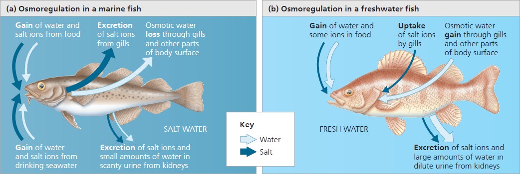 osmoregulation in marine and freshwater bony fishes
Transport Epithelia in Osmoregulation
Description
although the ultimate function of osmoregulation is to control solute concentrations, most animals do this indirectly, managing instead the solute content of the fluid that bathes the cells
in insects and animals with an open circulatory system, this fluid is called hemolymph
in vertebrates and animals with a closed circulatory system, this fluid is a mixture of solutes controlled indirectly by blood
maintaining the composition of such fluids depends on individual cells that regulate solute movement and complex organs such as the kidney
Transport Epithelia
one or more layers of epithelial cells specialized for moving solutes in controlled amounts in specific directions
typically arranged into tubular networks with extensive surface areas
some transport epithelia face the outside environment directly whereas others line channels
Osmoregulation in Freshwater Animals
Descriptions
the osmoregulatory problems of freshwater animals are the opposite of those in marine animals
the body fluids of freshwater animals must be hyperosmotic because animal cells cannot tolerate the low salt concentrations in freshwater
water balance relies on excreting large amounts of very dilute urine and drinking almost no water
salts lost by diffusion and in urine are replenished by eating or by taking up salts across their gills
Salmon
When living in freshwater
produces large amounts of dilute urine
takes up salt from the dilute environment through their gills
When living in saltwater
produce the steroid hormone cortisol which increases the number and size of salt- secreting cells
other physiological changes enable salmon to excrete
connected to the outside by an opening on the body surface
in most animals, osmoregulation and waste disposal rely on transport epithelia
“The Albatross Experiment”
reserachers gave captive birds only seawater to drink
although very little salt appeared in the birds’ urine, the fluid dripping from the tip of their beaks was a concentrated solution of salt
the source of this solution is a pair of nasal salt glands found in sea turtles and marine iguanas that use active transport to secrete a fluid much saltier than the ocean
the salt gland enables marine vertebrates to achieve a net gain of water
in contrast, humans who drink seawater musts use a greater volume of water to extract the salt out of their system
transport epithelia that function in maintaining water balance also often function in disposal of metabolic wastes
excess salt from their gills and produce only small amounts of urine
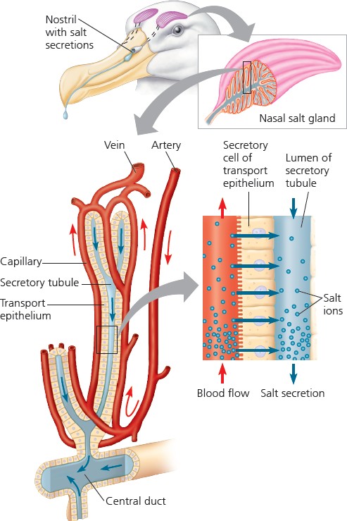
salt secretion in the nasal glands of a marine bird
Osmoregulation in Animals that Live in Temporary Waters
Descriptions
dehydration or desiccation is fatal for most animals however, a few invertebrates that live in temporary ponds can lose almost all their body water and survive
these animals enter a dormant state when their habitats dry up
tardigades, or water bears, in their hydrated state, contain about 85% water by weight but they can
dehydrate to less than 2% water and survive in an inactive state for a decade
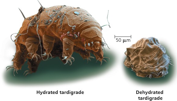
anyhdriobiosis
Anhydrobiosis
the dormant state involving loss of almost all body water
requires adaptations that keep cell membranes intact
studies of anyhdrobiotic roundworms (phylum Nematoda) show that desiccated individuals contain large amounts of trehalose
which seems to protect cells by replacing the water normally associated with proteins and membrane lipids
many insects that survive freezing in winter also use trehalose as a membrane protectant, as do some plants resistant to desiccation
Trehalose — a dissacharide that seems to protect cells from dehydration
An animal’s nitrogenous wastes reflect its phylogeny and habitat
Forms of Nitrogenous Waste
Definition
although some animal species excrete ammonia directly, others excrete alternative forms of nitrogenous waste
these forms of nitrogenous waste are ammonia, urea, and uric acid
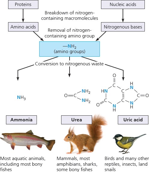
variations in forms of nitrogenous waste
Ammonia
Definition
a toxic metabolite produced by the dismantling of nitrogenous molecules, chiefly proteins and nucleic acids
in regulating and safeguarding internal fluids, animals must deal with ammonia
mechanisms like excretion serve to rid the body of ammonia and other waste products
because most metabolic waste must dissolve in water to be excreted, the type and quantity of waste has a large impact on water balance
Excretion — the disposal of nitrogen-containing metabolites and other waste products
Type 1 | Ammonia
is very toxic because its ion ( NH4+) can interfere with oxidative phosphorylation
can only be tolerated at very low concentrations, thus animals that excrete ammonia need access to lots of water
therefore, ammonia excretion is most common in aquatic species
soluble ammonia molecules (NH3 and NH4+),
which interconvert often, easily pass through membranes and are easily lost to surrounding water
in many invertebrates, ammonia release occurs across the whole body surface
Type 2 | Urea
ammonia is so toxic that it can only be excreted through large
Influence of Evolution and Environment on Nitrogenous Wastes
Definition
the type and amount of nitrogenous waste a species produces are matched to its environment
the availability of water is one key factor
terrestrial turtles excrete uric acid while aquatic excrete both urea and ammonia
the amount of nitrogenous waste produced is coupled to the animal’s energy budget and diet
endotherms, which use energy at high rates, eat more food and produce more waste as opposed to ectotherms
predators, which derive energy from protein (a nitrogenous molecule), excrete more waste than animals that rely on carbohydrates as
energy sources
Eggs
an animal’s egg pertains to the immediate environment relevant to the type of nitrogenous waste excreted
in an amphibian egg, which lacks a shell, ammonia or urea can simply diffue out of the egg
similarly, soluble wastes produced by an embryo are carried away by the mother’s blood
in birds and reptiles, the egg is surrounded by a shell that is permeable to gases but not to liquids
as a result, if ammonia or urea was released by the embryo, it could accumulate to
volumes of very dilute solutions
most terrestrial animals and marine species simply do not have access to sufficient water to secrete ammonia
instead, they secrete urea
the main advantages of urea is its low toxicity and high solubility in water
the main disadvantage of urea is its high energy cost since its production is a metabolic process
amphibians switch secreting ammonia from when they are tadpoles to
producing urea when they become land-dwelling
adults
Urea — a soluble nitrogenous waste
dangerous levels
for this reason, using uric acid which harmlessly solidifies inside the egg, conveys a selective advantage in reptiles
produced in the liver that combines ammonia with carbon dioxide
Type 3 | Uric Acid
insects, land snails, reptiles, and birds excrete uric acid
it is excreted as a semisolid paste causing very little water loss in the organism
it is however, more energetically expensive to create than urea, requiring more ATP
humans and some other animals generate a small amount of uric acid from metabolism
diseases that alter metabolism due to the insoluble property of uric acid may result to gout
gout can form uric acid
stones in Dalmatian dogs’ bladder, male human joints, and
dinosaur
joints as well
Gout — occurs when uric acid crystals or stones form in joints, restricting movement and causing inflammation
Uric Acid — a product of protein and purine metabolism that is relatively nontoxic and insoluble in water
Diverse excretory systems are variations on a tubular scheme
Filtration
Definition
excretory systems play a central role in homestasis as they are responsible for waste disposal and body fluid composition
excretory functions are built on a network of tubules that provide a large surface area for the exchange of water and solutes, including waste
excretory processes begin when body fluid is brought in contact with the selectively permeable membrane of a transport epithelium
hydrostatic pressure or blood pressure drives a process of filtration of micro and macromolecules
cells, proteins, and other large molecules remain in the body fluid
water, salts, sugars, amino acids, and nitrogenous wastes cross the membrane, forming filtrate
Filtrate — cell-free fluid extracted from body fluid by the excretory system
Process
Survey of Excretory Systems | Kidneys
Mammalian Excretory Organs
- Kidneys — filters fluids and produces urine (urea)
- Ureter — carries the urine from the kidney to the urinary bladder
- Urinary Bladder — a sac that acts as urine storage
- Urethra — where urine is expelled during urination
near the vagina for females through the penis in males
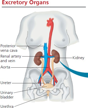
excretory organs
Kidneys
- Filtration — the excretory tubule collects filtrate from the blood, taking water and solutes from the capillaries
- Reabsorption — the capillary reclaims valuable solutes (glucose, vitamins, etc.) and water via the transport epithelium
- Secretion — the excretory tubule collects excess toxins and ions from the capillary and incorporates them into the filtrate via the transport epithelium
- Excretion — the altered filtrate leaves the system or body through urination
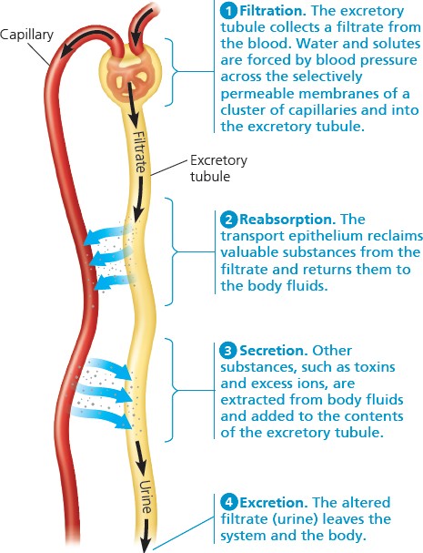
key steps of excretory system function
vertebrates and some chordates have excretory organs called kidneys
consist of tubules that are highly organized and are closely associated with the capillaries
also includes ducts that carry urine from tubules out of the kidney
vertebrate kidneys are typically nonsegmented
hagfishes however, have kidneys with segmentally arranged excretory tubules
because hagfishes and other vertebrates share a common ancestor, it is possible that the excretory structures of vertebrate ancestors were also segmented
kidneys function in both osmoregulation and excretion
Kidney Structure
- Renal Cortex and Renal Medulla
supplied by blood by a renal artery and drained by a renal vein
lie tightly packed excretory tubules with associated blood vessels
the tubules carry a filtrate produced from the blood
Survey of Excretory Systems | Protonephrdia
Protonephridia
flatworks (phylum Platyhelminthes) have excretory systems called protonephridia
also found in rotifers, some anneldis, mollusc larvae, and lancelets
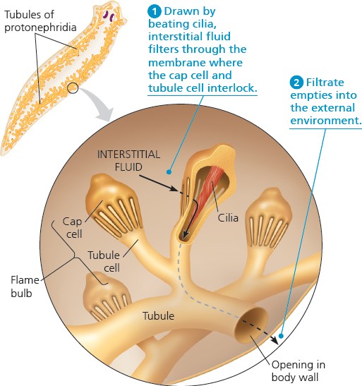
consist of a network of dead-end tubules capped by cellular units called flame bulbs that branch throughout the body
each flame bulb, consisting of a tubule cell and a cap cell, has a tuft of cilia projecting into the tubule which aid in excretion
protonephridia serves different functions in different types of flatworms
in freshwater flatworms, it serves chiefly in osmoregulation as most wastes diffuse out of the body across the body surface or are eliminated through the mouth
in parasitic flatworms, it serves chiefly for the disposal of nitrogenous wastes as the flatworm is isoosmotic to its environment
Process
nearly all of the fluid in the filtrate is reabsorbed into the blood vessels while the remaining fluid leaves the tubules as urine
- Renal Pelvis — where the urine is collected until it is allowed entry to the ureters
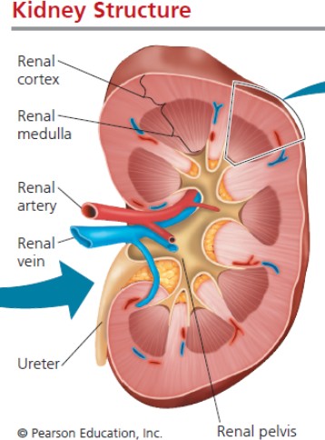
kidney structure
Nephrons
the functional units of the vertebrate kidney
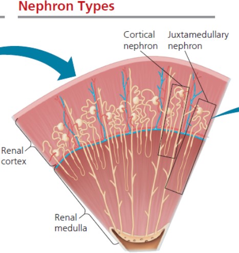
of 1 million nephrons, 85% are cortical nephrons while the rest are juxtamedullary nephrons
- Cortial Nephrons — reach only a short distance into the medulla
during filtration, the beating of the cilia draws water and solutes from the interstitial fluid via the flame bulb, releasing filtrate into the tubule network
the processed filtrate moves outward through the tubules and empties as urine via external openings
- Juxtamedullary Nephrons — extend deep into the medulla and are essential for the production of hyperosmotic urine
nephron types
Nephron Organization
Parts
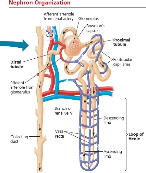
- Glomerulus — a ball of capillaries that carry blood flow
- Bowman’s Capsule — a cup- shaped swelling that surrounds the glomerulus which creates filtrate by taking water and solutes from the glomerulus capillaries
- Proximal Tubule — a tubule that connects the Bowman’s Capsule to the loop of Henle
- Loop of Henle — a hairpin turn with a descending limb and an
Survey of Excretory Systems | Metanephridia
Metanephridia
most annelids, such as earthworms have excretory systems called metanephridia
collects fluid directly from the coelom
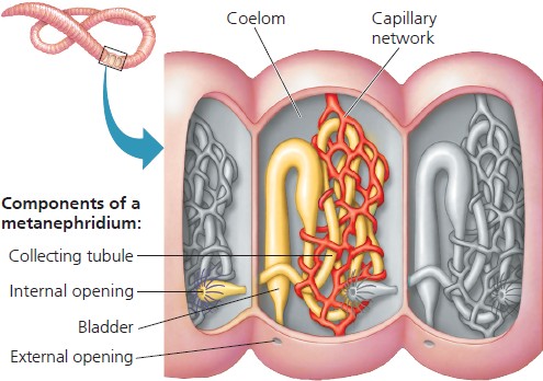
a pair of metanephridia are found in each segment of an annelid, where they are immersed in coelemic fluid and enveloped by a capillary network
the metanephridia of an earthworm serve both excretory and osmoregulatory functions
Process
a ciliated funnel surrounds the internal opening of each metanephridium which beats, causing waste to draw into a collecting tubule
the collecting tubule connects to a bladder that opens to the outside
earthworms inhabit damp soil and usually experience a net uptake of water by osmosis through their skin
ascending limb
- Distual Tubule — a tubule that connects the Loop of Henle to the collecting duct
- Collecting Duct — receives processed filtrate from the Distal Tubule
Arterioles
- Afferemt Arteriole — supplies blood to each nephron and is the arteriole that supplies blood to the capillaries in the glomerulus
- Efferent Arteriole — the arteriole that leaves the glomerulus
- Pertibular Capillaries — branches of the efferent arteriole that surrounds the proximal and distal tubules
- Vasa Recta — branches of the efferent arteriole that are hairpin-shaped capillaries that serve the Loop of Henle
their metanephridia balance this by producing hypoosmotic urine which
causes the transport epithelium to reabsorb most solutes to return them to the capillaries
nitrogenous wastes remain in the tubule and are excreted to the environment
Coelom — the fluid-filled body cavity of an animal that contains the internal organs
metanephridia of an earthworm
nephron organization
Survey of Excretory Systems | Malphigian Tubules
Malphigian Tubules
insects and other terrestrial arthropods have excretory systems called Malphigian tubules
removes nitrogenous wastes that also function in osmoregulation
extend from dead-end tips immersed in hemolymph to openings into the digestive tract
conserves water very effectively as almost all water is extracted out before urination
Process
the filtration step is absent in Malphigian tubules
instead, the transport epithelium that lines the tubules secretes nitrogenous wastes from the hemolymph into the tubule
water follows the solutes into the tubule by osmosis
as the filtrate passes from the tubules into the rectum, most solutes are pumped back into the hemolymph as water reabsorption follows
the nitrogenous waste in uric acid form are eliminated as
nearly dry matter along with the feces
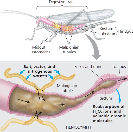
malphigian tubules of insects
The nephron is organized for stepwise processing of blood filtrate
From Blood, to Filtrate, to Urine
Process
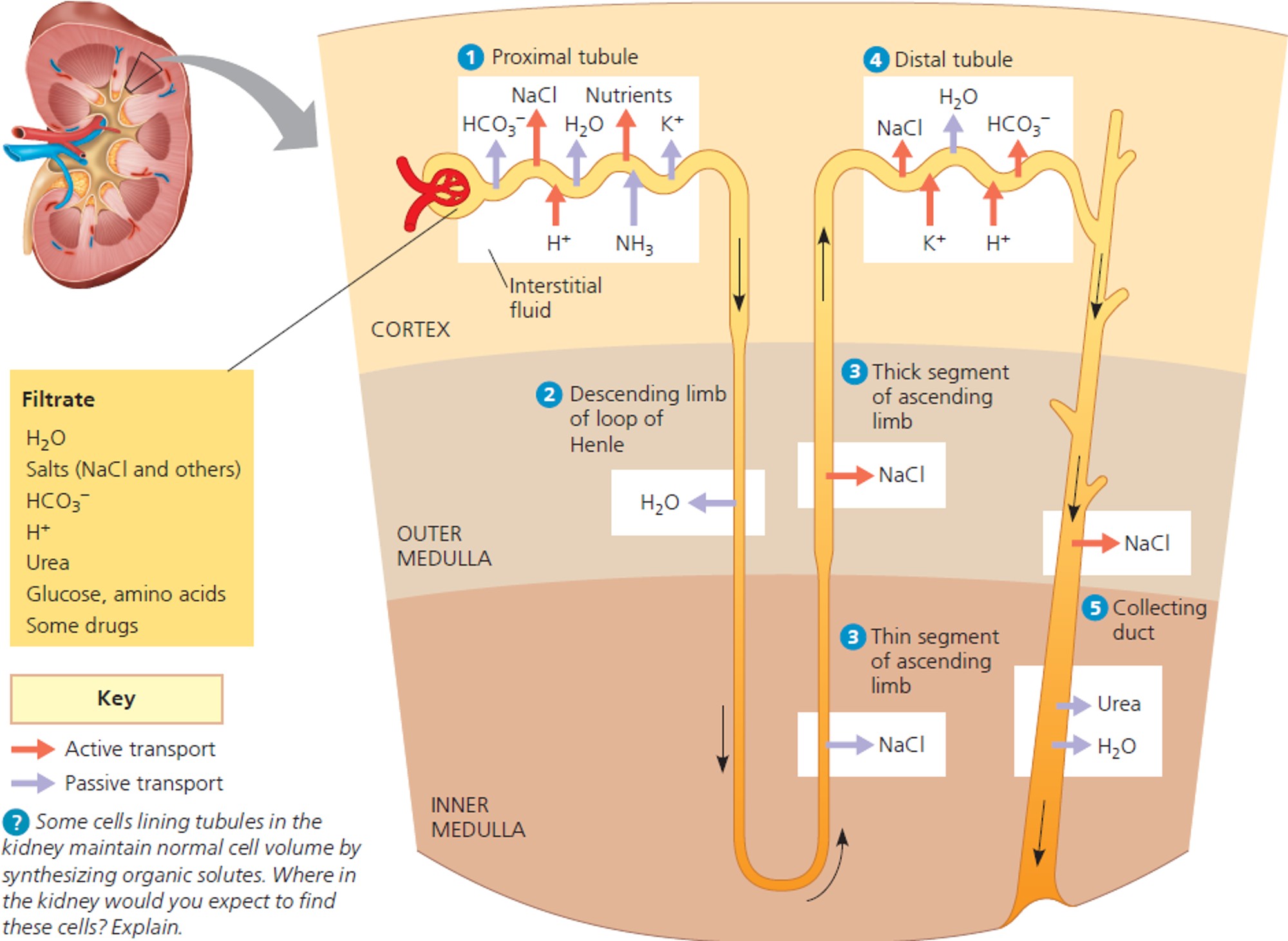
nephron function
Currently Contains | Will be Reabsorbed into the Capillaries | Will be Secreted into the Filtrate | Additional Notes | |
1. Water | ||||
2. Urea | ||||
3. Sodium | ||||
0. Glomerulus Filtrate | Chloride 3. Bicarbonate Ions | |||
5. Glucose | ||||
6. Amino Acids | ||||
7. Potassium Ions |
Tubule Filtrate |
|
|
|
|
2. Descending Limb of Henle Filtrate |
Ions (-1)
Chloride (-1)
|
|
| |
3. Ascending Limb of Henle Filtrate |
Ions (-1)
Chloride (-1)
4. Hydrogen Ions 5. Poisons |
|
| |
4. Distal Tubule Filtrate |
Ions (-1)
Chloride (-2) |
|
|
|
4. Hydrogen Ions 5. Poisons | potassium ions in filtrate regulate pH level | |||
5. Collecting Duct Filtrate |
Chloride (-3)
5, Poisons |
2. Urea |
| |
6. Final Filtrate |
Chloride (-4)
|
|
Introduction
Definition
in the human kidney, filtrate forms when fluid passes from the bloodstream of the glomerular capillaries to the lumen of Bowman’s capsule
the transport epithelium between the capillaries and lumen is permeable to water and small solutes but not to larger molecules
thus, the glomerular capillaries retain blood cells and large molecules
thus, the filtrate contains water, salts, glucose, nitrogenous wastes, vitamins, amino acids, etc.
the concentration of these substances in the initial filtrate are the same as those in blood plasma
1,600 L of blood flows through the kidneys everyday, yielding 180 L of filtrate
almost all of the water, sugars, amino acids, vitamins, and other nutrients are reabsorbed into the blood
this leaves only 1.5 L of urine to be transported into the
Adaptations of the Vertebrate Kidney to Diverse Environments
Description
vertebrates occupy various and differing habitats
comparing vertebrates from different environments reveals adaptive variations in nephron structure and function
the presence of
juxtamedullary nephrons is a key adaptation that enables
terrestrial animals to shed
salts and nitrogenous wastes without squandering water
differences in species among the length of the loop of Henle and in the numbers of juxtamedullary and cortical nephrons help fine-tune osmoregulation to particular habitats
Mammals
excrete the most hyperosmotic urine
long loops of Henle that extend deep into the medulla, maintaining steep osmotic gradients in the kidney resulting in very concentrated urine
Australian hopping mice, North American kangaroo
bladder rats, and other dessert
mammals
cortical nephrons make up most of the nephrons, resulting in a much lower ability to concentrate urine
beavers, muskrats, and other aquatic mammals
loops of Henle with intermediate length and the capacity to produce urine intermediate in concentration
terrestrial mammals living in moist conditions
Vampire Bat
the South American vampire bat illustrates the versatility of the mammalian kidney
it feeds on the blood of large birds and mammals during night
as the bat feeds, its kidneys extract large volumes of dilute urine, up to 24% of its body mass per hour, offsetting its increase in fluid amount
in the roost, the protein from the blood digests and produces nitrogenous waste, which causes their kidneys to shift to produce small quantities of highly concentrated urine
the vampire bat’s ability to alternate rapidly between large amounts of dilute urine and small amounts of very hyperosmotic
Solute Gradients and Water Conservation
Description
the ability of the kidney to conserve water is a key adaptation for terrestrial habitats
the production of hyperosmotic urine is only possible because considerable energy is expended for the active transport of solutes against concentration gradients
the primary solutes affecting osmolarity to exctract water out of the filtrate
NaCl which is concentrated by the loop of Henle
urea which is concentrated by the collecting duct
in humans, the osmolarity of blood is about 300 mOsm/L but the
kidney can excrete urine about up to 1200 mOsm/L
Australian hopping mice can produce urine with an
osmolarity of 9300 mOsm/L
Process of Concentrating Urine
urine is an essentail part of its adaptation
Birds and other Reptiles
most birds live in environments that are dehydrating
birds have kidneys with juxtamedullary nephrons, but their loops of Henle extend less far into the medulla than that of mammals
thus, bird kidneys cannot concentrate urine to high osmolarities achieved by mammalian kidneys
birds’ main water conservation adaptation is having uric acid as the nitrogenous waste molecule
reptiles only have cortical nephrons and they produce isoosmotic or hypoosmotic to body fluids
the cloaca of reptiles from which urine and feces leave the body conserves fluid by reabsorbing water from these wastes
reptiles’ main water conservation adaptation is exreting nitrogenous waste as uric acid
Freshwater Fishes and Amphibians
hyperosmotic to their surroundings, freshwater fishes
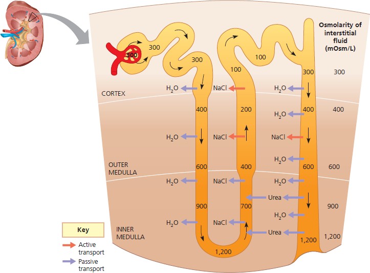
how the human kidney concentrates urine
produce large volumes of very dilute urine
their kidneys are packed with cortical nephrons produce filtrate at a high rate
salt conservation relies on the reabsorption of ions from the filtrate in the distal tubules
amphibian kidneys function differently according to their surroundings
when in freshwater, the kidneys excrete dilute urine while their skin accumulates salts from the water by active transport
when on land, they conserve body fluid by reabsorbing water across the epithelium of the urinary bladder
Marine Bony Fishes
compared with freshwater fishes, marine fishes have fewer and smaller nephrons
their nephrons also lack distal tubules and some lack or have small glomeruli
thus, filtration rates are low and little urine is excreted
the main function of kidneys in is to get rid of divalent ions (those with a charge of 2+ or 2-) such as
calcium (Ca2+), magnesium (
Mg2+), and sulfate (SO2−)
4
take in divalent ions by incessantly drinking seawater
they rid of ions by secreting them into proximal tubules of the nephrons and excreting them in urine
osmoregulation in marine bony fishes also rely on specialized chloride cells in gills
by establishing ion gradients that enable secretion of salt into seawater, the chloride cells maintain proper levels of monovalent ions in the fish
the generation of ion gradients and the movement of ions across membranes are central to salt and water balance in marine bony fishes
Hormonal circuits link kidney function, water balance, and blood pressure
Introduction
Definition
in mammals, both volume and osmolarity of urine are adjusted according to an animal’s water and salt balance and rate of urea production
urine volume and osmolarity are regulated by the Antidiuretic Hormone and the Renin- Angiotensin-Aldosterone System
high salt intake and low water intake
can excrete urea and salt in small volumes of hyperosmotic urine with minimal water loss
low salt intake and high water intake
the kidney an eliminate the excess water with little salt loss by producing large volumes of hypoosmotic urine
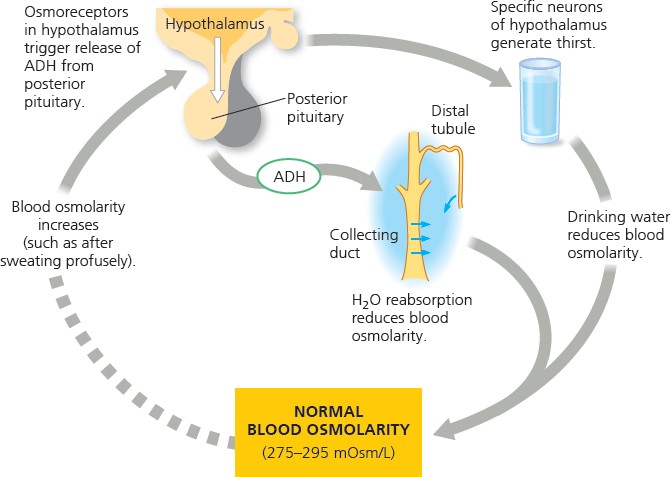
Thirst
plays an essential role in water and salt control balance
neurons in the hypothalamus are dedicated to regulating thirst as stimulating one set of neurons in mice causes intense drinking behavior
Antidiuretic Hormone
Definition
also called vasopressin or the anti- diuretic hormone
ADH molecules released from the pituitary activate membrane receptors on the surface of collecting duct cells
the activation initiates a signal that directs insertion of aquaporin proteins into the membrane lining the collecting duct cells
more aquaporin channels result in more water recapture, reducing urine volume and increasing urine concentration
blood osmolarity, ADH release, and water reabsorption are linked in a feedback circuit so anything that disrupts this circuit can interfere with water balance
alcohol inhibits ADH release, leading to excessive urinary water loss and dehydration
contrary to common belief there is no connection between caffeinated drinks and increased urine production
Diuresis — a high level of urine production by volume
stimulating a second set causes an immediate halt in water consumption
follow-up studies are focused on identifying the cellular pathways linking these neurons to behavioral responses
Rise and Fall of Blood Osmolarity
when blood osmolarity rises, osmoreceptor cells in the hypothalamus trigger increased release of ADH from the posterior pituitary
the resulting increase in water reabsorption in the collecting duct concentrates urine, lowering blood osmolarity
when blood osmolarity falls, a negative-feedback mechanism reduces the osmoreceptor cell activity in the hypothalamus
the resulting decrease in water reabsorption in the collecting duct dilutes urine, increasing blood osmolarity
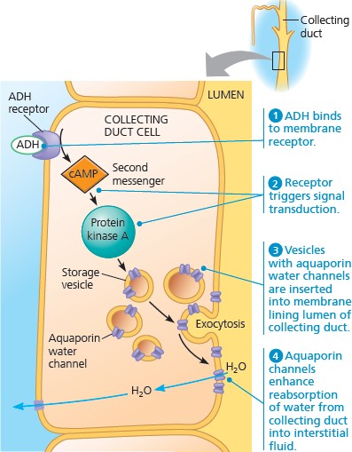
control of collecting duct permeability by ADH
The Renin-Angiotensin-Aldosterone System
Definition
release of ADH is a response to an increase in blood osmolarity, as when the body is dehydrated from excessive water loss or inadequate water intake
however, an excessive loss of salt and body fluids will reduce blood volume without increasing the osmolarity
ADH will not release as the receptors do not sense change in osmolarity
an endocrine circuit called the Renin-Angiotensin- Aldosterone System or RAAS responds to the drop in blood volume by increasing water and Na+ reabsorption
beacuse angiotensin II results in increased blood pressure, drugs that block angiotensin II production are used to treat hypertension or chronic high blood pressure
these drugs inhibit angiotensin converting enzyme to react to angiotensin
the RAAS operates as a feedback circuit, meaning that drops in blood pressure or volume will
regulation of fluid retention in kidney
 Aquaporin Experiment
Diabetes Insipidus — a disease that causes severe dehydration and solute imbalance
a mutation that prevents ADH production or that inactivates the receptor gene, disrupting homeostasis by blocking the insertion of additional aquaporin channels
researchers studied a patient with diabetes insipidus who had a normal ADH receptor gene but two mutant alleles
each mutation changed the protein sequence at a highly conserved position so to test the hypothesis that the changes affect function, reasearchers used frog oocytes
frog oocytes are cells that will express foreign messenger RNA
the results show that patient’s allele A and B oocyte do not take in water, meaning that the
eventually cause higher levels of blood pressure or volume, stopping the RAAS from operating
Process
- The juxtaglormerular apparatus or JGA will release the enzyme renin when blood pressure or volume drops
- Renin initiates cleaving of angiotensinogen, yielding angiotensin II
- Angiotensin II triggers vasoconstriction, increasing blood pressure and decreasing blood flow to capillaries in non-essential organs
- will also stimulate the adrenal glands to release aldosterone, causing the distal tubules and collecting ducts to reabsorb more Na+and water,
increasing blood volume and pressure
Juxtaglormerular Apparatus — specialized tissue consisting of cells around the afferent arteriole
Atrial Natriuretic Peptide (ANP)
a hormone that opposes the RAAS
the walls of the atria of the heart release ANP in response to an increase in blood volume and pressure
aquaporins do not activate when mutated
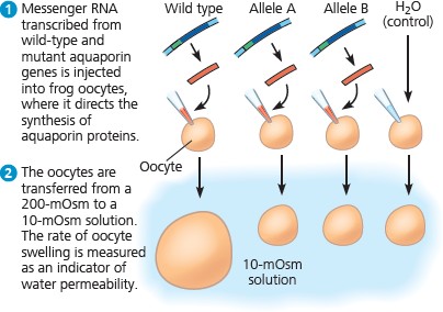
aquaporin experiment results
it inhibits the release of renin from the JGA and inhibits NaCl reabsorption by the collecting ducts, reducing also aldosterone release
Part II | Nervous System
Neuron structure and organization reflect function in information transfer
Neuron Structure and Function
Neuron
a nerve cell
the fundamental unit of the nervous system
has a structure that allow it to conduct signals by taking advantage of the electrical charge across its plasma membrane
Parts of a Neuron
- Cell Body — contains most of the neuron’s organelles
- Dendrites — one of usually numerous, short, highly branched extensions of a neuron that receive signals
- Axon — a long extension that transmits signals to other cells
- Axon Hillock — the cone- shaped base of an axon where signals that travel down the axon are generated
- Synapse — one of many branches of the axon that are junctions where a neuron communicates with another cell
- Synaptic Terminal — the part of each axon that transmits information
Information Processing
Description
occurs in three stages (using the cone snail as an example)
- Sensory Input — surveys the environment with sensors, sampling scents that might reveal fish
- Integration — networks of neurons in the snail brain process information to determine if a fish is present
- Motor Output — output from the brain initiates attack, activating neurons that trigger the harpoon-like tooth towards its prey
specialized populations of neurons handle each stage of information processing
- Sensory Neurons — transmit information about external and internal stimuli such as light, touch, blood pressure, etc.
- Interneurons — from local circuits connecting neurons in the brain or ganglia and are responsible for integration or analysis of sensory input
- Motor Neurons — transmits signals to muscle cells or
- Neurotransmitters — a molecule released by the synaptic terminal that passes information from the transmitting neuron to the receiving cell
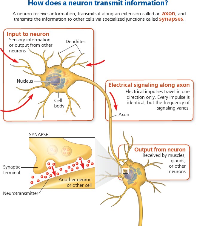
neuron transmission
glands, causing contraction and gland activity respectively
Other Info
all neurons transmit signals within the cell in an identical manner thus a neuron that detects an odor transmits signals the same as a neuron that moves a muscle
the particular connections made by the active neuron are what distinguish the type of information being transmitted
the shape of a neuron can vary from simple to complex with neurons of higher functions having more dendrites or axons
Nerves — bundles of the axons in neurons
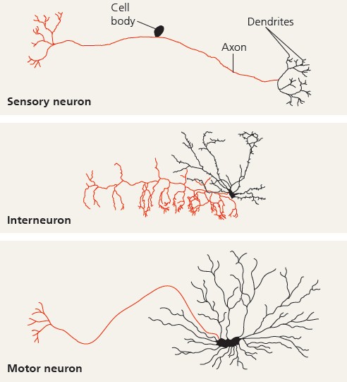
structurla diversity of neurons
Nervous System
Description
- Central Nervous System (CNS) — carries out sorting, processing, and integration which includes the brain or ganglia
- Peripheral Nervous System (PNS)
— carries information into and out of the CNS to other parts of the body
Glia — a supporting cell for neurons in the CNS and PNS that hold the neurons together
Ion pumps and ion channels establish the resting potential of a neuron
Introduction
Description
in neurons, ions are unequally distributed between the interior of the cells and the surrounding fluid
the inside of a neuron is negatively charged relative to its outside
this charge difference or voltage across the plasma membrane is called the membrane potential
when a neuron receives a stimulus, the membrane potential changes Action Potentials — rapid shifts in the potential of a neuron
Membrane Potential — the relatively negative charged voltage that crosses the plasma
membrane of a neuron
Resting Potential — the low voltage membrane potential characteristic of a resting neuron that is not sending a signal
Formation of the Resting Potential
Description
potassium ions (K+) and sodium ions (Na+) play essential roles in the formation of resting potential
these ions each have a concentration gradient across the plasma membrane of a neuron
in most neurons, K+ concentration is higher inside the cell while Na+
concentration is higher outside
the gradients are maintained by the sodium-potassium pump, which uses ATP
the concentration gradients of potassium and sodium ions represent a form of potential energy that can be harnessed for cellular processes
Selective Permeability — the characteristic of a membrane to only allow certain ions or substances to pass
Sodium-Potassium Pump — a transport protein that actively transports three sodium ions out of and two potassium ions into the cell
Ion Channels — pores formed by clusters of proteins that span the membrane that allow ions to diffuse back and forth across the membrane
Forming the Resting Potential
although the pumping generates a positive charge, it pumps quite slowly so the resulting change in the membrane potential is quite small
the membrane potential change comes mainly from ion channels, which supplies an electrical charge through the diffusion of ions in the membrane
the rapid movement of ions through ion channels also creates a current of positive or negative charge, generating a membrane potential
diffusion of potassium ions through potassium channels that are always open is critical for establishing resting potential
the potassium ion concentration is 140 mM inside the cell but only 5 mM outside, favoring a net outflow of potassium ions
a resting neuron has many open potassium channels but very few sodium channels
as more potassium ions leave the cell, while only few sodium ions come to replace them, this leaves a negative charge inside the cell
the buildup of negative charge within the neuron is the major source of membrane potential
the buildup of negative charge eventually reaches a critical point wherein the excess negative charges exert an attractive force that opposes the flow of additional positively charged potassium ions out of the cell
when too many of the potassium ions leave the cell, it makes some of the potassium ions don’t want to leave
the separation of charge thus results in an electrical gradient that counterbalances the chemical concentration gradient of K+
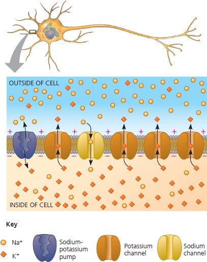
the basis of membrane potential
Modeling the Resting Potential
Gradients
- Chemical Gradient — the ion’s tendency to move to a less concentrated area
- Electrical Gradient — the ion’s tendency to move to an area electrically charged opposite to itself
Modeling
- Imagine a membrane contains many open potassium ion channels that allow for diffusion
- To mimic a mammalian neuron, we place 140 mM potassium chloride (KCl) in the inner chamber and 5 mM potassium chloride (KCl) in the outside chamber
- The potassium ions (K+) will diffuse down its concentration gradient to the outside chamber but because the chloride ions (Cl−) lack a means of crossing the membrane, there will be an excess of negative charge in the inner chamber
- When the model reaches equilibrium, the electrical gradient will balance the chemical gradient so that no further net diffusion of K+ will occur
- Equilibrium Potential — the magnitude of a cell’s membrane voltage at equilibrium; calculated using Nernst Equation
- for body temperature at 37°C and an ion with a net charge of 1+, Nernst Equation is
E = 62mV (log ionoutside )
ion
ioninside
- plugging the K+ concentrations into the equation reveals that the equilibrium potential for potassium ions is -90 mV
- this means that that potassium ions are at equilibrium when the inside of the membrane is 90 mV more negative than the outside
- while the equilibrium for potassium ions is -90 mV, the actual resting potential is less negative due to the small but steady movement of positively charged sodium ions inside the cell
- If we model a membrane in which the only open channels are permeable to sodium ions (Na+), we will find an equilibrium potential of +62 mV
- the actual resting potential (-60 to -80 mV) is much closer to the equilibrium potential of potassium (-90 mV) due to there being more open potassium channels than open sodium channels
- Neither potassium nor sodium is at equilibrium in a resting neuron so there is a constant net flow of each ion across the membrane
- however, the resting potential remains steady which means that the potassium and sodium ions have currents that are equal and opposite
- ion concentrations on either side also become steady because the resting potential arises from the net movement of far fewer ions than would be required to alter the concentration gradients
- If sodium ions are allowed to cross the membrane more readily, the membrane potential will move more towards +62 mV and away from the resting potential
- this happens in the generation of a nerve impulse
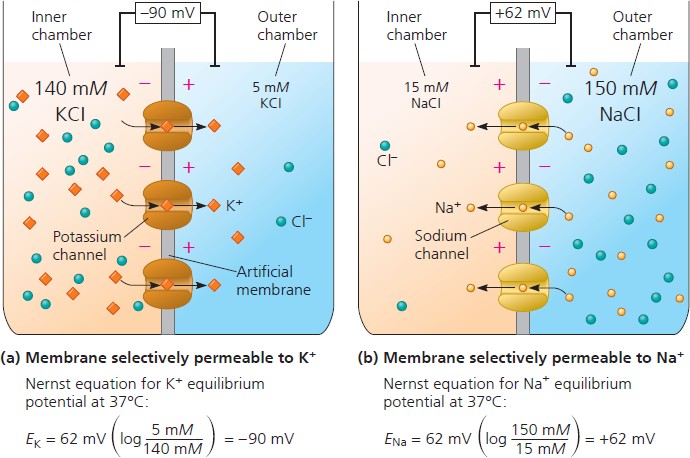
modeling a mammalian neuron
Summary of Findings
Summary
- Potassium (K+) and Sodium (Na+) in Neurons: Neurons have a concentration gradient of K+ and Na+ ions across their membrane. The concentration of K+ is higher inside the neuron, while the concentration of Na+ is higher outside. This gradient is maintained by ion channels in the neuron’s membrane.
- Resting Membrane Potential: When a neuron is at rest, it has a negative electrical charge, or voltage, across its membrane. This is due to the movement of K+ ions out of the neuron, which makes the inside of the neuron negatively charged. The resting membrane potential is typically between -60 and -80 millivolts (mV).
- Action Potential: An action potential is a rapid change in the electrical charge across the neuron’s membrane. It’s triggered when the neuron receives a signal. During an action potential, Na+ channels open, allowing Na+ ions to flow into the neuron. This makes the inside of the neuron positively charged, or depolarized.
- Equilibrium Potential: The equilibrium potential is the voltage that would be required to prevent an ion from diffusing across the neuron’s membrane. It can be calculated using the Nernst equation. For K+, the equilibrium potential is typically around -90 mV, and for Na+, it’s around +62 mV.
- Net Flow of Ions: In a resting neuron, there’s a constant movement of K+ and Na+ ions across the membrane. However, these movements balance each other out, so the overall electrical charge inside the neuron remains steady. If more Na+ ions are allowed to cross the membrane, the electrical charge inside the neuron will change, triggering a nerve impulse.
Action potentials are the signals conducted by axons
Research Method
Intracellular Recording in Measuring Membrane Potential
microelectrodes are inserted into neurons to measure voltage these are put outside and inside the cell
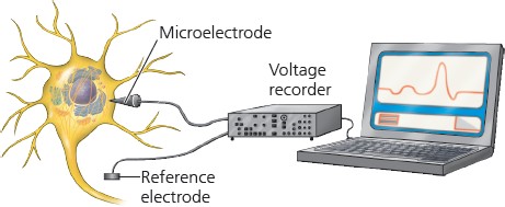
microelectrodes in a neuron cell
Introduction
Description
the membrane potential changes when it responds to a stimulus gated ion channels open or close in response to stimuli
when a gated ion channel opens or closes, it alters the membrane’s permeability to particular ions
results in a flow of ions across the membrane, altering membrane potential particular types of gated channels respond to different stimuli l
Voltage-Gated Ion Channels — open or close in response to a shift in voltage across the plasma membrane
Hyperpolarization and Depolarization
Hyperpolarization
a change in the cell membrane potential such that the inside of the cell becomes more negative relative to the outside
if gated potassium channels in a resting neuron open, the membrane’s permeability to potassium ions (K+) increases
as a result, net diffusion of potassium ions (K+) out of the neuron increases, shifting the membrane potential to -90 mV
Depolarization
a change in the cell membrane potential such that the inside of the cell becomes less negative relative to the outside
if gated sodium channels open, the membrane’s permeability to sodium ions increases (Na+)
as a result, net diffusion of sodium ions (Na+) into the neuron increases, shifting the membrane potential to +62 mV
Graded Potentials and Action Potentials
Graded Potential
an electrical response of a cell to a stimulus consisting of a change in voltage across the membrane proportional to the stimulus strength
sometimes, the response to hyperpolarization or depolarization is simply a shift in membrane potential
a larger stimulus = a greater change in membrane potential
generates small electrical currents that dissipate as it flows along the membrane thus, they decay with time and with distance from their source
Action Potential
an electrical signal that travels along the membrane of a neuron as a nongraded depolarization
this is a depolarization shift that has hit the membrane sufficiently, causing a massive change in membrane voltage
these have a constant magnitude and can regenerate in adjacent regions of the membrane
thus, these can spread along axons, making them well suited for transmitting signals over long distances
arise because some of the ion channels in neurons are voltage-gated
if a depolarization increases the membrane potential to a level called threshold, the voltage-gated sodium channels open
the resulting flow of sodium ions (Na+) result in further depolarization
the increase in depolarization will then cause more sodium channels to open, leading to a greater flow of current
the final result is a process of positive feedback that triggers rapid openings of many voltage-gated sodium channels
the positive feedback loop triggers an action potential when the threshold is reached, which is about -55 mV for mammals
it has a magnitude that is independent of the strength of the triggering stimulus because action potentials either occur fully or not at all (all or none response)
Threshold — the potential that a neuron cell membrane must reach for an action potential to initiate
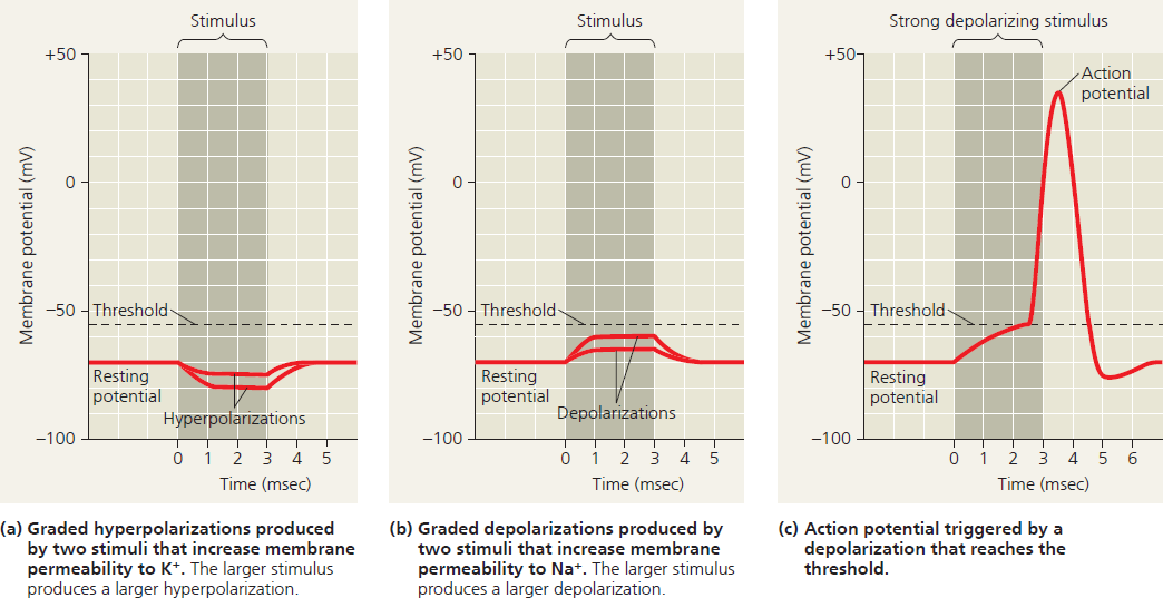
graded potentials and an action potential in a neuron
Generation of Action Potentials
Definition
rhe shape of an action potential graph reflects ion movement through voltage-gated sodium and potassium channels
depolarization opens both channels but they respond independently and sequentially sodium channels open first, initiating the action potential
as it proceeds, sodium channels remain open but become inactivated as proteins called inactivation loops block ion flow through the channel
sodium channels remain inactivated until the membrane returns to the resting potential
potassium channels open second and more slowly
potassium channels remain open and functional until the end of the action potential
the sodium channels remain inactivated during the fallingphase, and early undershoot
as a result, if a second depolarizing stimulus occurs during this period, it will be unable to trigger an action potential known as the refractory period
the refractory period is only due to the inactivation of sodium channels, not to a change in the ion concentration gradients
Refractory Period — a period during which a cell is unresponsive to further stimulation
Process
- When at resting potential, most voltage-gated sodium and potassium channels are closed
- When a stimulus sufficient for action potential is received, sodium channels open, allowing sodium ions to diffuse into the cell, increasing depolarization, opening more sodium channels that will lead to further depolarization
- Once the threshold is crossed and is approaching the equilibrium potential of sodium (this is called the rising phase), sodium channels inactivate and most potassium channels open, quickly bringing the membrane potential back toward resting potential (this is called the falling phase)
- Once it starts approaching the equilibrium potential of potassium as the membrane keeps releasing potassium ions (K+), the potassium channels close and it returns to the resting potential
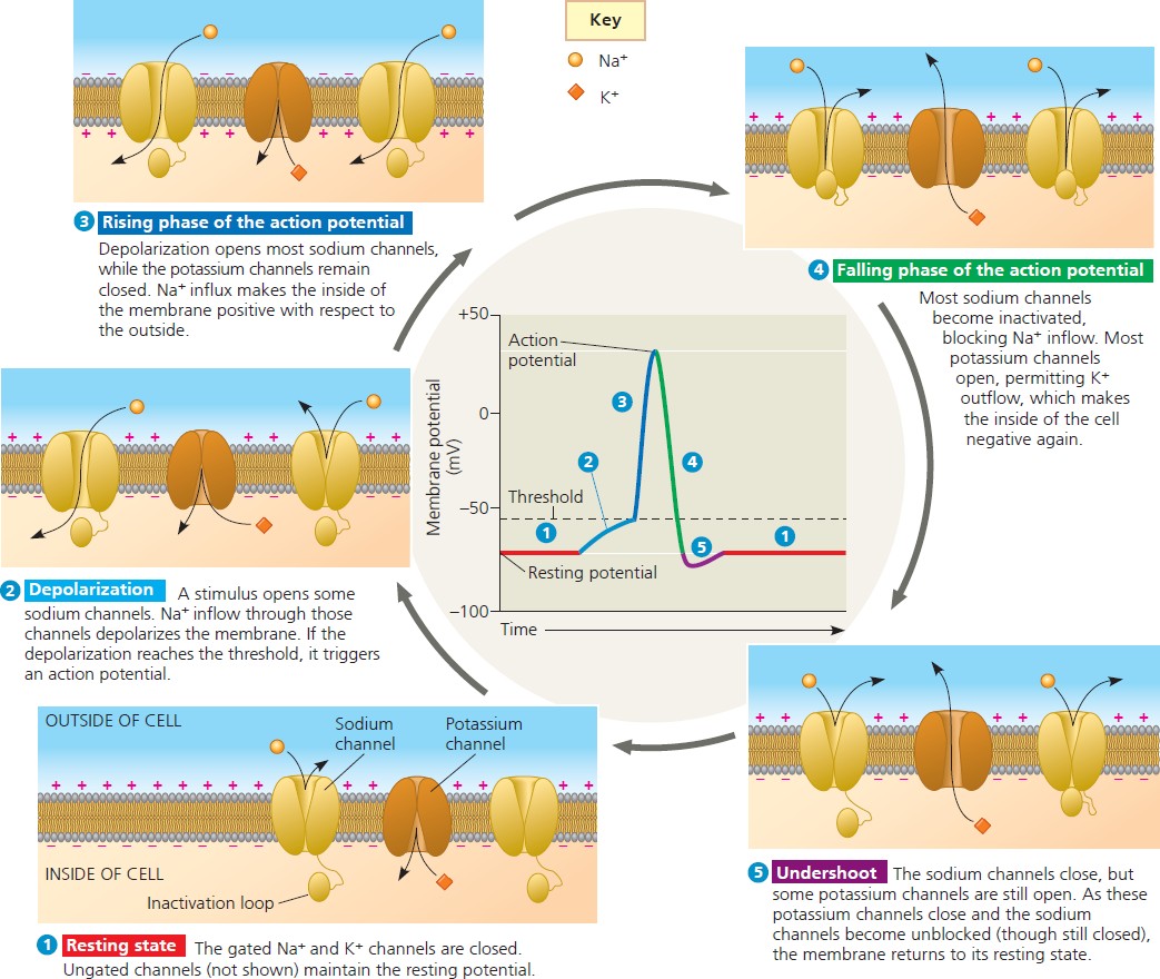
the role of voltage-gated ion channels in the generation of an action potential
Conduction of Action Potentials
Process
- At the axon hillock (where an action potential is usually initiated), sodium ion inflow depolarizes the neighboring region of the axon membrane
- If the depolarization is large enough to reach threshold, it causes an action potential in the neighboring region and this process is repeated along the length of the axon
since an action potential is an all-or-none event, the magnitude and duration of the action potential are the same at each position of the axon
- The net result is the movement of a nerve impulse from the cell body to the synaptic terminals
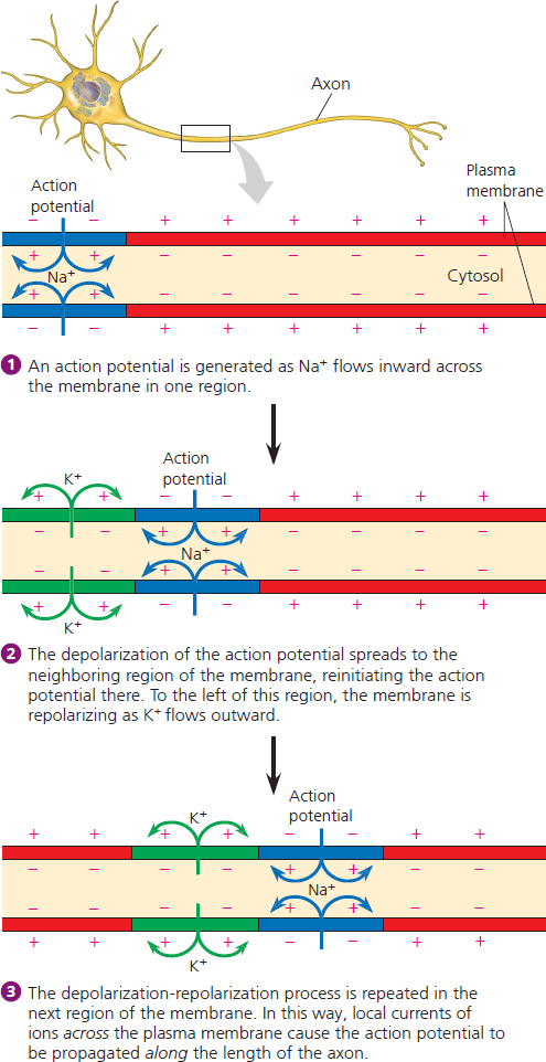
More Info
immediately behind the zone of depolarization, the sodium channels become inactivated, making the membrane refractory to further input
this disallows the action potential to return to the cell body since it cannot pass through inactivated sodium channels
consequently, the inward current that depolarizes the axon membrane ahead of the action potential cannot produce another action potential behind it
after the refractory period, depolarization of the axon hillock to threshold will trigger a new action potential
in many neurons, action potentials last less than two miliseconds
the rate at which action potentials are produced in a particular neuron is proportional to input signal strength
louder sounds in the ear result in more frequent action potentials between the ear and brain
Mutations and Diseases
mutations in genes that encode ion channel proteins can cause disorders affecting the nerves or brain
Myotonia — a mutation affecting voltage-gated sodium channels, causing periodic spasming of muscles
Epilepsy — a mutation affecting sodium channels in the brain in which groups of nerve cells fire simultaneously and excessively, producing seizures
Evolutionary Adaptations of Axon Structure
Description
the rate at which the axons within nerves conduct action potentials governs how rapidly an animal can react to danger or opporunity
natural selection often results in anatomical adaptations that increase conduction speed
one such adaptation is a wider axon, serving to lessen the movement resistance of action potentials
in invertebrates, conduction speed varies from several hundreds of m/sec in narrow axons to approximately 30 m/sec in the giant axons of arthopods and molluscs
in vertebrates, axons have narrow diameters but can still conduct action potentials at high speed
this is possibly only due to electrical insulation which causes the depolarizing current associated with an action potential to travel farther along the axon interior
this electrical insulation of lipids that surround vertebrate axons is called a myelin sheath
glial cells produce myelin sheaths and different types of glia produce in both the CNS and PNS
in myelinated axons, sodium channels are restricted to gaps in the myelin sheath called nodes of Ranvier
furthermore, interstitial fluid is only exposed to the axon at its gaps, thus, action potentials cannot be generated in the regions between nodes
Myelin Sheath — layers of lipids that surround vertebrate axons that serve as electrical insulation
Nodes of Ranvier — gaps in the myelin sheath where the axon comes in contact with Oligodendrocytes — glial cells that produce myelin sheaths in the CNS
Schwann Cells — glial cells that produce myelin sheaths in the PNS
/head