Term 1 revision
Bones and Joints: Things not to forget to revise
Long bone-one feature distinguish between superior and inferior, one feature to which way its pointing, one feature that tells if it’s front or back
vertebrae, ribs
Ligaments
Joints- type of joint is classification e.g. synovial hinge
name of joint is identification e.g. elbow joint, knee joint, elbow joint
primary cartilaginous will ossify, purely hyaline cartilage in primary, secondary cartilaginous will not ossify, fibrocartilaginous usually present in secondary
Histology
Development
Back Muscles
Mainly study intrinsic muscles of the back
-Splenius Muscles
-splenius capitis muscle
-splenius cervicis muscle
-Erector Spinae Muscles
-illiocostalis muscle
-longissmus muscle
-spinalis muscle
motor innervation from dorsal rami,
whole back innervated by 31 pairs of dorsal rami
how it works in vertebral column
how it helps
Nervous System
basic classification of nervous system
water is component
autonomic nervous system components, where they are located
somatic nervous system components, location
which is more complex
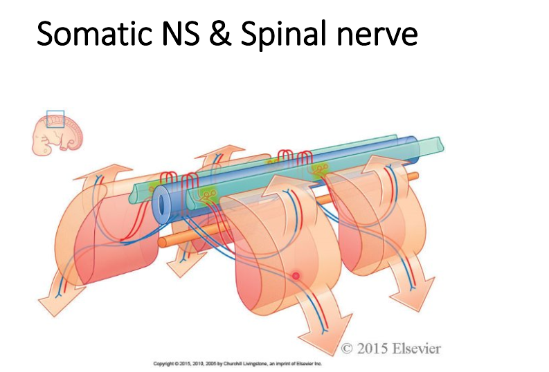
somites, mesenchymal origin
somites gives rise to myotome, dermatome, skelotome
Spinal segment and segmental innervation
Part of the spinal cord that gives rise to a pair of spine nerves on each side
Myotome
*Segmental innervation of muscles.
. Head and Neck: C1-C4 (Cervical Plexus) + Cranial nerves
. Upper Limb: C5-T1 (Brachial plexus)
. Intercostal muscles: T1-T12
. Abdomen: mainly T7-L1
. Lower Limb (+ pelvis): L1-L4 (Lumbar plexus), L4-S4/S5 (Sacral Plexus)
. Back intrinsic muscles: dorsal rami of spinal nerves.
. Anterolateral trunk + upper limbs + lower limbs muscles: ventral rami of spinal nerves
Sympathetic Nervous System:
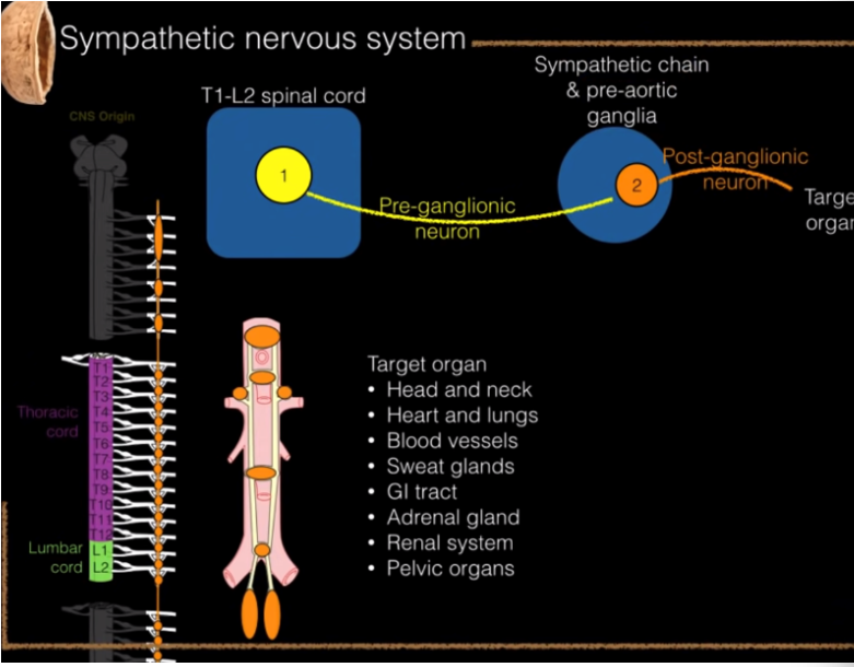
Where is location of preganglionic, sympathetic neuron, (motor)
ganglia are eggregors or cell bodies?
Dermatomes: L1, T3, T2, C5-T1 (upper limb) C4, T4-nipple, T5, T10-Belly button
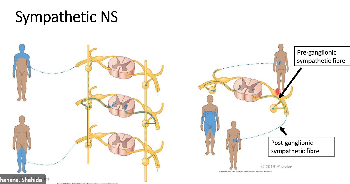
anterior horn of grey matter has cell bodies
lateral horn has pre ganglionic fibres which pass through white communicans
grey rami communicans are non myelinated
pre ganglionic are myelinated
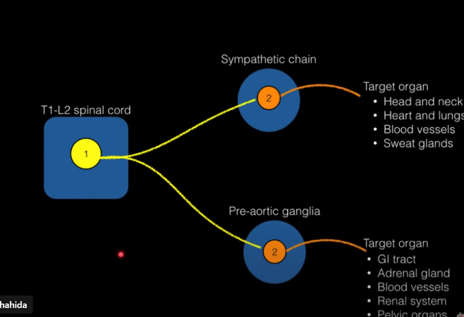
where they synapse onto and target organs
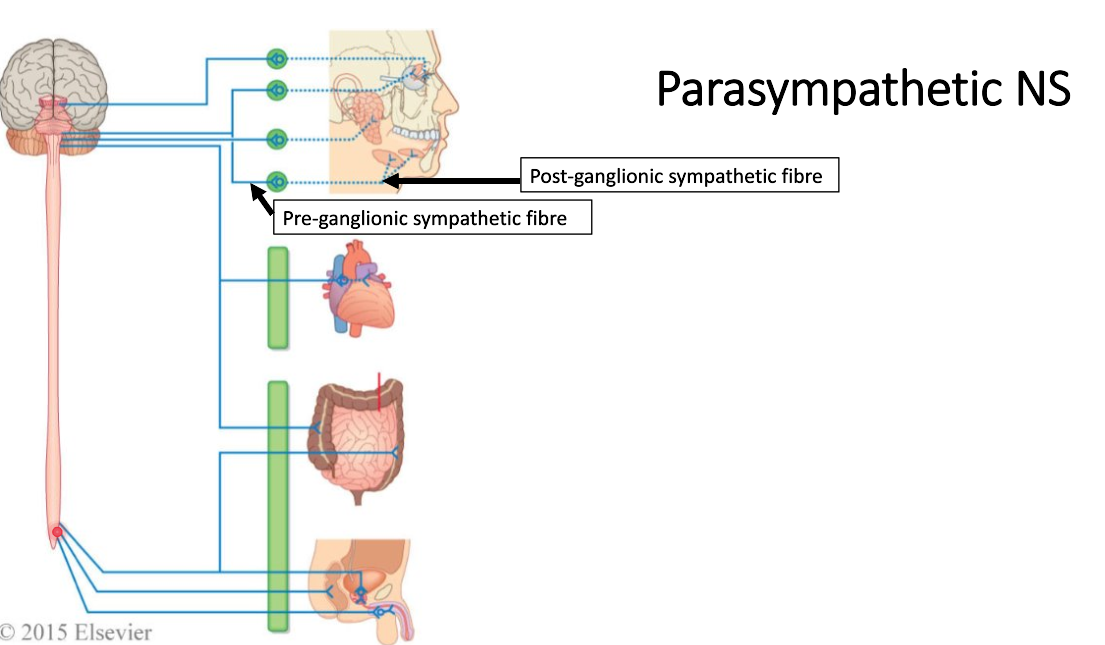
S2,3,4 segment-pelvic, splacnic nerve-sympathetic
Spinal cord and spinal meninges
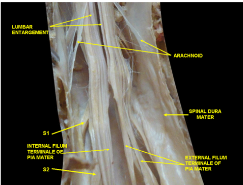
pia mater always covering spinal cord
dura mater quite fibrous
space outside dura is epidural space-fat and internal vertebral venous plexuses there
subdural space-between dura and arachnoid, underneath dura, outside pia mater which is surrounding spinal chord
subarachnoid space-lumbar puncture, epidural, spinal anaesthesia, to see haemorrhage, cancer, infection, meningitis
Dural sac ends S2
continues down to tip of coccyx-?
Lymphatic System
right side-right lympahtic duct, right jugular, right subclavian junction
thoracic duct formed in abdominal area, cisterna chyli, ends in left venous angle, left jugular and left subclavian junction
watershed line of lymphatic drainage of enteroabdominal wall divides drainage-above line is axillary nodes
below is superficial inguinal node
fluid from blood vessel comes out- interstitial fluid
majority of fluid returns to venous end
tiny stays in the space, lymphatic vessels clean up the little fluid that doesn’t return
-small vessels join up to form afferent vessels that go to lymph nodes
study histology too