Estrogen Receptor Agonists, Antagonists, and Endocrine Resistance
Estrogen Receptor Agonist and Antagonist Mechanisms
Estrogen Receptor (ER) Agonist vs. Antagonist:
The difference between agonist and antagonist action is most thoroughly studied for the estrogen receptor.
Agonist (Estradiol) Binding:
Estradiol binds to the Ligand Binding Domain (LBD).
It positions helix 12 (H12) in an active conformation.
This active H12 position is stabilized by Asp 351.
Asp 351 is located in the middle of the long H3 helix, opposite the N-terminal end of H12.
Three NH groups protruding from the helix end of H12 are about away from the carboxylate groups of the acidic residue Asp 351, stabilizing its negative charges.
Antagonist Binding:
Antagonists possess side chains that remain in the entrance channel after ligand binding.
These side chains prevent the proper closure of H12.
A basic group at the end of the antagonist's side chain is likely in a positively charged state.
This basic group forms a salt bridge with Asp 351, disrupting the H12 stabilization achieved by agonists.
Structural Basis of ER LBD Helix 12 Switch:
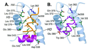
A study demonstrated the crucial role of H12 movement.
With agonist binding, H12 moves to open a groove, allowing for the binding of LxxLL peptide motifs found in coactivators.
With antagonist binding, H12 physically moves into the position where the LxxLL peptide would typically bind, thereby blocking coactivator interaction.
Estrogen Receptor Association with Coactivator Complex
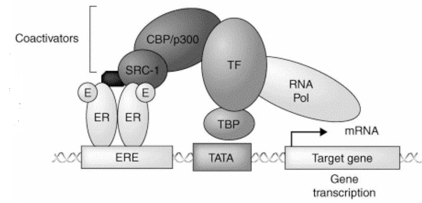
Primary Coactivators (SRCs):
Steroid Receptor Coactivators (SRCs) are primary coactivators interacting with Nuclear Receptors (NRs), including Estrogen Receptors (ERs) and Androgen Receptors (ARs).
Secondary Coactivators:
SRCs, in turn, recruit multiple secondary coactivators, such as the histone acetyltransferases (HATs) p300/CBP.
Transcriptional Activation:
These components form a minimal receptor-coactivator complex.
This complex can promote chromatin remodeling and facilitate transcriptional activation of target genes.
Binding of ER with Coactivators:
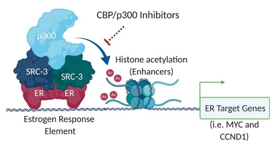
Electron cryo-microscopy (cryo-EM) was used to study the quaternary structure of the NR and coactivator complex.
The complex (~ kDa) included ER, SRC-3, p300, and CARM1, bound to a bp Estrogen Response Element (ERE).
This complex exhibited transcriptional activity.
The resolution of the cryo-EM map (~25\mathring{A}}) was lower than typical crystal structures of individual domains like the LBD.
Key Findings:
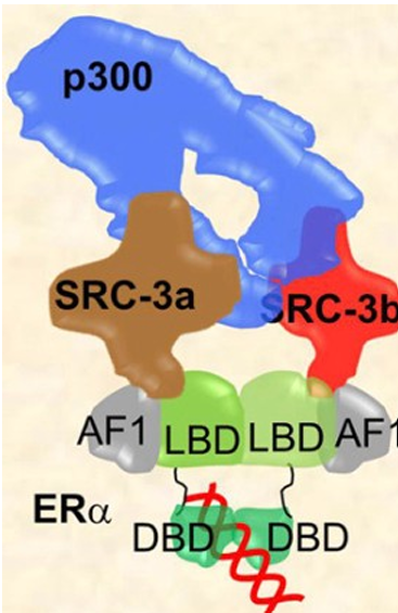
Multiple interactions were found between SRC-3 and p300.
Four distinct regions of p300 interact with SRC-3.
This interaction induces a conformational change in p300, which increases its histone H3 acetylation activity.
The N-terminal domain (NTD), also known as Activation Function 1 (AF1), is located near the LBD (AF-2).
AF1 participates in SRC-3 recruitment alongside AF-2.
Intrinsically Disordered Regions:
Both SRC and p300/CBP appear to contain intrinsically disordered regions.
These regions can convert to consistent structures upon interacting with other proteins, highlighting their dynamic nature in complex formation.
ER Functions Out of the Ordinary
Action Independent of ERE (Indirect Genomic/Non-classical Pathway):
ER can act as a co-regulator for other transcription factors, such as AP-1/c-Jun, ATF-2, NF-B, Sp1, and Sp3.
Sp-1 and AP-1 are observed to be dominant partners.
This pathway is significant because approximately of human primary estrogen-responsive genes lack ERE-like promoter sequences.
Caveat: Despite these ERE-independent actions, transgenic mice expressing ER mutants lacking DNA-binding ability show phenotypes similar to ER-null mice (e.g., loss of fertility in both males and females).
This indicates that multiple key functions of ER are still mediated via ERE.
Activation Without Ligand (Ligand-Independent Activation):
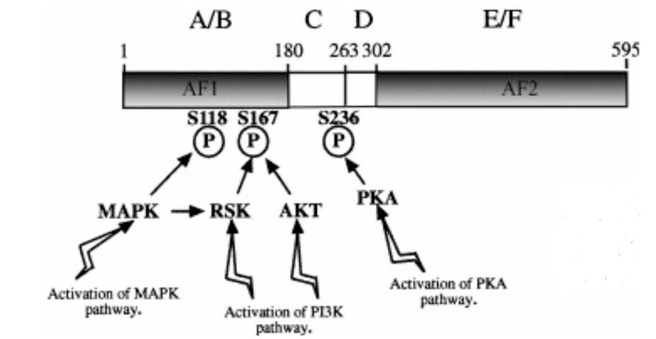
ER can be activated in the absence of its classical ligand, estrogen.
Mitogen-Activated Protein Kinase (MAPK) and Phosphoinositide 3-kinase (PI3K) Cascades:
Growth factors (e.g., EGF and IGF-1) activate these cascades.
These pathways phosphorylate cytoplasmic ER.
This phosphorylation leads to Estrogen-2 (E2)-independent nuclear import and subsequent ER regulation of transcription.
Specific Pathways:
RTK/c-Src Pathway: c-Src, a Receptor Tyrosine Kinase (RTK), can activate ER via the PI3K/Akt pathway.
This is mediated by phosphorylation of ER at Serine 167 (S167).
mTOR also contributes to increased Akt activation.
Blocking this pathway is a potential therapeutic route for breast cancer.
Ras/Raf/MEK/ERK Signaling Pathway: Growth factors binding to RTK receptors activate this pathway.
This promotes the phosphorylation of Serine 118 (S118) in the AF-1 domain of ER.
Phosphorylation at S118 activates ER-target gene transcriptional activity.
NTD Phosphorylation: Phosphorylation in the N-terminal domain (NTD) generally promotes transactivation.
Calcium Activation: Ca can activate ER via its LBD, inducing the expression of estrogen-responsive genes in the absence of ligand.
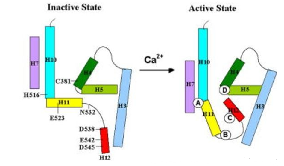
There are four potential calcium binding sites in the LBD.
Membrane Estrogen Receptors:
ERs can exist as plasma membrane-associated receptors, mediating rapid, non-genomic (independent of gene expression) pathways.
Membrane Localization:
Membrane localization of ER is mediated by Heat Shock Protein 27 (HSP27).
It associates with caveolae lipid rafts in the membrane through interactions with caveolin-1, Src, and striatin.
Asp-His-His-Cys (DHHC) domain palmitoyl acyltransferases (DHHC-7 and -21) palmitoylate ER and promote its plasma membrane localization.
Approximately of total ER is localized to the plasma membrane.
Signaling Pathways from Membrane ERs:
Estrogen-bound membrane-localized ER directly interacts with RTKs (e.g., the p85 regulatory subunit of PI3K, Src, and Shc).
These interactions activate PI3K/Akt/mTOR as well as RAS/RAF/MEK1/2-ERK1/2 signaling pathways.
These kinase pathways promote cell survival and proliferation.
They also phosphorylate ER and its coregulators, leading to the activation of nuclear genomic transcription.
Other Functions (Other Crazy Stuff):
Control of Mitochondria: ER localizes in the mitochondria and mediates the transcription of mitochondrial DNA (mtDNA) following E2 stimulation.
This is possible due to ERE-like sequences found in the D-loop of mtDNA.
mRNA Binding and Regulation of Translation: ER binds to many mRNAs, primarily at the Untranslated Region (UTR), to regulate translation.
It interacts with proteins that regulate RNA splicing, stability, and XPB1 (involved in unfolded protein response).
Endocrine Resistance
Loss of ER Expression:
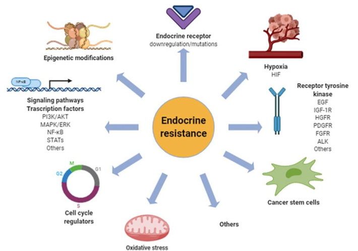
Approximately of breast cancer patients treated with endocrine therapy eventually lose ER expression over time.
Loss of ER is problematic because tumor growth becomes independent of ER, making the endocrine pathway ineffective for control.
Control of ER Expression (ESR1 Gene):
Methylation: ER has at least nine promoters and CpG islands for the A, B, and C promoters of ESR1, which are highly methylated in ER-negative breast cancer cases.
Methylation status is negatively correlated with ER expression and resistance to endocrine treatment in breast cancer cells.
Histone Acetylation: Histone acetylation is involved in gene control.
Histone deacetylase (HDAC) inhibitors, such as trichostatin A, have been shown to re-express ER in ER-negative breast cancer cells.
DNA demethylating agents and HDAC inhibitors restored tamoxifen sensitivity in ER-negative breast cancer cells.
RNA Stabilization:
AU-rich elements (AREs), typically AUUUA motifs, in the -UTR of ESR1 mRNA are critical for its stabilization.
ESR1 mRNA contains AUUUA motifs.
The ARE-binding protein HuR binds to ESR1 mRNA to stabilize it.
More than ten types of microRNAs (miRNAs) directly bind to ESR1 mRNA and repress its expression.
ER Post-Translational Modification (PTM):
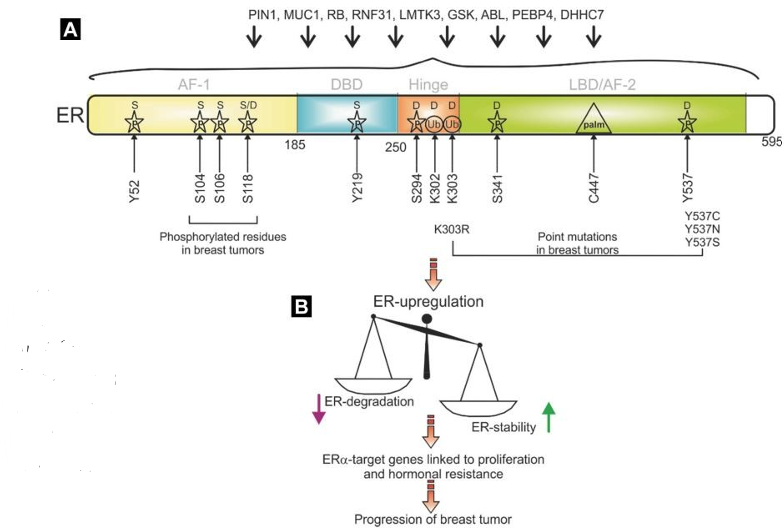
ER can be extensively modified, including phosphorylation, methylation, acetylation, SUMOylation, glycosylation, ubiquitination, palmitoylation, and isomerization.
ER has at least known phosphorylation sites.
These PTMs can control ER stabilization or activation.
Calcineurin Influence: Calcineurin, a Ca-dependent serine/threonine phosphatase, stabilizes and activates ER.
High calcineurin expression is associated with shorter recurrence-free survival in endocrine-treated ER-positive breast cancer patients.
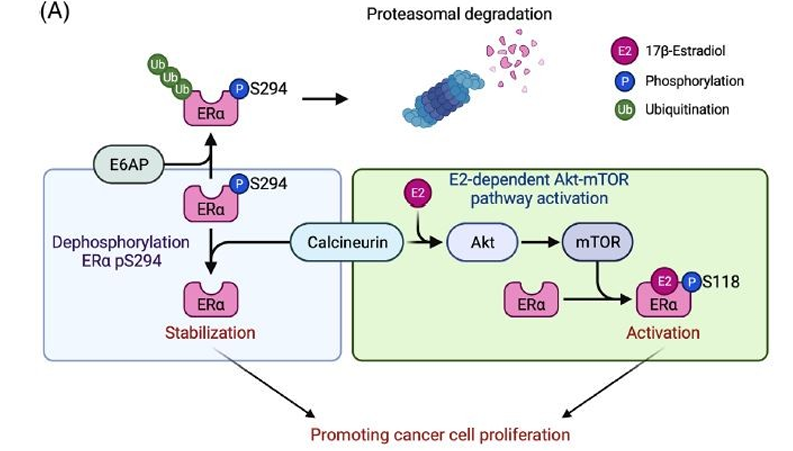
Ubiquitination:
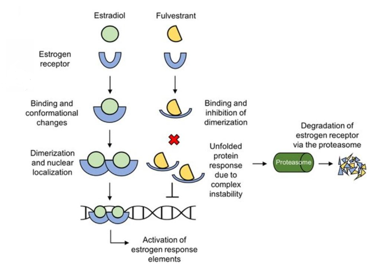
Fulvestrant, a Selective Estrogen Receptor Downregulator (SERD), exerts its tumor suppression by inhibiting ER through antagonistic action AND by promoting ER degradation via the ubiquitin proteasome system.
E3 ligases that polyubiquitinate ER (e.g., E6AP, CHIP, SKP2) generally promote its degradation.
E3 ligases that monoubiquitinate ER (e.g., BRCA1/BARD1, RNF8, RNF31, and TRIM11) tend to stabilize ER.
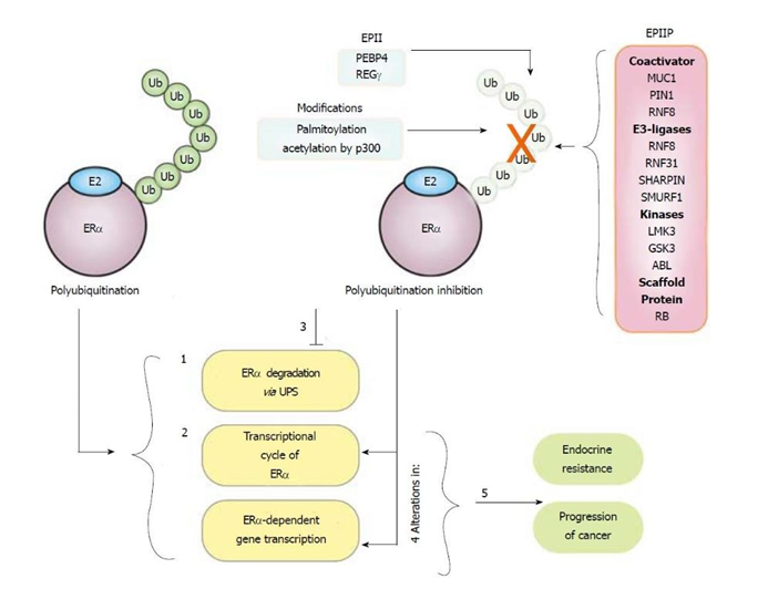
A detailed understanding of ER ubiquitination sites is not fully achieved, as ubiquitination is not always site-specific (e.g., mutation of a primary target lysine can result in ubiquitination of neighboring lysines).
K302 and K303 are listed as major monoubiquitination sites for BRCA1/BARD1 and major polyubiquitination sites triggered by fulvestrant.
ER Mutation (Hot Spot Mutations):
ER mutations are commonly found within the LBD.
The most common hot spot mutations are Y537S, Y537N, and D538G.
These mutations control the agonist state of the ER LBD, affecting conformational changes and protein stability.
Mutations at these residues alter the conformational dynamics of the loop connecting H11 and H12 in the ER LBD, leading to a stabilized agonist state that is unaffected by antagonists.
Y537 and D538 ER mutants are phosphorylated on S118 by the TFIIH kinase, cyclin-dependent kinase (CDK)7, in an estrogen-independent manner.
This phosphorylation may lead to endocrine resistance by increasing transcriptional activity.
Influence of Coregulators:
Cancer likelihood may increase if the balance or expression of coregulators is altered, affecting ER signaling and therapeutic response.