final exam study guide
Chapter 1:
Describe how an organism’s environmental adaptations result from evolution.
Environmental adaptions result from evolution is proven by natural selection and how, when climates or environmental situations change, only those that have the traits adapted for those circumstances survive to pass on those traits, eventually evolving over generations.
Identify (and give examples) of the different levels of the hierarchy of biology.
Biosphere: the earths total life and spaces where life exists
Environment: consists of all the living things in a particular area, along with all the nonliving components of the environment with which life interacts
Communities: The array of organisms inhabiting a particular ecosystem
Population: consists of all the individuals of a species living within the bounds of a specified area
Organisms: Individual living things
Organs: a body part that is made up of multiple tissues and has specific functions in the body
Tissue: a group of cells that work together, performing a specialized function
Cells: life’s fundamental unit of structure and function
Organelles: the various functional components present in cells
Molecules: a chemical structure consisting of two or more units called atoms
Describe the sexual competition hypothesis (in the context of giraffes’ long necks).
Giraffes evolved to have bigger necks because male giraffes use their long necks in intrasexual selection when fighting other males.
Those with longer necks often had more offspring because they could fight off more males and mate with more females, passing on their traits to the next generation.
Chapter 2-5
Compare and contrast covalent bonds, ionic bonds, and hydrogen bonds.
covalent bonds: sharing a pair of valence electrons by two atoms
ionic bonds: occurs between two ions, due to the attraction between cations and anions.
hydrogen bonds: occurs when hydrogen is bonded covalently with a more electronegative ion, which gives it a partial positive that can attract another more electronegative atom.
Describe the difference between polar covalent bonds and non-polar covalent bonds.
polar covalent bonds: occur when one atom is more electronegative than the other
non-polar bonds: occur when two atoms have equal electronegativity.
Describe how polar covalent bonds in water molecules result in hydrogen bonding.
since hydrogen has low electronegativity, when it bonds with an atom with greater electronegativity it gains a partial positive charge. Thus, it can attract other electronegative atoms that have partial negative charges. this is hydrogen bonding.
Predict the type of bond that will form based on electronegativities.
if the atoms are electronegative and not electronegative: polar covalent
if the atoms are equally electronegative: non-polar covalent
if the difference between electronegatively is too great, the highly electronegative atom might lose electrons and become a cation, and form ionic bonds with the anion that stole it’s electron.
If it comes across a hydrogen atom, it may be attracted to the partial positive charge and form a hydrogen bond.
The opposite is true with atoms that have relatively low electronegativity. They are less likely to form bonds with hydrogen, and ionic bonds would occur if they find electronegative atoms with a lot more electronegativity than them, causing either to lose or gain electrons and
Describe why (referring to bonding) ice is less dense than liquid water.
hydrogen bonds in water stabilize when it reaches low temperatures, which makes water expand when it solidifies.
hydrogen bonds in water are constantly forming and reforming due to the fast movement of water molecules. However, when water is lower than 4C, it begins to slow down, and then at 0C and below it freezes, each water molecule bonding to 4 partners that keep it an 'arm's length' away, which is why ice is expanded. This forms spacious crystal lattice structures as the hydrogen bonds are stable.
Explain why water is a polar molecule.
Water is polar because it has electronegative oxygen and less electronegative hydrogen.
Describe the link between CO 2 emissions and ocean acidification.
Increased CO2 in the atmosphere leads to it being dissolved in the ocean, reacting with water to form carbonic acid. This lowers the oceans pH.
Describe why ocean acidification is a threat to marine life.
Many marine organisms need carbonate to form calcium carbonate which is necessary to them, but with increased levels of carbonic acid, they aren’t able to access the carbonate. This is because when seawater acidifies, the extra hydrogen ions combine with carbonate to form bicarbonate ions, which is bad cuz it leaves less carbonate for marine life.
Describe the structure of a nucleotide.
Sugar phosphate backbone (pentose sugar, phosphate group)
Nitrogenous bases
Explain why the two strands in a DNA double helix are anti-parallel.
one goes in a 5’ → 3’ direction, and the other goes the opposite way.
they’re connected by hydrogen bonds between the nitrogenous bases.
Describe the molecular forces responsible for the stability of the DNA double helix.
hydrogen bonds btwn complementary base pairs and hydrophobic interactions among stacked pairs.
Explain why it is dangerous to not know the function of an isomer to a pharmaceutical product
isomers can have completely different properties and biological activities. Using the wrong isomer can lead to unexpected and harmful results, like in the case of using thalidomide, where using an isomer of a tranquilizer resulted in major birth defects.
Identify the behaviour (e.g., hydrophobic or hydrophilic) or an amino acid based on
its structure.An amino acid is hydrophobic when it has hydrophobic, nonpolar side chains
lack of oxygen, mostly carbon and hydrogen (less electronegative)
An amino acid is hydrophilic when it has hydrophilic, polar side chains.
lots of OH or oxygens
acidic & basic side chains (having carboxyl or amino groups in side chain) are hydrophilic
Describe how a change in primary structure can affect a protein’s structure and
function.Altering the sequence of amino acids in the primary structure can impact the protein’s folding pattern (secondary and tertiary structures), leading to changes in its overall shape and function.
Describe secondary and tertiary protein structure (including the bonds that stabilize
the structure.)Primary structure: its sequence of amino acids.
held together by peptide bones
Secondary structure: segments of the polypeptide chain repeated coiled or folded in patterns.
a helix: coil held together with hydrogen bonding between every fourth amino acid
b pleated sheet: two or more folded sefments lying side by side, held together by hydrogen ponds.
Align sickle-cell disease to the hierarchy of biology.
at the molecular level, it involves a single amino acid substitution (glu to val) that affects protein structure. at a cellular level, it forms a sickle shaped cell that isn’t able to carry oxygen throughout the body, which affects the tissues and organs of the individual. the organism experiencing sickle cell disorder experience clogging of blood vessels. It is 1/12 carriers of african descent. There’s a heterozygote advantage where malaria is common.
Chapter 6:
Describe the differences between prokaryotic and eukaryotic cells
prokaryotic cells:
lack a true nucleus; dna is located in nucleoid region
lack membrane-bound organelles
typically smaller in size
have a simple structure
eukaryotic cells:
have a true nucleus containing DNA
contain membrane-bound organelles
larger in size
have more complex structure
Describe structure and function of individual organelles
nucleus: contains genetic material (DNA), controls cell activities
mitochondria: site of cellular respiration, produces ATP energy
endoplasmic reticulum:
rough ER: studded with ribosomes, involved in protein synthesis
smooth ER: synthesizes lipids and detoxifies substances
Golgi apparatus: modifies, sorts, and packages proteins for secretion or use within cells
lysosomes: contain digestive enzymes, break down cellular waste
vacuoles: storage organelles for nutrients, waste, or pigments (larger in plant cells)
chloroplasts: found in plant cells, sites of photosynthesis, contain chlorophyll
cytoskeleton: provides structural support, aids in cell movement
centrioles: involved in cell division
Describe what is part of the endomembrane system.
endoplasmic reticulum (ER): Rough ER involved in protein synthesis, smooth ER involved in lipid synthesis
Golgi apparatus: modifies, sorts, and packages proteins
Vesicles: transport materials within and outside the cell
Plasma membrane: regulates traffic in and out of the cell
Nuclear envelope:
structure: double membrane surrounding the nucleus
function: regulates the passage of macromolecules (like proteins and RNA) between the nucleus and cytoplasm. Provides structural support and protection to the nucleus.
lysosomes:
structure: membrane bound vesicles containing digestive enzymes
function: break down cellular waste, debris, and foreign materials through hydrolytic enzymes. Essential for cellular waste recycling and maintaining cellular homeostasis.
phagocytosis: lysosome digesting food
autophagy: lysosome breaking down damaged organelles
Describe the structure of the plasma membrane.
phospholipid bilayer: double later of phospholipids with hydrophilic heads and hydrophobic tails
proteins: integral proteins span the membrane, peripheral proteins are on the surface.
cholesterol: provides membrane stability
carbohydrates: found on the outer surface, involved in cell recognition.
Describe the evidence that supports the endosymbiont theory of the origin of mitochondria and chloroplasts.
endosymbiont theory: states that an early ancestor of eukaryotic cells engulfed an oxygen-using nonphotosynthetic prokaryotic cell, which eventually formed a relationship with the host cell in which it was enclosed, becoming an endosymbiont. It eventually merged into a single organism—a eukaryotic cell with a mitochondrion.
similarities in structure: mitochondria and chloroplasts resemble bacteria in size and structure.
double membrane: both organelles have a double membrane
own DNA: mitochondria and chloroplasts have their own circle DNA, similar to bacterial DNA
reproduction: both replicate independently of the host cell by binary fission.
ribosomes: similarities in ribosomal structure to bacterial ribosomes
Chapter 12:
Draw the chromosomal structure & number throughout the cell cycle.
Interphase: (2n chromosomes)
Includes G1, S, G2
G1: cell prepares for division, here you have 46 single chromosomes, each in pairs of 2 (one from each parent). DNA in a cell is 46 double-stranded molecules.
S Phase: Once cell prepares, it replicates each chromatid into 2 sister chromatids (still considered 1 chromosome). CAN’T BE SEEN UNTIL PROPHASE, when they condense.
G2: still 46 chromosomes, cell just grows & organelles/proteins develop.
Mitotic Phase: (2n In PM, 4N in AT, 2N in C)
Cell division: includes prophase, metaphase, anaphase, telophase.
Prophase: Still 46 chromosomes
Metaphase: 46 chromosomes
Anaphase: Chromosomes are divided into 2 groups of 46 (92 total)
Telophase: Chromosomes are in 2 groups of 46 (92 total)
Cytokinesis: cell separates into 2, each cell has 46
Identify the stage of the cell cycle based on chromosomal structure & number.
If it’s long, thin, chromatin fibre: cell isn’t dividing or it’s replicating in preparation
Thick and coiled, condensed chromatin: after DNA replication
Can be seen in a light microscope
2 sister chromatids: occur once duplicated (S phase)
sister chromatids are joined copies of the og chromosome, containing the same dna molecule
attached to each other by protein complexes called cohesions
each has a centromere which is where theyre attached to eo
later in cell division (anaphase & telophase) the chromatids move into two new nuclei at each end of the cell. now theyre individuals. this step doubles the amt of chromosomes in the cell (from 2n to 4n)
mitosis division of genetic material in the nucleus (divorce) is usually followed by cytokinesis, which is like moving houses.
Describe what a replicated and un-replicated chromosome looks like.
replicated: two chromatids joined together, shaped like an X
un-replicated: chromatin fibre, thin strand of genetic material. not visible by light microscope.
Describe the cell cycle control system.
checkpoints are control points in the cell cycle where stop and go ahead signals can regulate the cycle. Three are found in G1, G2, and M phases.
Regulatory molecules main cell cycle events
kinases maintain a steady concentration throughout the different phases of the cell, but cyclins flucuate, depending on the maturation promoting factor.
The G1 checkpoint (restriction point) is usually the most important. If it receives the go-ahead, it will usually complete G1, S, G2, and M phases and divide.
If it doesn't, it may exit the cycle and switch to a nondividing state called the G0 phase, where most human cells are. But they can be called out by external cues like growth factors.
Internal signals occur at the 3rd checkpoint in M phase. Anaphase doesn’t occur untill all the chromosomes are properly attached to the spindle at the metaphase plate, if the kinetochores are unattached, anaphase will be delayed. Protein complex only activates then, which activates the enzyme seperase which cleaves the cohesins. This prevents missing or extra chromosomes.
A checkpoint in S phase stops cells with DNA damage from proceeding in the cell cycle.
there's also evidence that another checkpoint between anaphase and telophase that ensures anaphase is completed and the chromosomes are well separated before cytokinesis can begin, thus avoiding chromosomal damage.
In most mammalian cells, cells only divide if growth factor stimulates it.
density-dependent inhibition is when crowded cells stop dividing, by noticing a single layer of cells is formed on the inner surface. If there’s an empty spot, more will divide to fill it.
anchorage dependence is when cells need to be attached to a substratum to divide.
cancer cells display neither density-dependent inhibition or anchorage dependence.
Chapter 13:
Explain why meiosis is a “reduction division”.
during meiosis, the number of chromosomes are reduced by half. the set of chromosomes goes from 2 sets to one in gametes. because of meiosis, each human sperm and egg cell is haploid (n=23)
Explain how meiosis can increase genetic variation.
because of independent assortment in meiosis I, crossing over in meiosis I, and random fertilization. the combinations of possibilities when producing gametes is huge and then it increases during fertilization (as it depends on the other parent as well) and results in limitless changes that could effect the genes of any offspring.
Describe the three mechanisms that contribute to genetic variation. (Independent assortment of chromosomes, crossing over, random fertilization.)
independent assortment of chromosomes:
At metaphase I, there’s a 50% chance whether a particular daughter cell will get the maternal or paternal homologue of a chromosome.
Each pair sorts its maternal and paternal homologues into daughter cells independently of every other pair— independents assortment. each daughter cell represents an outcome of all possible combinations of maternal and paternal chromosomes.
The combination of possibilities is 2^n
crossing over
occurs in early prophase I
Crossing over produces recombinant chromosomes, individual chromosomes that carry genes derived from two different parents. Randomly happens in chromosomes resulting in different gene combinations as the alleles swap in the process.
in metaphase II, since sister chromatids aren’t identical, chromosomes with one or more recombinant chromosome can be oriented into two alternative ways. increases nymber of genetic types of daughter cells that can occur.
random fertilization:
each male and female gamete represents on of about 8.4mill(2²3) possible chromosomes combinations.
The fusion of a male & female gamete will produce a zygote with any of 70 trillion (2²3 × 2²3) diploid combinations.
Predict how many different combinations of maternal and paternal chromosomes can
be packaged in gametes (when given a specific diploid number).2^n, n being the haploid number.
Compare and contrast meiosis and mitosis.
meiosis:
forms gametes
produces genetically variable offspring
reduces amount of chromosomes sets from 2n to n
divides twice
occurs in germ cells when producing gametes
because of crossing over in meiosis i, sister chromatids aren’t identical
synapsis & crossing over
alignment of homologous pairs at the metaphase plate
separation of homologues.
mitosis
forms somatic cells
produces genetically identical offspring.
chromosome sets double and then are divided to 2 cells (so number overall stays the same)
divides once
occurs in all other cells including zygotes
genetically identical sister chromatids
sister chromatids separate in anaphase
positioned on the metaphase plate as individual chromosomes
the sister chromatids separate in anaphase
Chapter 14:
Describe the difference between an allele and a gene.
Genes are DNA sections that code for specific proteins or functional RNA, whereas alleles are variations of these genes. E.g., a gene exists that codes for a specific eye colour, and an allele are the different versions of these genes (e.g., allele for blue eyes, allele for brown eyes).
An individual’s combination of alleles is known as their genotype.
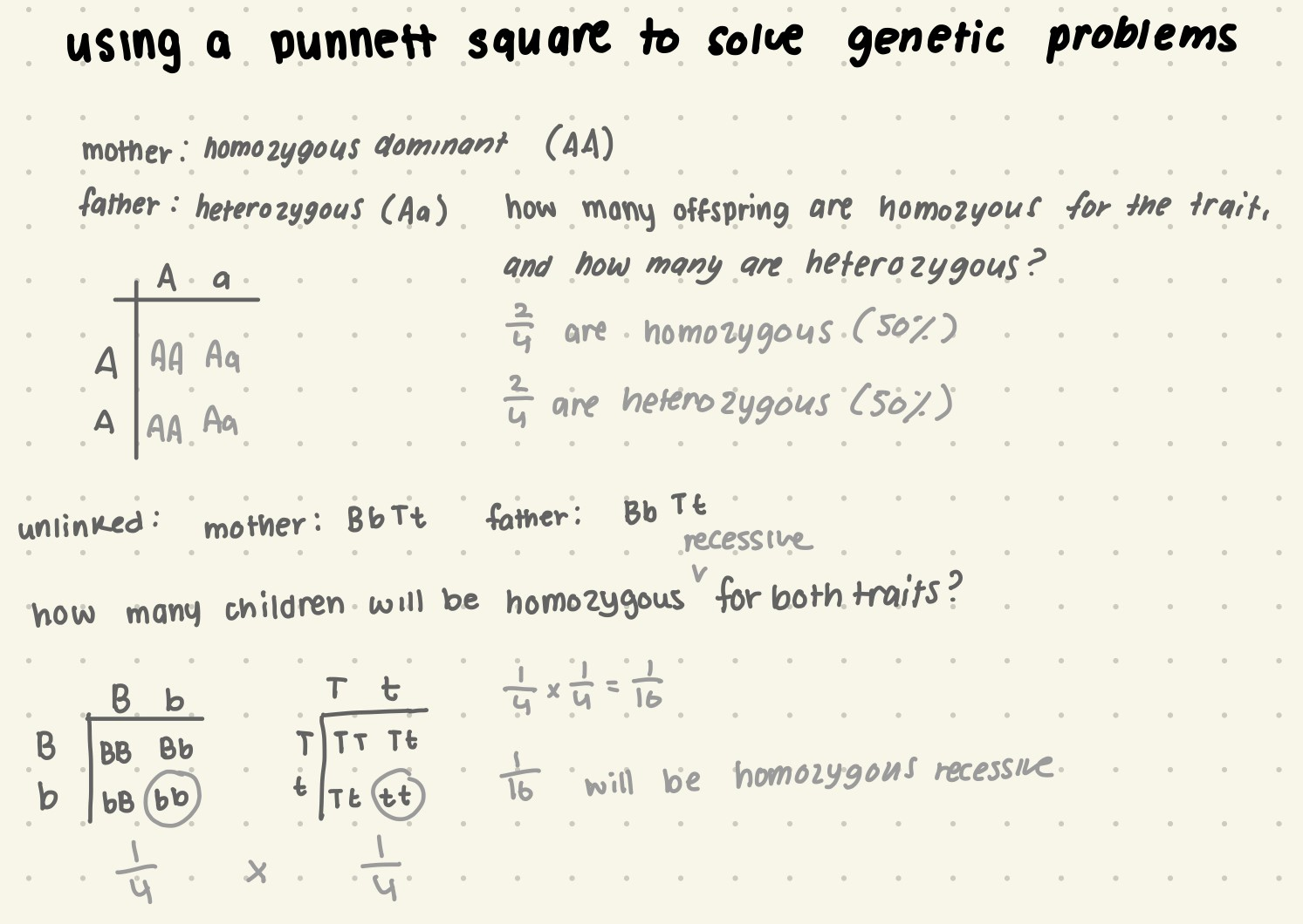
Use a Punnett square to solve genetic problems.
Describe when and why a testcross is used.
A test cross is used when the genotype of an organism is unknown.
To test cross, you mate an unknown organism with a known recessive true-breeding organism. If all offspring are dominant heterozygous, the organism was dominant homozygous. If some offspring are recessive, then the organism was heterozygous.
Predict potential gametes based on parental genotypes.
Organisms inherit two alleles for each gene, one from each parent. Each pair of allele segregates independently of any other pair.
So if a parent cell has AaBb, the gametes could be AB Ab aB ab
If there are more than one genes being passed down, they can be calculated with 2^n, n being the amount of different alleles (etc, AaBb is 2^4 , or 16 different possible genotypic outcomes of both parents gametes combining).
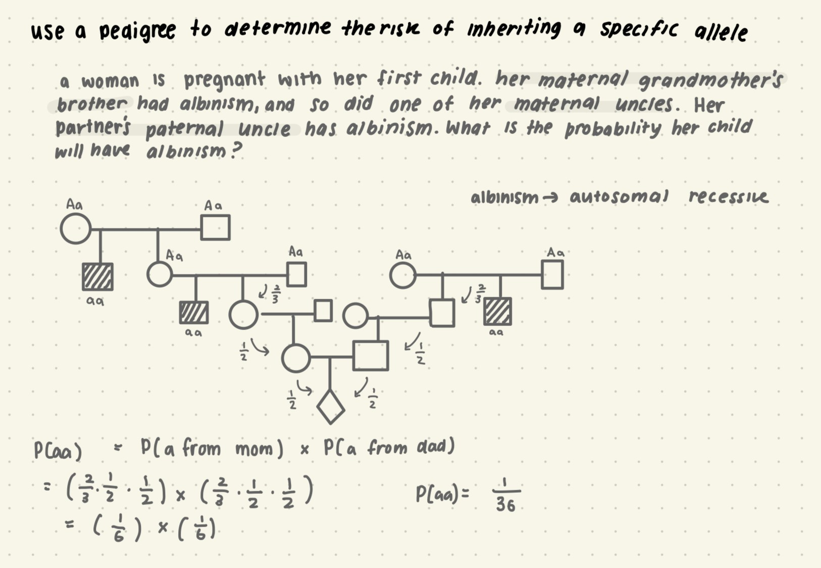
Calculate the percentage of progeny expected to have specified traits based on
parental genotypes.Use a pedigree to determine risk of inheriting a specific allele.
Determine the inheritance pattern by examining a pedigree.
inheritance pattern
identifiers
examples
autosomal recessive
equal likelihood of males and females affected
the parents of an affected individual must be heterozygous
goes to 25% of offspring
tay sachs diseas
cystic fibrosus
sickle cell anemia
albinism
autosomal dominant
one of the parents must have the trait, dominant traits do not skip generations.
equal likelihood of males and females affected
the parents of an affected individual must be heterozygous
goes to 50% of offspring
achondorplasia
huntington’s
y-linked
males only (dads to sons)
never skips generations
x-linked recessive
more common in males
if mother is a carrier sons can have it
if mother & fathers are carriers, daughters may have it
males can never be carriers, they must show the trait if they are affected
hemophilia A
colour blindness
duchenne muscular dystrophy
x-linked dominant
one parent must have the trait, dominant traits do not skip generations.
males can never be carriers, they must show the trait if they are affected
CGH - Congenital Hypertrichosis
mitochondrial
female transmits to all offspring
males do not transmit
mitochondrial myopathy
Leber’s hereditary optic neuropathy
Describe why inheritance by a single gene may deviate from simple Mendelian patterns.
When alleles aren’t completely dominant or recessive
When a gene has more than two alleles
When a gene produces multiple phenotypes
Explain the difference between complete dominance, incomplete dominance, and
codominance.Complete dominance: heterozygous phenotype same as homozygous dominant
Incomplete dominance: heterozygous phenotype intermediate between two homozygous phenotypes
Codominance: both phenotypes expressed in heterozygotes
Describe how the alleles resulting in the ABO blood group system are both completely dominant AND codominant.
They are completely dominant because A and B are dominant to O— the genotype AA or AO both result in blood group O
They are codominant because when A and B are together they have a separate phenotype— AB.
Chapter 15:
Explain the outcome of X inactivation.
X inactivation occurs because females do not need both x chromosomes, so in each cell one of the x chromosome is randomly inactivated. When the x chromosome has alleles that differ from one another (she is heterozygous for the gene), the x that remains active shows the trait. In tortoiseshell cats, this shows up as random cells being either coding orange or black fur, showing up as a mosaic.
Describe sex-linked inheritance
sex-linked genes are genes that are located on either sex chromosome.
X chromosomes have genes for many characters unrelated to sex, whereas the y chromosome mainly encodes genes related to sex determination. X-linked genes follow specific patterns of inheritance. For a recessive x-linked trait to be expressed:
Female needs two copies of the allele (homozygous)
Male needs 1 copy of the allele (hemizygous)
Predict which parent nondisjunction occurred in, based on an individual’s karyotype or family tree genetic disease history.
X0 : can come from mom or dad
XXX: can come from mom OR dad
XXY: can come from mom OR dad
XYY: can come from dad only
You can use the phenotype or genotype of the offspring and family to determine which parent it came from.
A boy with CGH is born with Klinefelter syndrome. His mother does not have CGH, but his father also has CGH. In which parent did nondisjunction occur?
In Klinefelter syndrome (XXY), the extra x could’ve come from the dad or the mom.
Since this is an x-linked disorder, we know that the father must’ve given both x AND y chromosomes to the son, since the mother had no way of transmitting this disorder. This means that nondisjunction occurred in the father, not the mother.
Chapter 16:
Describe the structure of DNA.
DNA (deoxyribonucleic acid) consists of the sugar-phosphate backbone and the nitrogenous bases. The backbone consists of phosphate groups that are bonded with a pentose sugar with phosphodiester bonds (covalent bonds), and the nitrogenous bases pair together (A —> T & C —> G).
A pairs with T with 2 hydrogen bonds
C pairs with G with 3 hydrogen bonds.
Describe the process of DNA replication.
DNA replication starts at the origin of replication, which are points in the DNA where the strands begin to separate as catalyzed by helicase. This creates a replication bubble, which has y-shaped areas on either end that are replication forks. Helicase separates the parent strands, and then SSB proteins stabilize the now separated template strand. In front of helicase, topoisomerase unwinds and corrects the position of the strands by breaking a single strand, correcting its position, and putting it back together. Now that the strands are prepped, an RNA primer is attached to the strand using primase, and then DNA polymerase can come along and start attaching nucleotides from the 3’ end (which has the OH group). DNA replicates from the 5’ to 3’ direction. This happens in the leading strand, it’ll eventually replicate throughout the
Explain how DNA replication of the lagging strand is different from the leading strand.
Along one template strand of DNA, the DNA polymerase synthesizes a leading strand continuously, moving towards the replication fork from the origin of replication.
To elongate the other new strand, called the lagging strand, DNA polymerase must work in the direction away from the replication fork.
As the bubble expands and the fork opens, small sections will be elongated away from the replication fork. Its still moving 5’ to 3’, but it starts away from the replication fork of that template strand. As the replication fork expands from the other side (from the other dna building and helicase opening it up) therna primer attaches and then polymerase repeats this over, building into the previous one that was there.
The lagging strand is synthesized as a series of segments called Okazaki fragments which are joined together by DNA ligase.
Dna ligase glues the fragments together following the direction of the strand.
DNA polymerase I removes the RNA pieces, and once they are replaced by DNA ligase comes and glues them to each other.
Identify and describe all the proteins involved in the DNA replication complex.
helicase: separates nitrogenous bases
ssbs: go through separated strands to prevent them from rejoining
topoisomerase: corrects overwinding ahead of the helicase by breaking, swiveling, and rejoining DNA strands
primase: starts rna chain & adds nucleotide primers
Describe the evolutionary significance of altered DNA nucleotides.
when nucleotides are altered they can result in mutations which increase genetic variability
Chapter 17:
Explain why the “central dogma” of biology is an oversimplification.
Explain how transcription and translation are different in prokaryotes vs eukaryotes.
Use the codon table to predict an amino acid sequence from a coding sequence of DNA
Identify the template strand and coding strand of DNA, based on protein primary sequence.
Describe the stages of transcription and translation.
Describe the different types of RNA processing that occurs in eukaryotes.
Predict a protein sequence from the mRNA sequence.
Describe how proteins are targeted to the ER.
Describe how polyribosomes enable cells to make many polypeptides very quickly.
Describe the different types of mutations and the potential impact they have on protein structure and function.
Describe how the CFTR mutations affect protein structure and function. Align this mutation to the hierarchy of biology.
Describe how the CCR5delta32 mutation affects protein structure and function. Align this mutation to the hierarchy of biology.
Describe how the sickle cell mutation affects protein structure and function. Align this mutation to the hierarchy of biology
Chapter 22, 23, 24:
Describe why it isn’t possible for an individual to evolve in its lifetime.
Describe what is meant by the phrase “descent with modification.”
Describe why natural selection is NOT a random process.
Describe what is required in order for natural selection to occur.
Describe the four types of data that document the pattern of evolution.
Describe how Galapagos finch beak depth change is an example of evolution.
Describe the difference between homologous features and features that are similar due to homoplasy.
Describe the fossil evidence for the transition from land to sea for whales.
Describe the mechanisms that cause allele frequency change.
Explain why variation in heritable traits is a prerequisite for evolution.
Describe what average heterozygosity is, and explain how it is an indicator of variation within a population.
Explain the following statement, and give examples: “Not all variation is genetic.”
Describe what the sources of genetic variation are.
Describe the conditions for Hardy-Weinberg.
Calculate allele frequencies and genotype frequencies from phenotypic data.
Use the Hardy-Weinberg equation to determine if a population is evolving.
Compare and contrast natural selection, genetic drift, and gene flow.
Explain the statement “Only natural selection can lead to adaptive evolution.”
Describe directional selection, disruptive selection, and stabilizing selection.
Compare and contrast intersexual selection and intrasexual selection.
Describe microevolution and macroevolution.
Describe the biological species concept.
Explain why the biological species concept only is applicable to certain types of species.
Describe what is meant by “reproductive isolation”.
Describe the five prezygotic barriers discussed in class, and give examples for each type of barrier.
How do prezygotic barriers block fertilization?
Describe the three postzygotic barriers discussed in class, and give examples for each type of barrier.
How do postzygotic barriers prevent the hybrid zygote from developing in a viable, fertile adult?
Describe the limitations of the biological species concept.
Compare and contrast sympatric speciation with allopatric speciation.
Describe how polyploidy can lead to rapid speciation.
Be able to interpret phylogenetic trees, including identifying monophyletic groups,
common ancestors, and closest living relatives
Short Answer:
It has been observed that that elephant tusk size has decreased in areas of historically heavy poaching. Propose a hypothesis to explain this observation and design an experiment to test your hypothesis.
Hypothesis, Prediction:
H: Poaching leads to elephants with smaller tusks having greater fitness than those with large tusks.
P: If elephants outside of sanctuaries have larger tusks, they are more likely to be poached, so they can’t survive to reproduction and pass on their traits to offspring.
Control(s):
Multiple populations observed (to reduce confound effects)
Same measurement levels
Independent and Dependent Variables:
IV: Protected vs unprotected populations
DV: Tusk size in new generations
How IV & DV are manipulated / measured:
IV: One population of elephants will be kept in a sanctuary, whereas the others will remain in the wild.
DV: Tracking the parents and counting their offspring over long periods of time.
Hold other variables constant:
Habitat should be similar.
Same species of Elephants (African elephants)
Replicates (n#):
Experiment to be repeated:
Awareness that one can never prove a hypothesis
Ethics:
Ethical issues with this is allowing elephants to be poached in order to prove a hypothesis.
Confounding effects / bias:
The possibility of other predators targeting elephants in the wild.
A new type of bacteria (call it “Bacteria X”) has been discovered. You suspect that this bacterium is a common cause of ulcers. Propose a hypothesis to explain this connection and design an experiment to test your hypothesis.
Advertisements for a herbal product, Gingko biloba, claim that it promotes memory. To determine if the claim is fraudulent and prior to accepting this claim, what type of evidence would you like to see? Propose a hypothesis to explain this connection and design an experiment to test your hypothesis.
Your biology professor has a room that houses turtles. While waiting for the dozens of incubating turtle eggs to hatch in the room, she leaves for a two-week vacation. When she returns, she notices that all of the turtle eggs have hatched but that there is something strange about the hatched turtles. Instead of the turtles being roughly half males and half females, all of the hatched turtles are males! Your professor also happened to notice that the temperature in the room is about 5ºC cooler than normal, and thinks this may have something to do with the phenomenon. Your professor then asks for your help to solve this mystery. Propose a hypothesis and design an experiment that helps to explain why all of the hatched turtles are all males.
Professor Maydianne Andrade researches sexual cannibalism in the redback spider and in closely related black widows. Recently, it has been observed that male spiders of the species Latrodectus hasselti (a type of black widow) can copulate with immature females (who haven’t had their final moult – meaning they haven’t shed their immature exoskeleton) by piercing the female’s exoskeleton to access her newly developed sperm storage organs. Propose a hypothesis to explain this behaviour and design an experiment to test your hypothesis.