Reproductive System
The reproductive system is responsible for producing gametes and hormones necessary for reproduction.
Male reproductive system includes the testes, seminal vesicles, and prostate gland.
Female reproductive system consists of the ovaries, fallopian tubes, uterus, and vagina.
Topic 1: Anatomy and Physiology of the Male Reproductive System
Consists of the scrotum, testes, epididymis, ductus deferens, uretha, penis, and accessory sex glands. The function of the male reproductive system is to work with the female reproductive system for the purpose of sexual reproduction. The male reproductive system primarily serves to produce sperm and hormones, as well as to maintain and transport the male gamete and protective fluid (semen). It is also responsible for discharging spem within the female reproductive tract during sexual intercourse and also for producing and secreting male sex hormones.
Structures of the Male Reproductive System
Scrotum: the scrotum is a small, muscular external sac that encloses and supports the testes. The scrotum supports the testes at a temperature lower than the body’s temperature, which is necessary for the development of sperm.
Testes: The testes, or testicles, are the male sex glands where sperm and testosterone are produced. The testes are suspended in the scrotum by the spermatic cord and are comprised of dense connective tissue that contain the seminiferous tubules.
Epididymis: is a tube at the upper part of each testicle that connects the testes to the ductus deferens. It secretes part of the semen, stores semen before ejaculation and provides a passageway for the sperm. It also serves as the area where sperm become capable of motion. The epididymis is composed of a head, body and tail and connects to the ductus deferens.
Ductus Deferens: are muscular tubes that connect to the epididymis and function to carry sperm from each testis to the ejaculatory ducts. These excretory ducts are encase in the spermatic cord.
Accessory Sex Glands: add secretions to the sperm and flush urine from the urethra prior to the arrival of sperm. They include the seminal vesicles, prostate gland, and bulbourethral glands. Not all of these glands are preseent in all species.
Urethra: is a tube by which semen leaves the body. It serves reproductive and urinary functions.
Penis: is a male sex organ and is composed of erectile tissue. it carries reproductive and urinary products outside of the body.
The Testes
the word is a plural; a testical is one of the paired glands. At the most basic level, sexual reproduction requires that a single spermatozoon from a male fuses with the ovum, or egg of a female of the same species. The actual process of reproduction is much more complicated, but with the testes, this process wouldnt be possible. The testes are one of the most essential organs in the system because they produce spematozoa.
Each testical is an oval-shaped male sex gland enclosed within the scrotum and secured at either end by the spermatic cord. Each testicle is composed of connective tissue and divided into compartments that contain coiled tubes called the seminiferous tubules. Leydig’s cells make up the space between the compartments containing these tubules. These cells have endocrine function. Cells within the testes that support sperm growth are referred to as setoli cells. The seminigerous tubules serve as areas that produce sperm as well as the pathway sperm use to leave the testes.
The male gamete is not the only thing produced in the testes. Male hormones are also produced here. Testosterone produced in the testes is responsible for the regulation of pituitary hormone initiation and maintenance of sperm production. Testosterone is also instrumental in the development and fucntion of the accessory glands, sex organs, male sex characteristics and behavior.
Spermatozoon
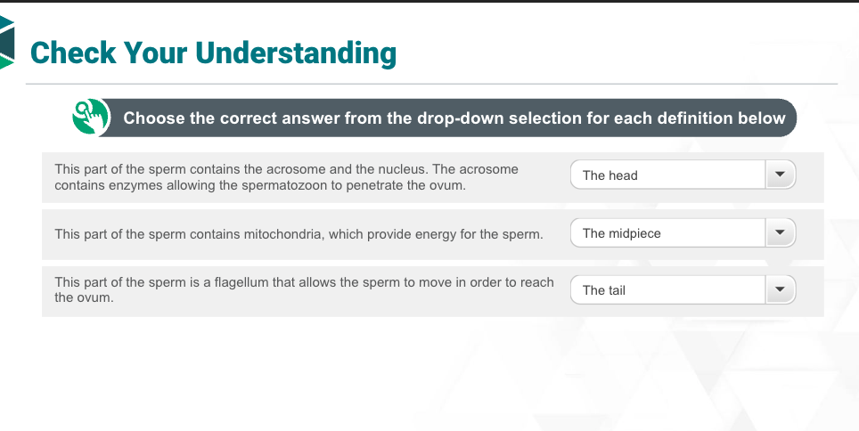
We know where spermatogenesis takes place, but let’s look at the parts of an individual spermatozoon. You will remember that spermatozoa is the plural form pof spermatozoon. Spermatozoa is also known as sperm which are the male sex cells. A sperm has three main parts.
Head: the head contains the acrosome and the nucleus. The acrosome contains enzymes allowing the spematozoon to penetrate the ovum.
the midpiece contains mitochondria, which provide energy for the sperm.
The tail is a flagellum that allows the sperm to move in order to reach the ovum.
Accessory Sex Glands
these glands add fluid to semen. There are three main accessory structures. Not all of these accessory structures are present in all species of animals.
Seminal Vesicles/Vesicular Glands: Are paired accessory sex glands that open into the ductus deferens at the intersection with the urethra. The seminal vesicles secrete a clear fluid that adds volume and nutrients to semen.
Prostate Gland: surrounds the urehtra. It is responsible for secreting prostactic fluid, which facilitiates the movement of sperm.
Bulbourethral Glands: also called cowpers glands are a pair of glands located on either side of the urethra. These glands secrete a thick, protein-rich compound that acts as a lubricant for sperm. The secretion from these glands also serves to flush acific urine from the urethra before semen enters.
Not all accessory sex glands are present in every species. The prostate gland is present in all of the species listed, but let’s take a look at some of the animals that do and do not have seminal vesicles and bubourethral glands:
Dogs have neither
Cats only have bulbourethral
Pigs have both
Ruminants have both
Horses have both
The Penis
the penis is the primary sex organ of the male reproductive system. It carries reproductive and urinary products out of the body through the urethra. The glans penis is the distal part of the penis where the opening of the urethra is located. The prepuce is the retractable fold of skin that covers the glans penis and is sometimes called the foreskin. The penis is comprised of erectile tissue, which is a collection of blood sinusoids, separated by connective tissue. This erectile tissue fills with blood upon sexual stimulation, resulting in an erection

The Anatomy and Physiology of the Female Reproductive System
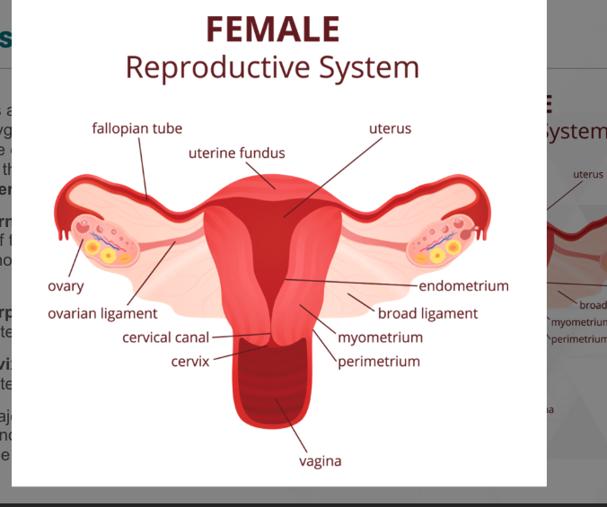
consists of the ovaries, uterine tubes, uterus, cervix, vagina, vulva, and mammary glands. The function of the female reproductive system is to create and support new life. This system produces eggs, transports them for fertilization and implantation, supports the developing embryo, provides a birth canal for the fetus, and produces necessary hormones.
Ovaries: The ovaries are a small pair of organs responsible for the production of the ova (egg) and secreting the hormones estrogen and progesterone.
Uterine Tubes: the uterine tubes are also referred to as oviducts or fallopian tubes. These tubes acquire the ova, or egg, from the ovaries and transport the zygote to the uterus once fertilization has taken place. The secretions of the uterine tubes also provide an environment that is suitable for the survival of sperm traveling from the vagina and the uterus.
Uterus: is a thick-walled, hollow organ that supports the developing embryo and fetus during gestation in pregnant females
Cervix: the cervix is made up of ring-like smooth muscle sphincters that provide a barrier to prevent foreign substances and infection from entering the uterus. The cervix remains closed except during estrus, when it relaxes to allow the entry of sperm.
Vagina: the vagina is a muscular tube lined with mucosa that serves as the passage from the opening of the vulva to the cervix or the uterus for coitus and childbirth.
Vulva: the vulva is also known as the pudendum, or the external female sexual organs
Mammary Glands: are milk producing glands in the female reproductive system. The number of glands is dependent upon the species and varies from 2 to 6 or more. In large animals, these glands are referred to as udders.

The Ovaries
Just like the testes in the male reproductive system are responsible for production of the male gamete, the ovaries are responsible for the production of the female gamete. The ovum, or egg is an essential part of the process of reproduction, because it fuses with a spermatozoon in order to create a fertilized egg, or zygote. The ovaries are indispensable to this process, because they are responsible for the production of the ova, along with estrogen and progesterone hormones.
The size and location of the ovaries varies depending on the species, but an ovary is comprised of a medulla and a cortex. The medulla is made up of connective tissue, while the the cortex contains small sacs called follicles. Each of these ovarian follicles contains a developing ovum. The primary function of the ovaries is to prepare and develop the ova to be expelled, or ovulated, when each ovum matues. Once an egg is released, it travels down the uterine tubes to the uterus.
Estrogen and progesterone are hormones produced by the ovaries to help regulate the reproductive cycle. The ovaries also produce hormones that are responsible for stimulating the growth that are responsible for stimulating the growth of graafian follicles; these are called follicle-stimulating hormones.
The Uterus
the uterus serves as the site of implantation for the fertilized egg, or zygote and supports the development of the embryo during pregnancy. The uterus consists of three parts: the cornus, the corpus, and the cervix.
The cornus: or horns of the uterus, are the cranial end of the uterus. This segment of the uterus has two horns that connect to the uterine tubes.
Corpus: the middle segment of the uterus.
Cervix: the cervix, or neck, is the caudal segment of the uterus that extends into the vagina.
There are three major tissue types that make up the uterus. A membranous outer layer of connective tissue known as the perimetrium surrounds the uterus. The muometrium make up the middle layer of the uterus and has two muscle layers divided by the stratum Vasculare. This is a later of connective tissue that contains the blood vessels and nerves of the uterine wall. The endometrium is the inner later of the uterus and has a mucosa and submucosa layer that contains endometrial glands, which assist in the development and survival of the embryo in the uterus.
The Mammary Glands
These glands are comprised of connective tissue and adipose tissue and contain internal compartments known as lobes. These lobes are divided into lobules, which are comprised of connective tissue and milk-producing cells called alveoli. Each mammary gland is associated with one nipple, or papilla, and the lobes of the mammary glands drain toward the teat via a lactiferous duct. These ducts come together and form a lactiferous sinus, which is made up of a gland cistern and a teat cavity. Milk travels from the gland cistern to the teat cavity and empties into the papillary ducts. The milk exists the body at the streak canal.
The mammary glands are responsible for lactation and the production of milk after parturition.


Topic 3: The Estrous Cycle, Mating, and Pregnancy
At puberty, an animal’s reproductive system begins a process to prepare the uterus to accept a fertilized egg. This process is known as the estrous cycle or heat cycle, and it is a cycle that will continue, unless interrupted by pregnancy, at a rate that is dependent upon the species. Monestrous species will experience one heat cycle a year, while polyestrous species will have multiple estrous cycles each year.
Ova develop in ovarian follicles until they reach mature size, at which time they are referred to as a graafian follicle. Ovulation is the rupture of a graafian follicle, which results in a ovum being expelled from the ovary and into the uterine tube. The ruptured follicle, now referred to as the corpus luteum, continues to grow in the ovary and produces progesterone, which prevents future estrous cycles. if the ovum is not fertilized, the corpus luteum will shrink and progesterone and secretion will slow to allow for a new estrous cycle to begin.

Now that we know what the estrous cycle is, lets take a look at the individual phases that divide the estrous cycle.
Phase 1: Proestrus: During this phase, the follicles in the ovary begin to mature and the endometrial lining of the uterus thickens in preparation for potential implantation of a fertilized ovum.
Phase 2: Estrus: This is the period when the female is receptive to mating, marked by heightened levels of estrogen and the presence of a mature ovum ready for fertilization.
Phase 3: Metestrus: In this phase, also known as diestrus, the ovary forms a corpus luteum from the remnants of the follicle, which secretes progesterone to support the endometrial lining and maintain the conditions suitable for pregnancy.
Phase 4: Diestrus: During the diestrus phase, if pregnancy does not occur, the corpus luteum degenerates, leading to a decrease in progesterone levels and the eventual shedding of the endometrial lining, resulting in menstruation.
Phase 5: Anestrus: This phase represents a period of inactivity in the reproductive cycle, where the female does not exhibit estrus behavior and ovarian activity is minimal. During anestrus, hormone levels are low, and the body prepares for the next reproductive cycle.
Terms related to mating used in the vet office
Copulation or coitus are terms used to describe sexual intercourse
Mount is a term used to describe the initial step in animal mating; it involves one animal climbing on top of another animal
Tie is a period of copulation between a male and female canine during which the animals are locked together by penile erectile tissue. This is a normal phenomenon; during tie dogs can stay locked together for 15-20 minutes.
Conception is the beginning of a new animal resulting from fertilization; fertilization is the term for the formation of a viable zygote through the combining of a male sperm and female ovum
Zygote is a fertilized egg
Meiosis is the division of sex cells in which the cell receives half of the chromosomes of each parent
implantation is the process by which the zygote attaches to and embeds in the uterus
Embryo- a zygote becomes an embryo after the implantation process
Monoestrous - is an animal that has only one estrous cycle each year
Polyestrous is an animal that has more than one estrous cycle each year
The Placenta and Fetal Membranes
the placenta is an organ that function to join the mother to the developing fetus during gestation. This organ provides oxygen and nutrients to the developing fetus and helps to remove waste products from the blood. The umbilical cord is the structure that connects the mother to the developing fetus. The umbilical cord connects to the fetus at the umbilicus or navel.
“fetal membranes'“ is a term used to describe the placenta and its associated structures. The amnion or amniotic sac, is the innermost membrane that surrounds the embryo and forms the amniotic cavity. This cavity is filled with amniotic fluid and protects the fetus. The allantois is the innermost layer of the placenta. Fetal waste products accumulate in the sac that is formed between it and the amnion. The outermost layer of the placenta is called the chorion.
Terms Related to Pregnancy
nulligravida - refers to a female that has never been pregnant
primigravida refers to a female in their first pregnancy
multigravida refers to a female who has had multiple pregnancies
multiparous is a female who has had multiple offspring during the same gestation
Viviparous refers to a female who is bearing live young
Oviparous refers to a female who is bearing eggs
Ovoviviparous refers to a female who produces eggs that hatch within the body, so that live young are born within the body but without placental attachment, snakes are an example.
Birth and Stages of Parturition
Parturition is the act of giving birth. Antepartum is the term used to define the period of time before the onset of labor. Parturition itself can be divided into three stages:
Stage 1: Dilation of the cervix
Stage 2: Uterine contractions and expulsion of the fetus
Stage 3: Separation of the placenta from the uterus
Postppartum is the term used to refer to the time period immediately following labor. This period begins after the delivery of the fetus. Once the fetus is delivered, the mother’s uterus begins to return to normal size in a process called uterine involution.


Terms for Testing, Pathology and Procedures of the Reproductive System
Common Diagnostic Procedures
Aminocentesis: a surgical procedure used to obtain a sample of amniotic fluid for testing, often performed to assess fetal health and detect genetic conditions.
Radiography: a diagnostic imaging technique that uses X-rays to visualize the structures of the reproductive system, often employed to identify abnormalities such as tumors or fractures.
Ultrasound: a non-invasive imaging technique that uses high-frequency sound waves to create visual images of the developing fetus and assess the condition of the reproductive organs, commonly used in prenatal care.
Diseases and Disorders of the Reproductive System
Dystocia: a complication during labor characterized by difficult or abnormal delivery, often requiring medical intervention such as the use of forceps or cesarean section.
Fetal Defects in the Reproductive System: abnormalities present at birth affecting the structure or function of the reproductive organs, which can lead to complications in sexual development and fertility later in life.
Cryptorchidism: a condition in which one or both of the testes fail to descend into the scrotum, increasing the risk of infertility and testicular cancer if not treated.
Mastitis: an infection of the breast tissue that results in breast pain, swelling, warmth, and redness, and can lead to flu-like symptoms. It often occurs during breastfeeding due to blocked milk ducts or bacteria entering the breast.
Common Procedures of The Reproductive System
Assisted Delivery involves the use of hands or equipment to aid in the delivery of a fetus. In cattle, sometimes obstetric chains are placed around the cal’s legs and force is then applied to help extract the fetus from the mother
Cesarean Section or C-Section is the delivery of offspring through an incsion made in the mothers abdominal and uterine walls. A c-section is often preformed when a vaginal delivery would put the mother at risk.
Electroejaculation is a technique used to collect semen from male animals for breeding purposes, particularly when natural mating is not possible or practical. This method involves the use of an electrical stimulus to induce ejaculation, allowing for the harvesting of sperm for artificial insemination.
Episiotomy is a surgical incision made in the perineum and vaginal wall to facilitate childbirth. This procedure is often performed to prevent tearing during delivery or to expedite the delivery process in cases where the fetus is in distress.
Fetotomy is a surgical procedure performed during labor, typically in cases of fetal death, where the fetus is too large to be delivered naturally. This technique involves making incisions to remove portions of the fetus to facilitate safe delivery.
Mastectomy: A surgical procedure to remove one or both breasts, typically performed as a treatment for breast cancer or as a preventive measure for those at high risk of developing the disease.
Oophorectomy: A surgical procedure that involves the removal of one or both ovaries, usually performed to treat ovarian cysts, tumors, or other ovarian diseases, and can also be done as a preventive measure in women at high risk for ovarian cancer.
Orchectomy/Neuter: A surgical procedure that involves the removal of one or both testicles, commonly performed to prevent reproduction and reduce the risk of certain diseases in male animals.
Ovariohysterectomy: A surgical procedure that entails the removal of both the ovaries and the uterus, commonly performed in female animals to prevent unwanted pregnancies and certain health issues.
Vasectomy: A surgical procedure that involves surgically removing the ductus deferens.
Abbreviations Related to the Reproductive System
ACT - American College of Theriogenologists
AI - Artifical Insemination
BPH - Benign Prostatic Hypertrophy
C- Castrated
C-Section - Cesarean Section
CL - Corpus Iuteum
CMT - California Mastitis Test
N- Neutered
OB - Obsetrics
OHE - Ovariohysterectomy
OVH - Ovariohysterectomy
pg - pregnant
RP - Retained Placenta
S - Spayed
TVT - Transmissible Venereal Tumor