Integumentary System (Skin) - Vocabulary Flashcards
Integumentary System (Skin) – Comprehensive Study Notes
Overview
The skin has two distinct regions: the epidermis (superficial) and the dermis (underneath the epidermis).
The hypodermis (subcutaneous layer) lies deep to the skin and is not part of the skin proper.
Epidermis is epithelial tissue and serves as the protective shield of the body; dermis is dense irregular connective tissue and provides strength and elasticity. Only the dermis is vascularized; nutrients reach the epidermis via diffusion from the dermis.
The hypodermis is primarily adipose tissue and acts as insulation, shock absorption, and a superficial fascia that anchors the skin to underlying muscles.
Glands and other structures (hair follicles, sebaceous glands, sudoriferous glands) are derived from epidermal tissue and reside in the dermis.
Quantitative facts about the skin
If the skin were removed and laid flat, the surface area would be about .
The skin weighs about , which is roughly 7 ext{%} of total body weight.
Skin thickness varies from roughly or more in different parts of the body.
Epidermis thickness is generally comparable to a sheet of paper; the variation exists across the body.
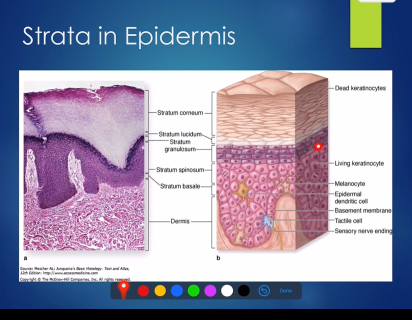
Epidermis: structure and cells: Keratinized stratified squamous epithelium
Epithelium type: The epidermis is a stratified squamous epithelium with multiple cell layers; the surface layer consists of flat, dead cells. Keratinization is highlighted today as a key concept in the epidermis.
Epidermis layers (four in thin skin, five in thick skin): stratum basale, stratum spinosum, stratum granulosum, stratum lucidum (only in thick skin), stratum corneum.
Cells in the epidermis (major cell types):
Keratinocytes – main cell type; synthesize keratin, which provides protection. They are tightly joined by desmosomes and originate in the stratum basale. By the time they reach the outermost layer, they have died and are sloughed off continuously.
Turnover: about 25 to 45 days from basale cell division to shedding.
Daily shedding: about 50 million cells per day (roughly 50 million cells/day).
Melanocytes – produce melanin, the pigment that colors the skin and helps shield against UV radiation. looks like a hand, palm sits in stratum basale
Location: primarily in the stratum basale.
Melanin travels to the keratinocytes at the surface to form a protective umbrella over the nucleus.
Melanin granules come in two forms: yellow-to-tan and reddish-to-black.
Percentage in the basale layer: approximately 10 ext{%} ext{ to } 25 ext{%} of cells are melanocytes; most are keratinocytes (~75 ext{%} ext{ to } 90 ext{%}).
Humans share the same relative number of melanocytes; skin color differences come from the amount and form of melanin produced.
Langerhans cells (dendritic cells) – macrophages of the skin; derived from bone marrow; they ingest foreign substances that penetrate the epidermis.
They are part of the immune defense of the skin.
Tactile cells (Merkel cells) – sensory nerve endings for fine touch; associated with specialized nerve fibers.
Epidermal layers and changes as keratinocytes move outward:
Stratum Basale – (strata means sheet) deepest layer; a single row of stem cells; actively mitotic; youngest keratinocytes; every time a new cell is made, one stays and the other migrates. all the way to the surface takes 25-45 days, melanocytes are 10/25% of the layer
Stratum spinosum – several cell layers; desmosomes create a spiny appearance; contains melanin granules and dendritic cells. web-like appearance due to the interconnecting processes of the keratinocytes, which provides strength and flexibility to the skin.
Stratum granulosum – (where cell appearance changes) Between 1–5 cell layers depending on body region; keratinocytes flatten as their nuclei/organelles disintegrate; keratinization begins; keratin granules form keratin; lamellar granules (water resistant glycolipid) secrete a water-resistant glycolipid to slow water loss. Cells above granulosum begin to die
Stratum lucidum – present only in thick skin (palms and soles); 2–3 rows of clear, dead keratinocytes.
Stratum corneum – most superficial; 20–30 rows of dead, flatten keratinized cells; accounts for about 75% of epidermal thickness; keratin and thickened plasma membranes protect against abrasion and penetration; glycolipid waterproofs the epidermal surface; cells are cornified and lack nuclei.
Visual analogy: cells look like shingles near the surface and appear horn-like due to cornification.
Consequence: dandruff and dander arise from shedding of corneocytes.
Additional details:
The epidermis originally receives nutrients via diffusion from the dermis since it is avascular.
The term “stratum” refers to a sheet or layer.
The image in class shows the dermis with the epidermis and the various cell types embedded in layers.
Dermis: structure and components
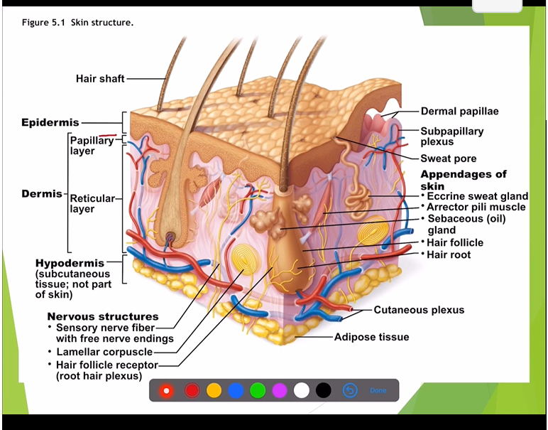
The dermis is a strong, flexible connective tissue layer composed of dense irregular connective tissue. highly vascularized
Cells in the dermis include fibroblasts, macrophages, and mast cells, which play essential roles in providing structural support, immune defense, and inflammatory responses.
Two sublayers:
Papillary layer (superficial, thin): made of areolar connective tissue; contains dermal papillae that project into the epidermis. Because it is lose it allows phagocytes (immune system cells) to roam
Dermal papillae ( hill looking ) contain capillary loops and small sensory nerve endings; they increase surface area for exchange with the epidermis and contribute to fingerprints when they project into the epidermis in thick skin.
In thick skin, dermal papillae sit on dermal ridges, forming distinct fingerprints.
Reticular layer (deeper, thick): makes up about 80%of the dermis; dense irregular connective tissue with collagen and elastic fibers. Create cleavage lines because most collagen fibers parallel to skin surface
Collagen fibers provide strength and water-binding capacity; elastic fibers provide resilience and recoil.
The elastic network decreases with UV exposure, contributing to leathery skin and loss of elasticity.
Glands and appendages: hair follicles, sebaceous glands, and sweat (sudoriferous) glands are associated with the dermis and are derived from epidermal tissue.
Nerve fibers and blood vessels: the dermis is highly vascularized and richly innervated; contains lymphatic vessels.
Cleavage (t) lines: collagen fibers run in various directions but tend to align parallel to the skin surface; incisions made parallel to these cleavage lines heal with less gap and scarring.
Hypodermis (Subcutaneous layer)
Location: deep to the skin; not technically part of the skin.
Composition: primarily adipose tissue (fat) and some areolar connective tissue.
Functions: insulation, cushioning/shock absorption, energy storage; anchors the skin to underlying muscles (superficial fascia).
Skin color and pigmentation
Major pigment: Melanin
Produced in melanocytes and transferred to keratinocytes; provides color and UV protection.
Two forms of melanin granules: yellow-to-tan and reddish-to-black; color differences arise from the amount and form of melanin and the number of melanocytes.
Melanocytes comprise roughly of cells in the stratum basale; the majority of cells in this layer are keratinocytes (about ).
All humans have about the same relative number of melanocytes; skin color differences are due to melanin production and distribution, not large differences in melanocyte numbers.
Prolonged sun exposure stimulates melanin production, darkening the skin (tanning) as a protective umbrella for keratinocytes.
Other pigments:
Carotene: a yellow-orange pigment found in carrots and sweet potatoes; accumulates in the stratum corneum and the hypodermis; more visible in the palms and soles where the stratum corneum is thick; can be converted to Vitamin A (retinol) for vision.
Hemoglobin: the oxygenated form imparts a red/pink hue; more visible in light-skinned individuals where the epidermis is more translucent to reveal underlying blood (rosy complexion).
Clinical color cues and interpretations:
Cyanosis (blue): potential hypoxemia or poor oxygenation.
Erythema (red): embarrassment, fever, hypertension, or inflammation due to increased blood flow.
Pallor (pale): anemia, low blood pressure, fear, or shock.
Jaundice (yellow): liver disorders in adults; in newborns it can indicate an inability to process bilirubin or other issues; will be discussed later.
Bronzing: associated with Addison's disease (adrenal insufficiency).
Bruises: clotted blood beneath the skin.
Practical implications and takeaways
The skin is an organ with multiple tissues and functions, including protection, sensation, temperature regulation, metabolism (Vitamin D synthesis), and immune defense.
Understanding the layered structure (epidermis and dermis, plus the hypodermis) helps explain not only normal skin function but also clinical observations such as sun damage, aging, and color changes.
The epidermis’ keratinization and the presence of a waterproof glycolipid layer are critical for preventing water loss and maintaining barrier integrity.
Cleavage lines influence surgical approaches and healing outcomes due to the organization of collagen fibers in the dermis.
Quick review of key terms
Epidermis: stratified squamous epithelium; avascular; four to five layers depending on skin type.
Dermis: dense irregular connective tissue; papillary and reticular layers; contains hair follicles, glands, nerves, and blood vessels.
Hypodermis: subcutaneous layer; adipose tissue; insulation and cushioning.
Keratinocytes, Melanocytes, Langerhans (dendritic) cells, Merkel (tactile) cells.
Strata: basale, spinosum, granulosum, lucidum (thick skin only), corneum.
Dermal papillae: contribute to fingerprints and surface area.
Cleavage lines: orientations of collagen fibers for optimal wound healing.
Pigments: Melanin, Carotene, Hemoglobin.
Notes on references and future topics
The epidermis receives nutrients via diffusion from the dermis due to its avascular nature.
Later discussions will cover physiology of integumentary structures (e.g., hair, glands) and deeper dives into skin diseases, aging, and dermatologic conditions.