Lecture 2 and 3 - Restriction Enzymes
A phenomenon called host-controlled variation was described by various groups: Luria and Human (1952), Anderson and Felix (1952) and Bertani and Weigle (1953)
Bacteriophages varied in their ability to grow on different host strains
Once growth was achieved on one strain, the bacteriophages could continue to grow on this strain but were restricted in their ability to grow on other strains
The finding of host-controlled variation provided the first evidence that:
Bacteria applied an identification (modification) tag to their DNA
DNA lacking this tag (upon transfer from one strain to another) was recognised as ‘non-self’ and restricted
In the 1960s, Werner Arber and Daisy Dussoix demonstrated that the phage DNA carried the host range imprint
The imprint was conferred by methylation of DNA and host-controlled variation came to be known as bacterial restriction-modification system
Bacterial restriction-modification system
Restriction enzymes protect bacteria from infection by viruses
Non-host DNA is cleaved whilst the cell’s own DNA is protected from digestion by site specific methylation by DNA methyltransferases
Restriction enzymes occur in combination with DNA methyltransferases, both recognising the same DNA sequence
Methylated DNA poses a hinderance to restriction
In 1971, Kathleen Danna and Daniel Nathans demonstrated that:
Endonuclease R (discovered by Hamilton Smith and Kent Wilcox) could be used to produce specific fragments of simian virus 40
Electrophoresis provided a good tool to separate the DNA fragments
In 1978, Arber, Smith and Nathans were awarded the Nobel Prize
Restriction enzymes are needed as naturally occurring DNA molecules (chromosomes and some plasmids) are too large to be manageable in the laboratory eg.
Human genome → 3038 Mb
Escherichia coli chromosome → 4.64 Mb
Streptomyces coelicolor chromosome → 8.6 Mb
What are the properties of restriction enzymes?
Hydrolyse phosphodiester bonds in sugar-phosphate backbone of each strand of DNA at specific nucleotide sequences
Generate discrete, gene size DNA fragments suitable for manipulation
The fragments generated can be re-joined in the lab
Mechanism of action
All restriction enzymes catalyse the hydrolysis of DNA phosphodiester bonds, leaving a phosphoryl group attached to the 5’ end and a 3’ hydroxyl group
Cleavage takes place by in-line displacement of 3′ oxygen from phosphorus by magnesium-activated water
Four types of restriction enzymes
Type I → multisubunit, restriction-modification enzymes (dual activity) that randomly cut DNA away from recognition sequence
Type II → cut DNA at defined positions close to or within the recognition sequence, recognise symmetric DNA sequences or palindromes (sequence in 5’-3’ direction on one strand same in the 5’-3’ direction on the other)
Type III → large, combination restriction and modification enzymes, recognise specific 5-7 bp sequences and cut DNA outside the recognition sequence
Type IV → methylation-dependant restriction enzymes, cleave at variable distance from recognition site
Type I
Identified in E.coli as barriers to foreign DNA entry and have several sub-families (A-E)
Present in almost one-half of Bacteria and Archaea
Consist of three different subunits
HsdM → modification
HsdR → restriction
HsdS → sequence recognition
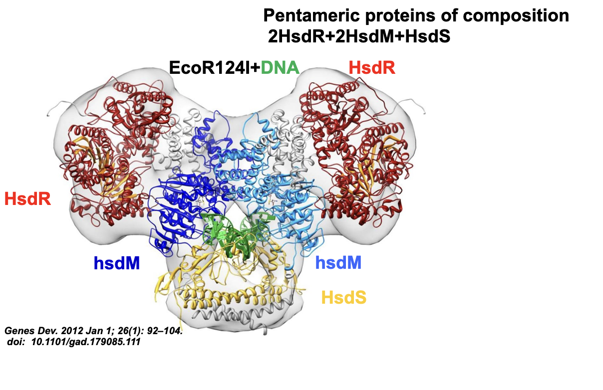
Mechanism of Type I
Require ATP, Mg2+ and S-adenosylmethionine for activity
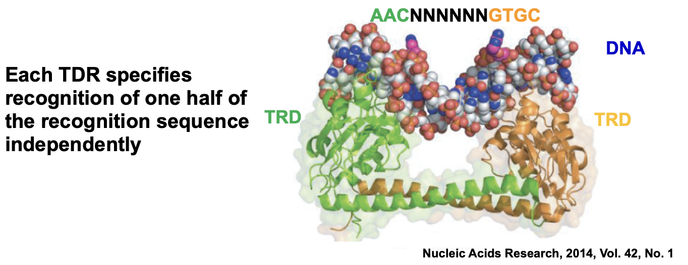
They interact in general with two asymmetrical bi-partite recognition sites → AACNNNNNNGTGC
Translocate the DNA in an ATP-hydrolysis dependent manner and cut the DNA distal to the recognition sites, approximately half-way between two sites
Specific plasticity
HsdS subunits of Type I enzymes are responsible for DNA sequences recognition
Duplicated organisation comprising of two target recognition domains (TRDs) in tandem
Each TDR specifies recognition of one half of the recognition sequence independently
Genetic rearrangements between hsdS genes results in hybrid HsdS subunits with novel specificity
Type III
Recognise two sequences in opposite orientations within same DNA and cleave 25-27 nucleotides away from their recognition site
Examples are EcoP1I and EcoP15I
Found in most sequenced bacterial genomes
Require ATP, Mg2+ and S-adenosylmethionine as cofactors although they do not necessarily need S-adenosylmethionine
Number of nucleotides between two sequences is variable
Type III cleavage mechanism
Type III restriction enzymes consist of two subunits:
Mod (homodimeric) → responsible for DNA recognition and methylation
Res → responsible for ATP hydrolysis, DNA translocation and cleavage
Form a hetero-tetrameric Res2Mod2 complex
Subunit composition
Type III restriction enzymes comprise two
modification (Mod) subunits, each containing a target
recognition domain (TRD) to bind to the target sequence
A MTase catalytic domain to monitor the methylation
status of an adenine in the target and transfer –CH3 group
to N6 of adenine
And two restriction (Res) subunits each
containing a DNA helicase and ATP-hydrolysing domain,
and an endonuclease domain (6, 8)
Type II
Cleaves specifically within or close to its recognition site
No requirement of ATP or GTP for its activity
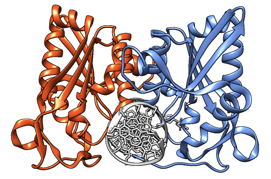
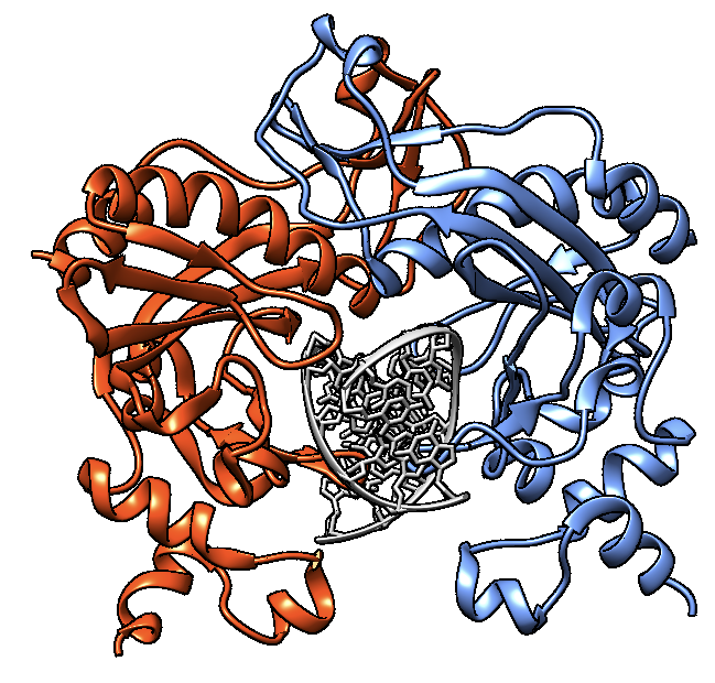
Form homodimers of 2 × 30 kDa molecular mass
Recognises palindromic sequences of 4-8 bp in length
Requires Mg2+ for its activity
Cleaves the bond between the 3’-OH and the 5’-phosphate in DNA
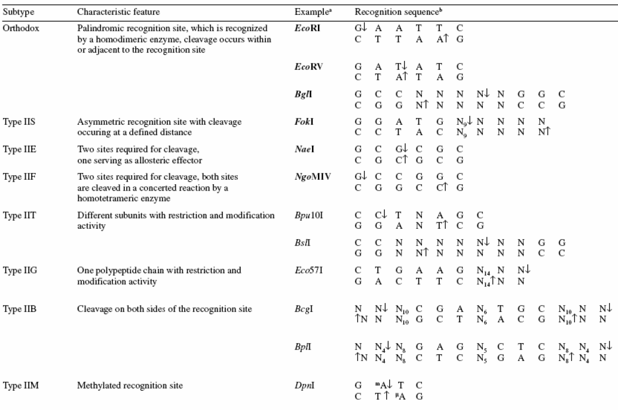
Classified based on enzymatic behaviour rather than evolutionary relatedness
Type II subtypes
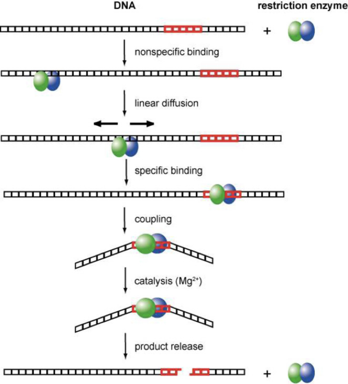
Target Site Location
Binds to DNA non-specifically and then slides along DNA randomly until it encounters the target sequence
Star Activity
Restriction enzymes are capable of cleaving sequences which are similar, but not identical, to their defined recognition sequence
This altered specificity has been termed star activity
For example, EcoRI’s recognition sequence is GAATTC and has star activity at N/AATTN (GGATTT, AAATTT, GAATTT and GAATTA)
What are the causes of star activity?
Low ionic strength and alkaline pH
Presence of organic solvents, such as glycerol or DMSO
When Mg2+ is replaced by Mn2+
High enzyme concentrations under optimum buffer conditions
Among the most accurate enzymes known:
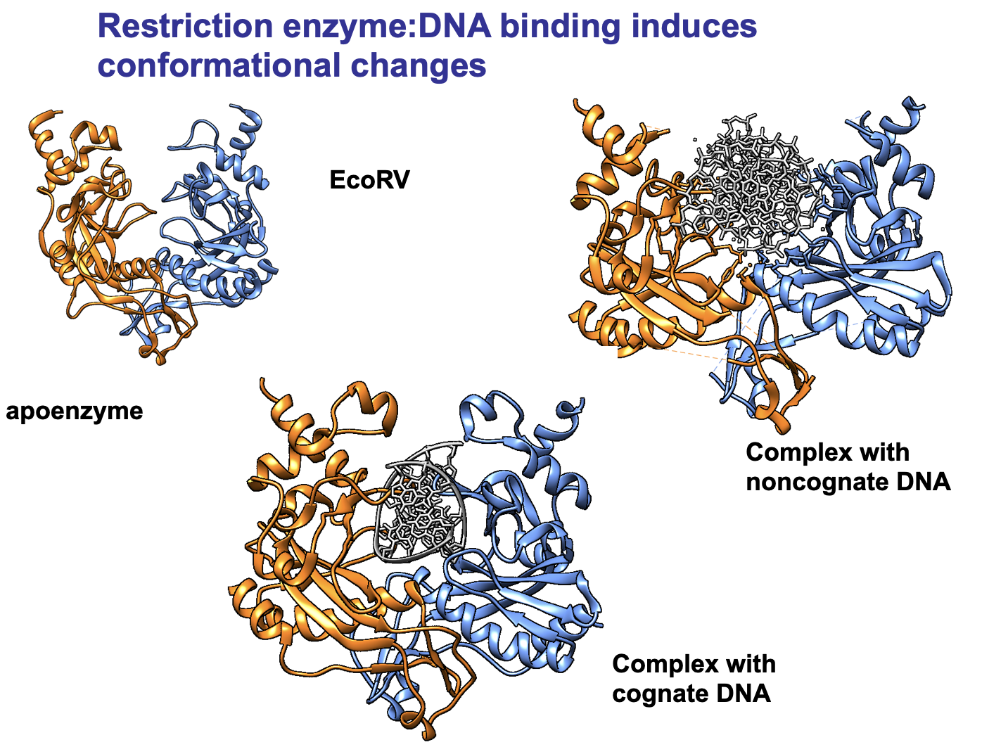
Cleavage of non-cognate substrates is at least five orders of magnitude slower than cleavage of the cognate substrate
Different types of DNA ends after restriction
5’overhang ends
Blunt ends
3’overhang ends
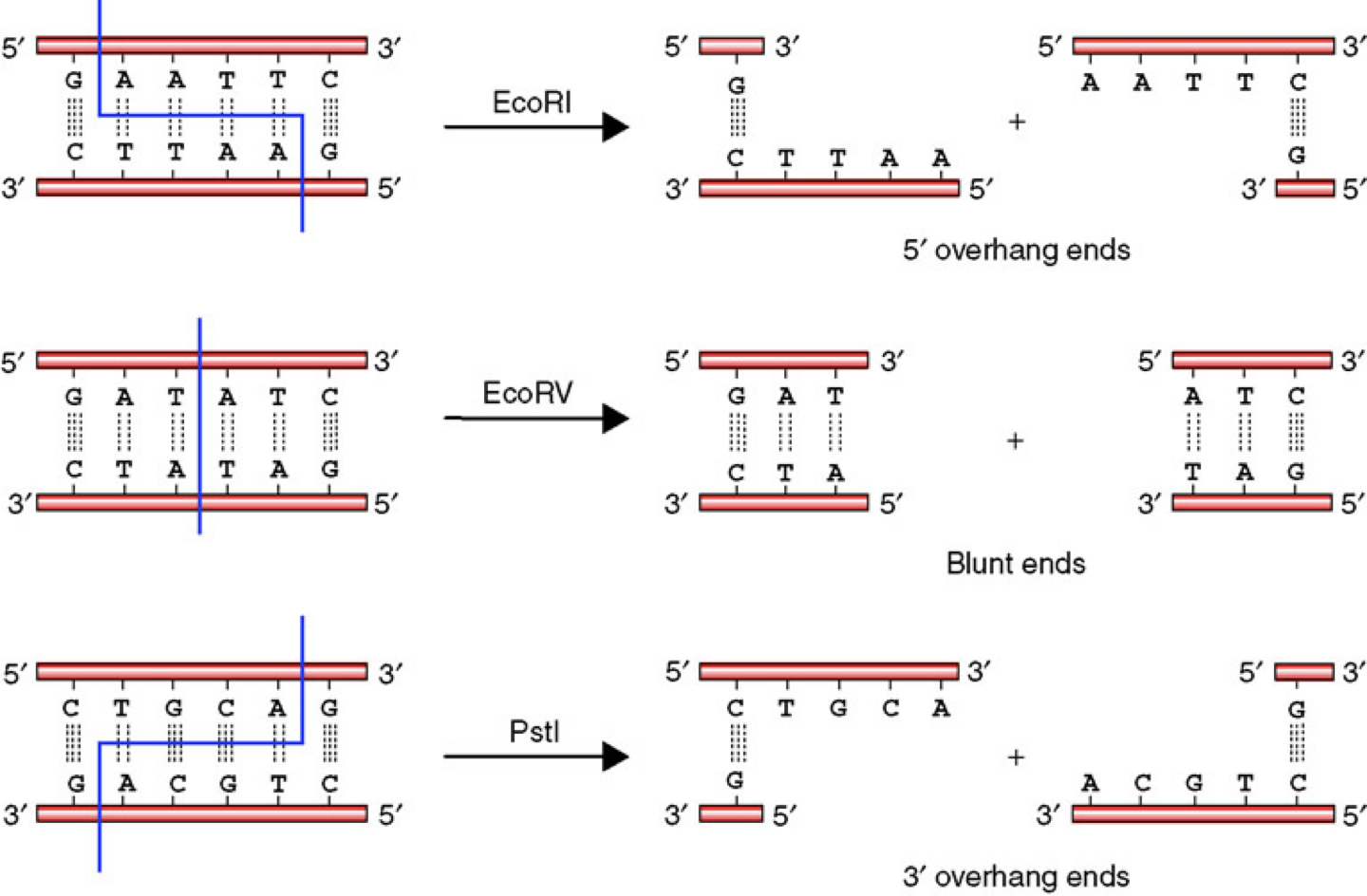
Why is it important to have different DNA ends?
Blunt ends are not sequence-specific and can be joined to any other blunt-ended DNA fragment, regardless of its sequence
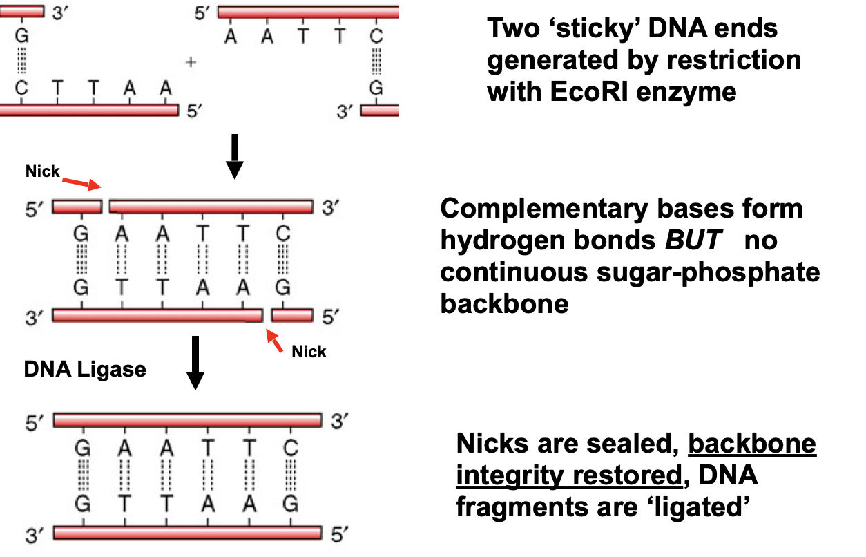
Sticky ends are useful for cloning because the complementary overhangs can temporarily hold two different DNA fragments together, allowing DNA ligase to permanently join them
Nomenclature of Restriction Enzymes
The name reflects origin → species name of the host organism where the restriction enzyme was isolated from
The number written as Roman numeral indicates the order in which they were isolated
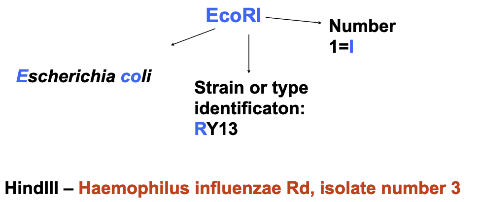
A restriction enzyme will always cut at a specific nucleotide sequence no matter the source of the DNA
Recognition sequences can be continuous or discontinuous
Continuous sequences → the bases are all adjacent and the enzyme cuts within this sequence eg. Kpnl 5’-GGTACC-3’
Discontinuous sequences → the recognised bases are separated by one or more non-specific (spacer) nucleotides and enzyme identifies two short half-sites eg. Xmnl 5’GAANNNNTTC-3’
Recognition sequences can vary in length eg. EcoRI 5’GAATTC-3’ and Notl 5’-GCGGCCGC-3’
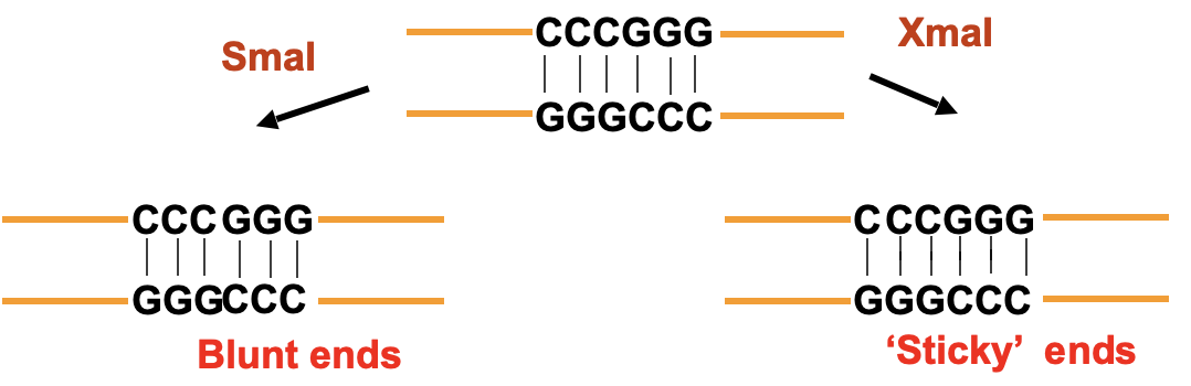
Isoschizomers can recognise the same recognition site but cut at a different position
DNA ligases
Catalyse the formation of phosphodiester bond between two DNA chains that are part of a double helix
Requires a free hydroxyl group (OH) at the 3’ end of one DNA chain and a phosphate group (Pho) at the 5’ end of the other
Can ligate ‘sticky’ or blunt ends
Their natural role is to seal nicks in the backbone of double-stranded DNA after replication, recombination or repair processes
Energy sources for Ligase activity:
Escherichia coli → NAD+
Animal cells and bacteriophages → ATP
T4 DNA Ligase from bacteriophage T4 is the most commonly used in the lab
DNA ligase mechanism of action
Ligase reacts with ATP to form a covalent enzyme-ADP complex
Ligase-AMP complex activates phosphate group at 5’end of DNA
Nucleophilic attack by OH group at 3’end of DNA which results in a phosphodiester bond being formed and AMP being released
Energetic cost → hydrolysis of two phosphate bonds at step 1 to form one phosphodiester bond at step 3
DNA cloning
Clone → A copy or imitation of something ( eg. organism genetically identical to another, two or more identical DNA molecules)
Cloning → Ability to construct recombinant DNA molecules and maintain them in cells, generating clones
What is needed for this?
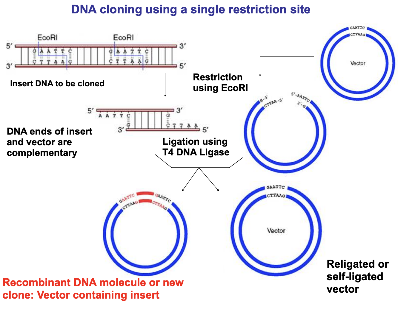
Insert DNA → A DNA fragment generated by enzymatic activity (eg. restriction enzymes) or physical treatment (eg. mechanical fragmentation of a large DNA molecule)
Vector → A DNA molecule that provides the information necessary to propagate the cloned DNA fragment, commonly a closed-circular double-stranded plasmid DNA molecule
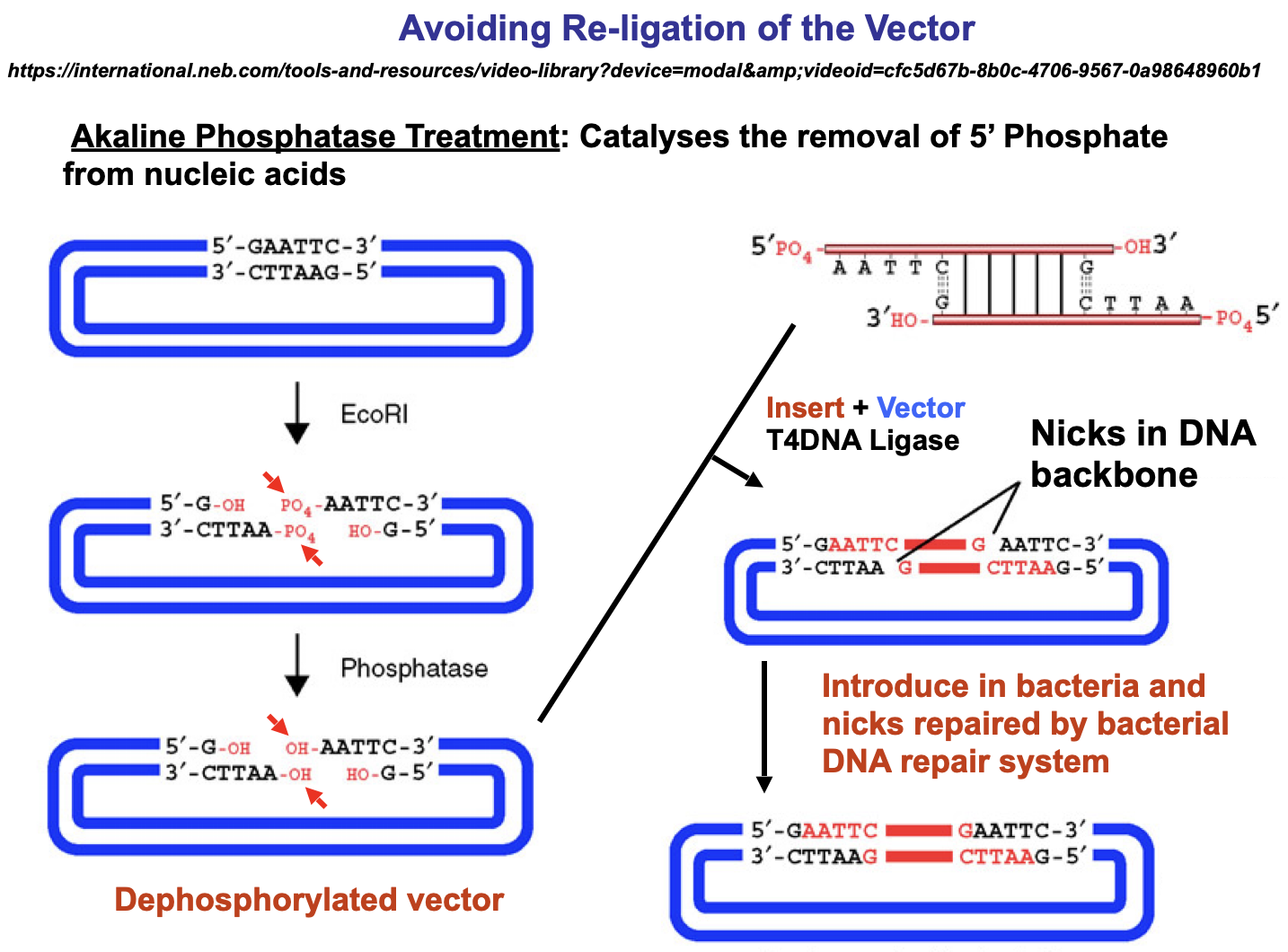
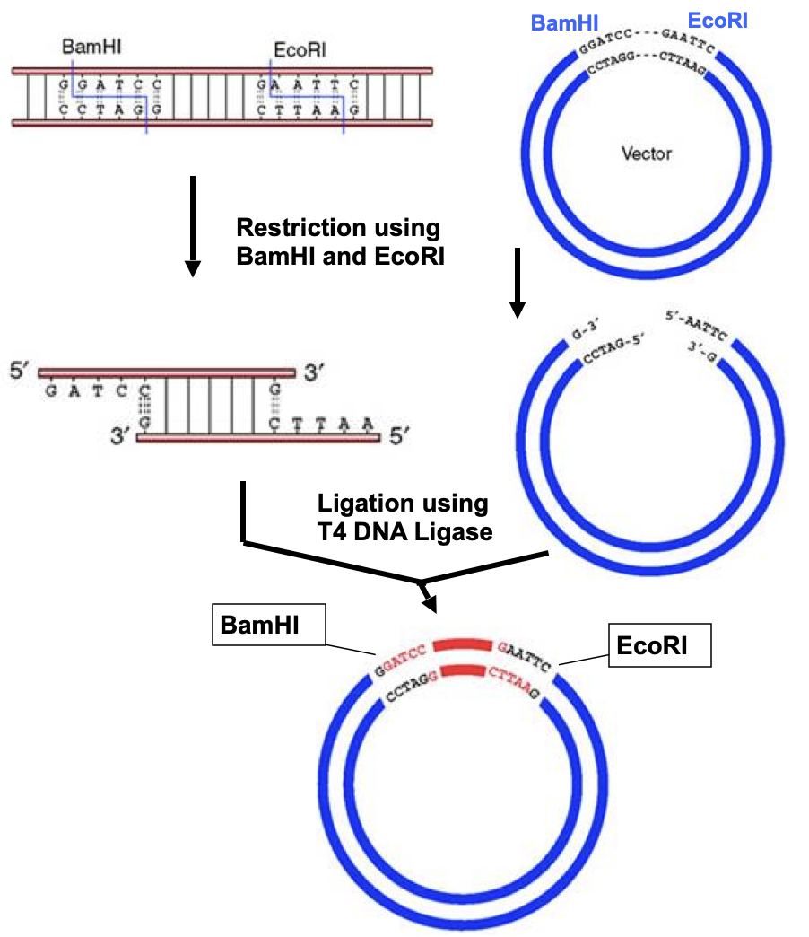
The use of phosphatase can be avoided by the use of two different, non-compatible restriction enzymes
Ends of the vector cannot anneal as the overhang bases are not compatible
Ligation only occurs with the fragment
The fragment can only insert in one direction for the same reason
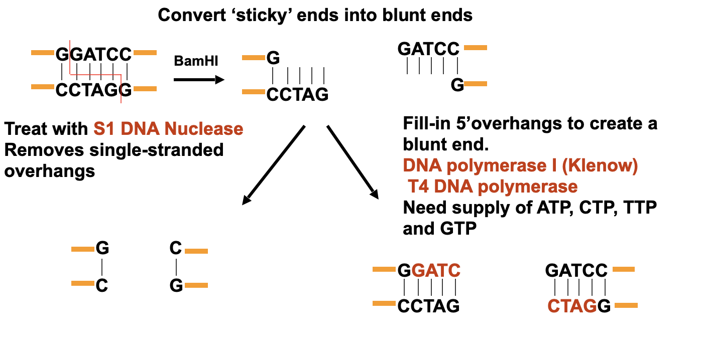
What happens when restriction sites do not occur at the right place in the fragment you want to close?
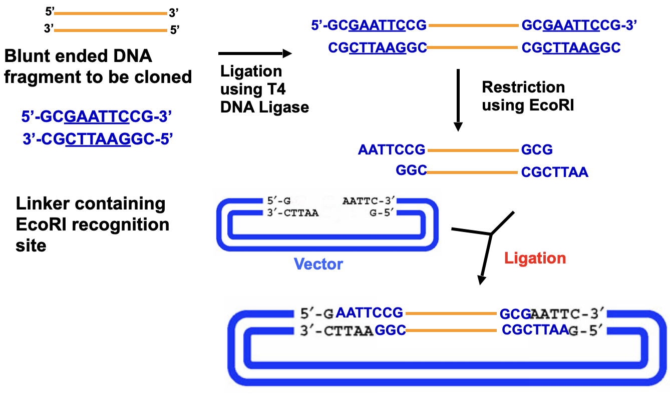
Any blunt end can be ligated to another blunt end, even if generated with different restriction enzymes
Ligation of blunt ends is less efficient than ligation of sticky ends
Oligonucleotide Linkers
Linkers are short duplex oligonucleotides that contain a restriction endonuclease cleavage site and can be ligated to a double stranded DNA fragment in order to create a new restriction site
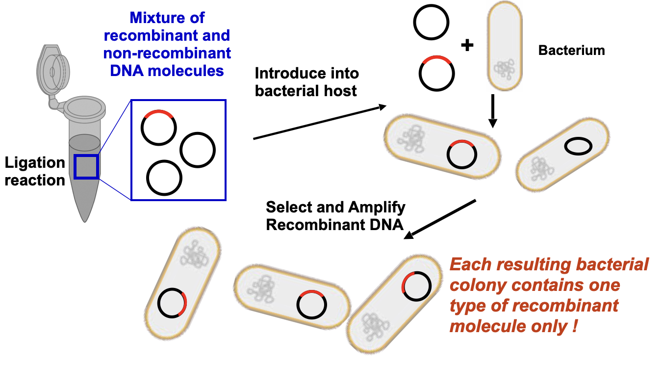
Introducing DNA into bacteria to propagate and maintain cloned DNA fragments
Introduction of DNA (vectors or ligation reactions) into the cell
Allows isolation of individual clones from complex mixtures of recombinant DNA
Mixed population of DNA is segregated into its individual components
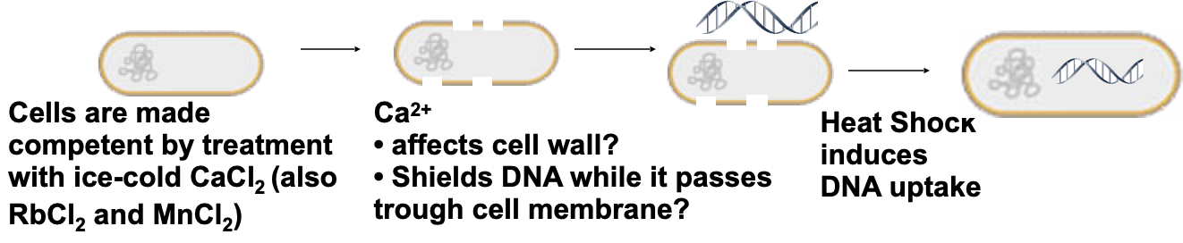
Basic Methods to introduce DNA into bacteria
Chemical transformation of competent cells:
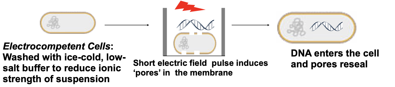
Electroporation
Protoplasts transformation

Main features of E. coli strains used in molecular cloning
Partially or totally deficient in native E. coli DNA restriction systems
Exonuclease and recombination activities reduced or abolished
Activity of proteases is abolished
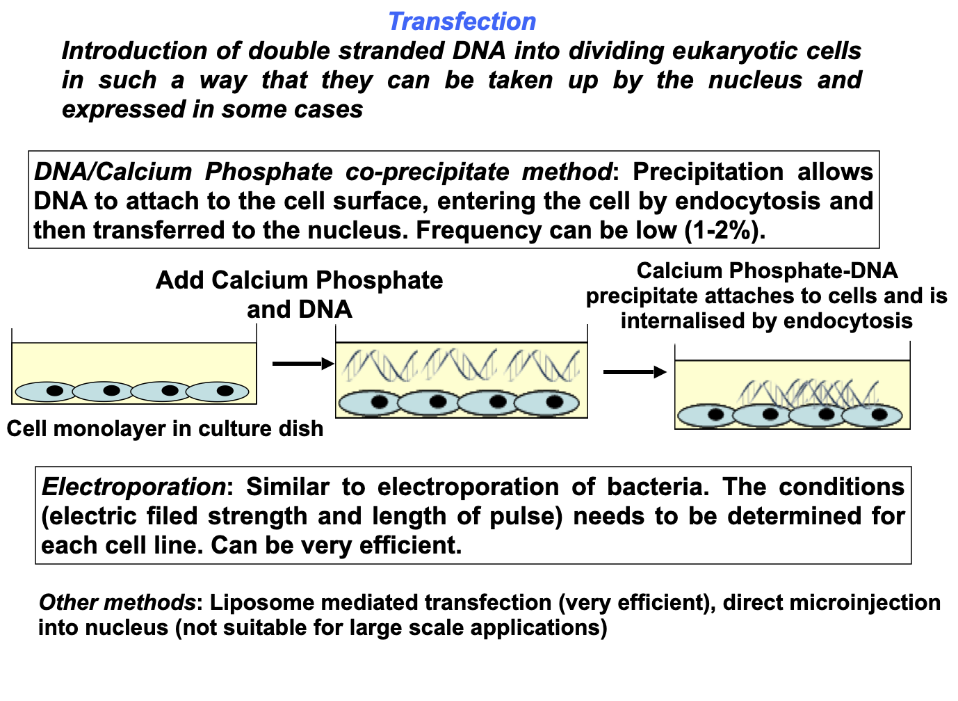
Introducing DNA into Mammalian cells