Brain and Cognition
Term 1
Week 8 - Intro
Intro
Cognitive psych - The study of mental processes related to sensory input, memory, perception, attention, and thought (Neisser, 1966). Involving understanding the internal processes in normal and pathological contexts.
Looking - visual perception, listening - auditory perception, paying attention - attention, understanding - language skills, learning - memory encoding, remembering - memory retrieval
We learn about cognitive because it provides insight about attributes that help make us human (thought, emotion, sensation, memory, language and consciousness). It will help us understand the behaviour of those with neurological and psychiatric problems. It teaches you about the strengths and weaknesses of the scientific method
Historical Context: 1950s-1980s: Growth of interest in the mind, psycholinguistics, and disillusionment with behaviorism. Key Developments include The mind-computer metaphor for brain processes.
Approaches in Cognitive Psychology:
Experimental Cognitive Psychology: Studies cognitive processes in healthy individuals.
Cognitive Neuropsychology: Research on brain-damaged patients.
Computational Cognitive Science: Development of models to explain cognition.
Cognitive Neuroscience: Investigates how cognition is implemented in the brain.
Common Research Questions: Is the brain modular or holistic? How are different brain regions involved in cognition?
Cognitive Research Settings:
Naturalistic: Ecologically valid but harder to control variables.
Quasi-Experimental: Useful but correlation does not imply causation.
Experimental: Controlled but low ecological validity.
Applications of Cognitive Psychology:
Commercial: Product design and advertising.
Engineering: Instrumentation and signage design.
Judicial: Improving face recognition in law enforcement.
Education: Effective learning techniques.
Clinical: Cognitive tests to diagnose and intervene in neurocognitive diseases.
Research Methods in Cognitive Psychology:
Reaction Time (RT): Measures mental processing speed.
Donders' Subtraction Method (1868): Time taken for each stage of a cognitive process.
Sternberg's Additive Factor Logic (1969): Time to process multiple components (e.g., short-term memory scanning).
Laboratory Methods: Reaction times, accuracy, and brain imaging techniques (fMRI, EEG). Study of brain regions and neural processes involved in cognition.
Week 9 - Anatomy and Physiology of the Nervous System
The structure of neurons, the cells of the nervous system
The use of animals in research: Most biological psychologists and neuroscientists are interested in the human behaviour, but why animals?
Mechanisms of behaviour are similar across species – and sometimes easier to study in non-human species
We are interested in animals for their own sake
Animal studies shed light on human evolution
Legal or ethical restrictions prevent certain kinds of research on humans
Degrees of opposition to animal research:
Minimalist –reduction, replacement, refinement
Abolitionist – “We have no moral option but to bring this research to a halt. Completely” (Regan, 1986)
The human nervous system is comprised of two kinds of cells: neurons and glia. The human brain contains approx 100 billion individual neurons. Neurons are specialised cells that can receive information, integrate information, and transmit it to other cells. Behaviour depends upon the communication between neurons:
“sum more than the parts” – one cell is pretty helpless, together they form a working system (you!)
“chain reaction” – information is passed from neuron to neuron, without a decay in the strength of the signal
Santiago Ramon y Cajal (1852-1934):
One of the first to study cell design and produced detailed drawings
Showed, for example, that nerve cells remain separate rather than merge into another
Neurons contain the following structures → Membrane, Nucleus, Mitochondria, Ribosomes, Endoplasmic reticulum
Cell body/Soma → contains the nucleus, mitro, ribo and other structures found in other cells. Also responsible for the metabolic work of the neuron
The membrane- a structure that separates the inside of the cell from the outside environment
The nucleus – a structure that contains the chromosomes
The mitochondrion - structure that performs metabolic activities and provides energy that the cells requires
Ribosomes - sites at which the cell synthesizes new protein molecules. Proteins provide building materials for cell and facilitate various chemical reactions
Endoplasmic reticulum – network of thin tubes that transport newly synthesized proteins to their location
BUT, neurons are different from other cells because they:
Have specialised extensions called dendrites and axons
Communicate with each other through an electrochemical process
Contain some specialised structures (e.g., synapse) and chemicals (e.g., neuro-transmitters)
Dendrites are branching fibers with a surface lined with synaptic receptors responsible for bringing information into the neuron. Some dendrites also contain dendritic spines that further branch out and increase the surface area of the dendrite. Shape of dendrites vary and depend upon varying inputs
Axon - thin fiber of a neuron responsible for transmitting nerve impulses towards other neurons, organs, or muscles. Can be afferent (“admit”) or efferent (“exit”). Can be covered with insulating material called the myelin sheath, with interruptions in the sheath known as nodes of Ranvier
Myelin sheaths and Nodes of Ranvier are part of a mechanism that helps electrical impulses travel rapidly along a neuron. Myelin prevents charge leakage and helps action potential “jump” from node to node
Neuronal Communication - Electrochemical Process: Neurons send electrical signals down their axons and release neurotransmitters across the synaptic gap to activate post-synaptic receptors on other neurons.
Neuron Types:
Motor Neurons: Conduct impulses from CNS to muscles/glands.
Sensory Neurons: Transmit impulses from sensory organs to the CNS.
Neurons:
Specialized cells that receive, process, and transmit information.
Key to behavior via communication between neurons.
Variability in size, shape, and function (e.g., Purkinje cells, pyramidal cells).
Glia (Glial Cells):
Support neurons, smaller but more numerous (50x more than neurons).
Astrocytes: Help synchronize axons' activity by absorbing chemicals.
Microglia: Remove waste, viruses, and harmful microorganisms.
Oligodendrocytes (CNS) & Schwann Cells (PNS): Produce myelin to insulate axons.
Radial Glia: Guide neuron migration and axon growth during embryonic development.
Grey vs. White Matter:
Grey Matter: Contains cell bodies and dendrites.
White Matter: Composed of myelinated axons for signal transmission.
Gross anatomy of the nervous system
Central Nervous System (CNS):
Brain: Major control center for thoughts, emotions, behavior, and processing sensory information.
Spinal Cord: Connects the brain to the body, transmitting sensory and motor signals. It consists of grey matter (cell bodies) and white matter (myelinated axons).
Peripheral Nervous System (PNS): Divided into:
Somatic Nervous System: Controls voluntary movements, conveying sensory information to the CNS and sending motor commands to muscles.
Autonomic Nervous System: Regulates involuntary functions (e.g., heart rate, digestion):
Sympathetic: "Fight or flight" response, increases arousal and stress responses.
Parasympathetic: "Rest and digest," responsible for calming the body and conserving energy.
The structural components of the brain
Brain Structure:
Hindbrain:
Medulla: Controls vital reflexes (breathing, heart rate); damage can be fatal.
Pons: Works with the medulla to regulate arousal and readiness.
Cerebellum: Coordinates motor movement and balance; also involved in attention shifts.
Midbrain:
Tectum: Includes superior colliculi (visual processing) and inferior colliculi (auditory processing).
Tegmentum: Contains nuclei controlling eye movements and part of the reticular formation.
Forebrain:
Cerebral Cortex: Outer layer involved in complex thinking, sensory processing, and voluntary movements.
Limbic System: Emotions and memory (includes hippocampus, amygdala)
Thalamus: Sensory relay
Hypothalamus: Drives and homeostasis (controls pituitary gland)
Basal Ganglia: Motor planning, linked to Parkinson’s and Huntington’s diseases
Ventricles: Cerebrospinal fluid, supports brain function
Hippocampus: Critical for memory formation.
Week 10 - Plasticity
Brain Development
Embryo Development:
Begins with a zygote, which forms the brain around week 2/3.
Neural plate forms the neural tube → differentiates into fore-, mid-, and hindbrain.
Neuronal Development Processes:
Proliferation: Production of new cells.
Migration: Movement to target destinations (guided by chemicals like immunoglobulins).
Differentiation: Formation of axons and dendrites.
Myelination: Glia cells produce myelin, continues through adolescence.
Synaptogenesis: Formation of synapses, continues into adulthood.
Axon Pathfinding:
Axons use chemical gradients to find targets (e.g., Sperry’s 1943 visual system study in newts).
Neural Competition:
Synapses compete, and only the most appropriate survive.
Neurotrophins like nerve growth factor (NGF) promote axonal survival; lack of NGF leads to apoptosis.
Brain Maturation
Grey matter: Rapid increase in early childhood, volume decreases but becomes denser over time.
White matter: Increases with age, peaking around age 30.
Effects of Experience: Enriched environments lead to thicker cortex and more dendritic branching.
Plasticity after brain damage
Plasticity After Brain Damage:
Brain reorganizes after injury, with surviving areas taking over.
Diaschisis: Decreased activity in surviving neurons due to damage in other areas.
Regrowth of Axons: Occurs in the peripheral nervous system, limited in the CNS.
Collateral Sprouting: Healthy axons sprout to fill in for damaged ones, but mismatched connections can interfere with recovery.
Super sensitivity: Increased neuron response due to inactivity in nearby neurons (e.g., denervation super sensitivity).
Phantom Limb: After amputation, areas of the brain previously connected to the lost limb are taken over by nearby sensory regions, leading to sensations in the missing limb.
Effects of Experience on the Brain
Sensory Loss: Reorganization occurs, e.g., in blind individuals, the visual cortex may respond to touch, auditory, or olfactory stimuli.
Critical Periods: Certain brain developments, like language learning, must occur within specific periods (e.g., Lenneberg’s hypothesis from 2 years to puberty).
Expertise: Structural changes are observed in experts (e.g., London taxi drivers have larger hippocampi).
Lateralisation of function
Hemispheric Specialisation:
Left Hemisphere: Language (in right-handed people), controls right side of the body.
Right Hemisphere: Emotion processing, spatial awareness, controls left side of the body.
Corpus Callosum: Connects the two hemispheres, transferring information between them.
Split-Brain Patients: After the corpus callosum is severed (for epilepsy), patients struggle with hemispheric coordination, particularly in tasks requiring cross-hemispheric communication.
Language and Aphasia:
Broca’s Aphasia (Non-fluent aphasia): Difficulty producing language; speech is broken, omits small words (e.g., pronouns, prepositions), but comprehension is generally intact.
Wernicke’s Aphasia (Fluent aphasia): Fluent but meaningless speech, difficulty in comprehension, unaware of the condition.
Lateralisation of Emotion:
Right Hemisphere: Processes most emotions.
Valence Hypothesis: Left hemisphere for positive emotions; right for negative.
Approach-Withdrawal Model: Left hemisphere governs approach-related emotions (e.g., anger), right hemisphere governs withdrawal-related emotions (e.g., fear).
Spatial Cognition and Handedness
Right Hemisphere: Dominant for spatial functions (e.g., navigation, object manipulation).
Handedness: Right-handed people show stronger left hemisphere language dominance, while left-handed individuals have more variable Lateralisation.
Week 11 - Evolution and Genetics
The genetic of behaviour: Behaviour - product of interaction between genetics and enviro.
Basic concepts in genetics
DNA (deoxyribonucleic acid) - double stranded molecule containing genetic instruction. 4 chemical bases where info is stored:
Adenine (A)
Guanine (G)
Cytosine (C)
Thymine (T)
Each base us cinnected ti 1 phosphate molecule and 1 sugar molecule. The sugars and phosphates link together to form a ‘backbone’. Each base also is linked/bonded to a diff base (A~T and G~C).
Around 99% of these bases are the same in both you and me, around 98.7% of these bases are the same in me and chimpanzees, Around 50% of these bases are the same in me and bananas. The order of DNA base pairs is very important. DNA is ‘read’ in predetermined ‘chunks’, much like our words.
Each point or ‘locus’ on a strand of DNA means something specific to the body, if we know the specific role of a locus its called a gene, a range of base-pair combinations found at that gene, these are ‘alleles’.
Transcription – the process of making an RNA (ribonucleic acid) copy of DNA:
An enzyme (RNA polymerase) attaches to a specific location of the DNA
DNA helix unwinds
RNA synthesis begins (generating mRNA)
Transcription continues for one whole gene
New RNA strand separates from DNA
RNA strand is used as a template to make specific proteins
Most humans have 46 chromosomes, 22 pairs called autosomes and 2 sex chromoses (XY and XX). Sperm and egg give us 23 chromosomes each and at fertilisation they join to make pairs
Trisomy condiitons → missing or 1+ extra chromosomes,
Down’s syndrome → extra copy of genetic material on 21st chromosmes. Cause: error in cell division causing reproductive cell with an abnirmals number of chromosomes
Monosomy condition → only one of given chromosome than a pair, very rare in autosomes (body cells without pair don’t tend to survive), but can occur in the sex chromsome
Turner syndrome → Characterised by one X and no 2nd chromosome (X or Y) on the sex chromosome, affects 1/2500 girls, no known cause
Genes (and Alleles) come in pairs, one of each of the paired chromosomes. Which allele asserts influence depends on:
Whether the alleles are homozygous or heterozygous
Whether the particular alleles is dominant or recessive
Homozygous: a person has an identical alleles on the two chromosomes
Heterozygous: a person has an unmatched alleles on the two chromosomes
Alleles can be:
Dominant: allele shows strong effect in both homozygous and heterozygous conditions (capital letters)
Recessive: allele shows its effect only in homozygous condition (lower case)
Intermediate: both alleles are expressed with equal force
Genotype: What alleles are found at the gene-locus for a specific trait
Phenotype: What you look like/ behave like as a physical entity
Types of genetic conditions: Autosomal dominant, Autosomal recessive, Sex linked dominant, Sex linked recessive
Autosomal dominant genetic disorders: Only one abnormal gene from one of the parents is needed for the child to inherit and manifest the disorder. Children who do not inherit the gene will not develop the disorder, and will not pass it on. Can occur in either sex E.g. Huntington’s disease
Huntington’s diease → incurable neurodegenerative disease affecting muscle coordination and some cognitive factors
Autosomal recessive genetic disorders: Two copies of the abnormal gene must be present for the child to develop the disorder. Children who inherit one abnormal gene will be carriers. Children who inherit two affected genes will display symptoms. Can occur in either sex E.g. Phenylketonuria (PKU)
Phenylketonuria (PKU) → Deficiency in enzyme that metabolises phenlyalanine. Inhibits brain development and causes progressive loss of cognitive function, brain damage, seizures
X-linkage: genes carried on the X chromosomes. Men only have 1 X chromosome, so any recessive genes will be expressed. Women have 2 X chromosomes so recessive genes on one may be suppressed by dominant on the other. X-linkage is why males are prone to disorders linked with recessive genes on the X chromosome
Sex linked dominant genetic disorders → Single abnormal gene on the X chromosome E.g. Fragile X syndrome. Affected males pass disorder to all daughters but no sons. Affected heterozygous mothers pass the disorder to half of their children
Both matching genes must be abnormal to produce the disease
Many more men than women will show the disorder (e.g. haemophilia)
If the father has the gene and the mother does not
100% chance of healthy son and 100% chance of carrier daughter
Females can get X linked recessive disorders but this is very rare
If the mother is a carrier and the father has the disease: 25% chance of each
Basic concepts in evolution and natural selection
Premise 1: individual members of a particular species show variation in their phenotype (Principle of variation)
Premise 2: Part of this variation is heritable (genetically or socially) and some will be passed from one generation to the next (Principle of inheritance)
Premise 3: Individual members of a species compete for resources and sometimes variation makes them more successful (Principle of adaptation)
Animal and plant species are dynamic
Evolution is a gradual and continuous process
All organisms evolved from one ancestor
Natural selection is the concept that those best suited to their environment will pass on their alleles to the next generation
Some behavioural characterisitics are closely associated with specific biological feathures → infer some psych characterisitcs have been inhrited/evolved, do they give us an adaptive advantage?
Studying genetics
Molecular genetics
Cause: factor that increases the chance of an event occurring, Usually multiple factors
Single gene disorders → some cases, the mutation is a sufficient condition. Other cases the genetic mutation is a necessary condtition for the disease to occur. BUT… this tells us nothing about when/how badly the diease will manifest
Multiple genes in combination with lifestyle/environment. Often cluster in families but no clear-cut inheritance. Which genes contribute to which traits → difficult to determine because traits are continous - no present or absent, environmental factors.
Variations in genotype systematically associated with varitions in environment
Passive correlation - child is passive recipient of enviro which fits genotype
Active correlation - genetic tendency becomes correlated with particular enviro
Evocative correlation - child’s genetic disposition evokes reponses from people in enviro
Genes or prenatal influence → Increase of some tendency ↔ enviro that faciltates
Difficulties in determining gene contribution:
More than one gene usually contributes
Genetic factors can interact
Multiple environmental factors to consider
Environmental factors can also interact!
Genetic factors can influence which environmental factors contribute
And vice versa
Some genetic and environmental characteristics occur together – gene-environment correlation
Genes do not have continuous effects
Quantitatve genetics
Estimate of proportion of trait variance due to differences in genetics. Heritability estimates: 0-1 (0 = no genetic influence, 1 = variance entirely genetic).
Estimates vary! → Population, Timing, Environment. Heritability is not: How genes influence trait, A percentage Heritability is: About variation, .60 heritability – for a set population that varies on a trait, 60% of variation is down to difference in genetic make up
Twin studies compare MZ and DZ twins:
Genetic influence: suggested if MZ twins correlate more strongly than DZ
Environmental influence: suggested if MZ and DZ twins have similar correlation on a trait
Key assumption: Environmental differences do not make MZ twins more similar than DZ twins
Sources of error:
Genotype-environment interaction increases variance more for DZ than MZ
Treatment of MZs and DZs differ – violates assumption of equal environments
MZ twins share embryonic membrane – more similar prenatal environment
Gender effects?
Measuring heritability - adoption studies: compare adopted children at birth and adoptive families (correlation with bio and adopt families). Key assumption is enviornment before adoption has no systematic effect on behaviour
Sources of error
Pre-natal environment: Smoking, Stress
Selective placement of adoptees: Generally in low-risk environments
Relative infrequency of adoption: Limited sample
Week 12 - Primal Human Drives
Primal drives - instintictive behaviour in animals, but we do things instincively too
Proximal – Happening right now. I feel hungry, thirsty or amorous
Distal – to eat/drink to survive; predisposed to want to pass on my genes.
Hunger
We need to eat to fuel our bodies and brian to survive, humans have evolved to feel hungry in the right wa to maintain a healthy body (not excessive or too little
Human signals - recieve hunger signals from our mouth, stomach, fat cells, intestines and others sent by the hypothalamus. 3 hypothalamus areas are involved:
Arcuate Nucleus,
Paraventricular Hypothalamus,
Lateral Hypothalamus
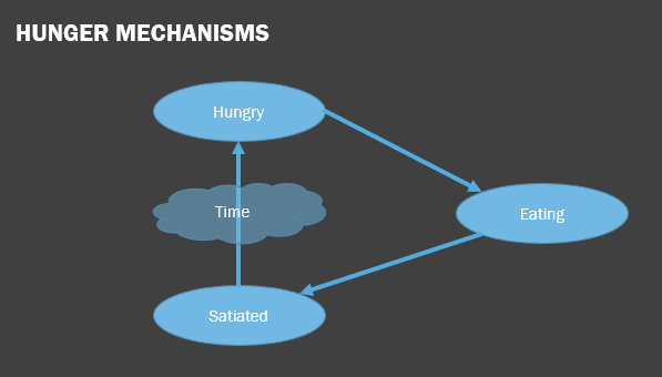
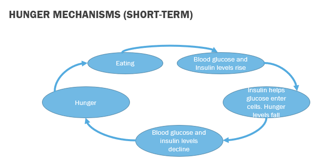
Balance between leptin and ghrelin to determin hunger and satiety. Ghrelin produced by cells the gastrointenial tract. Leptin is a hormone made by adipose cells
Pathways - the paraventricular hypothalamus recieves signals from the arcuate nucleus and de/increases appetite accordignly - also acts on the lateral ypothalamus to recieve insulin and affect appetite in other ways. Damage causes loss of appetite, stimulation cause a gain.
Short and Long term regulation - opposite pattern, tasting and swallowing without ‘eating’ doesn’t make us feel full. Rate with leaks in stomachs eat constantly.
The stomach signals satiety when it is distended by sending messages via the Vagus Nerve. Even in cases of severe spinal injury or in people without a stomach, satiety can still be felt due to hormonal signals from the duodenum.
Lateral hypothalmus projects to many other areas of the brain leading to food associated behaviours consistent with hunger and satiety levels
Prader-Willi syndrome - never feel full (hyperphagia) due to disruptions in their satiety network
Thirst
Osmotic thirst follows salty foods
Mammals maintain a constant solute level in the body. When salty foods are eaten, the solute level in the blood rises, increasing osmotic pressure. Water moves from cells to dilute the solute, triggering osmotic thirst as neurons detect water loss. The kidneys excrete concentrated urine to remove excess sodium while conserving water.
Krause et al (2001) found rats with strong osmotic thirst showed reduced anxiety, lower stress responses, and more social interaction with unfamiliar rats. These effects have not been tested in humans.
The OVLT and SFO are receptors that detect blood osmotic pressure and sodium levels. The OVLT also receives signals from the digestive tract, helping anticipate osmotic needs before they arise. Swallowing and stomach distension signal the brain when to stop drinking.
Hypovolemic thirst follows loss of gluids through belleding/sweating
Lost salts must be replaced like water. Neurotransmitter (Angiotensin II) released around hypothalamus leading to craving for water and salty tastes. Animals are suddenly attracted to salty water despite repulsion under normal circumstances
Leah Betts' death after taking ecstasy was caused by excessive water intake (7-8 liters in 90 minutes), not the drug itself. MDMA can also trigger SIADH, making urination difficult and leading to water intoxication and hyponatremia. Other causes of excessive drinking, like polydipsia, can indicate early diabetes.
Sex
Many species reporduce asexually
Sex isn’t necessarily for child rearing since many species that reporduce sexually don’t share childcare. Current thinking is we reproduce sexually because this makes us more adaptable to drastice environental changes and less susceptible to genetic disorders
Menstraul cycle - interaction between hypo, pituitary and varies. After the end of a cycle the anterior pituitary releases FSH (follicle-stimulating hormone). This hormone prompts the growth of a follicle in the overay which natures an egg. Sevel types of estrogen is produced by this
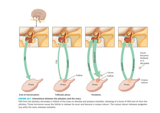
Pregnancy - production and development of a baby in roughly 38 weeks on average
Male cycle - Testosterone is crucial for male sex drive, increasing touch sensitivity to the penis. It binds to receptors in the hypothalamus, mainly in the ventromedial nucleus, MPOA, and anterior hypothalamus. In the MPOA, testosterone triggers dopamine release. Unlike in females, this system doesn't follow a fixed rhythm.
Castratio - removal of testicales. Greatly reduces sex drive, but doesn’t eliminate entirely. Castrated m rats produce normal amounts of dopamine in the MPOA but don’t release it in presence of a receptive female and don’t attempt to copulate
Stages of sex:
Desire/Excitement → Testosterone, in both sexes, plays a key role in driving excitement, with high levels making orgasm easier to reach. In women, luteinizing hormone also correlates with excitement. Dopamine release creates pleasurable sensations, while nitric oxide and noradrenaline increase blood flow to the genitals.
Plateau → Bodily responses to the sympathetic nervous system continues to increase, activity in the amygdala increases while the hippo decreases
Orgasm → Oxytocin released causing feelings of closeness and rhythmic muscle contraction and ejaculation. Nucleus accumbens releases a large amunt of dopamine making us feel relaxed
Resoution → After orgasm, dopamine surge causes relaxation and reduces sympathetic nervous system activity, slowing heart rate and relaxing muscles. Men need a refractory period to replenish neurotransmitters for another orgasm, while women, without this need, can experience multiple orgasms before resolution. The parasympathetic nervous system becomes more active during this phase.
Week 14 - Research Method
Reseach methods can:
Study structure vs. function (activity)
Study activity vs. lack of activity
Be invasive vs. non-invasive
Be spatially sensitive (where) vs. temporally sensitive (when)
Observing brain structure
Phrenology (Franz Joseph Gall 1758-1828) → study of individual differences in the anatomy of the skull. Find someone with unusual behaviour (looking for unusual brain features). 2 Key Assumption:
Different regions of the brain perform different functions and are associated with different behaviours.
The size of these regions produces distortions of the skull and correlates with individual differences in cognition and personality.
Limitations:
The shape of the skull does not correspond to brain anatomy
Limited participants (apparently it was mostly family and friends)
Inadequate classification for different brain regions (e.g., marvelousness)
Pathology → studying brain posthumously. Dependent on people donating their brains for scientific research - bias and size of the sample
Structural imaging techniques
Structural imaging techniques are based on the fact that different types of tissue (e.g. skull, white/grey matter…) have different physical properties
Different properties can be used to construct static maps of the physical structure
Similar to the way that an X-ray can see our bones or a sonagram can see a foetus
CT Scan (CAT scan) → computarised axial tomography. Based on the principle that bio mats varies in its density. Differebt types of tissue absorb different amounts of x-ray radiation. Inject a dye into the blood (increase contrast image)
Advantages → Extremely useful for identifying precise location or extent of damage or abnormalities, e.g.: Structural abnormalities, Tumours, Blockages in blood flow, Psychological disorders
Disadvantages → risks of radiation, Spatial resolution: 0.5-1.0 cm, not possible to discriminate two objects less than 0.5 cm apart, no temporal resolution
MRI scan (Magnetic Resonance Imaging) → Most human tissue is water-based, so body is full of hydrogen protons that are randomly spinning. When the magnet is switched on, these protons spin in alignment with the magnet. A radio pulse is then applied that knocks these protons off their axis by 90 degrees. The radio frequency is then switched off, and the protons revert to their original states, releasing energy
Advantages → Invaluable in determining damage or abnormalities in particular brain areas, Provide more detailed and clearer images of the brain than CT scans, Low intrusiveness, No use of x-rays or radioactive substances, High spatial resolution: 1-2 mm
Disadvantages → Can’t be used by people who have internal metallic devices (e.g. pacemakers or steel pins in bones), No temporal resolution, Like CT scans, only shows static brain structures/ anatomy, not activity, Risk of claustrophobia, Very expensive
Ultrasound → high-frequency sound pulses are transmitted into body
Advantages → Good temporal resolution (10-30 ms), real-time imaging, Good spatial resolution (< 2 mm), Low cost, No exposure to radiation, Can also detect blood flow, 3D ultrasound is also possible, by collecting many 2D ultrasound images
Disadvantages → Larger patients are difficult to ultrasound – more tissue weakens sound waves, Ultrasound waves cannot pass through bone, so cannot visualize brain once skull bones have grown together, Can only be used to visualise brain up to 18 months of age or during open-skull brain surgery.
Recording brain function
Functional approaches are based on the assumption that neural activity produces physioloigcal changes in certain areas of the brain
Electroencephalography (EEG) → continous recording of electrical activity in brain. Measure sleep. Oscillations at different frequencies are associated with different states of cognitive functioning:
Delta (0.5- 4 Hz)
Theta (4- 8 Hz)
Alpha (8- 13 Hz)
Beta (13- 30 Hz)
Gamma (35- 100 Hz)
Electrodes on the scalp record electrical actvity in the brain (raw EEG). Raw EEG signals are not sensitive enough to detect subtle changes in brain activity. It is difficult to differentiate between the relevant brain signals and irrelevant noise. To address this, researchers average EEG signals across multiple presentations of the same stimulus. This averaging process helps to reduce the impact of random noise and enhances the visibility of the desired brain signal.
Electrical activity at the onset of a stimulus recorded → filter and amplify → average across trials and individuals → collapsed to form a “grand average” or mean of means
Tomography vs Topography:
Topohraphy → “Topos” = greek meaning place and “Graphein” = to write.
In neuroscience refers to projection of a sensory surface
Advantages:
Equipment is relatively cheap,
Can be used on wide range of participants
Can be used to study brain activity over an extensive length of time
Can also be used to study brain activity during long, complex tasks
Low invasiveness
High temporal resolution (1 msec)
Disadvantages:
Signal can be contaminated by artefacts (e.g. blinks, muscle etc.)
We can only allude to which brain areas are active and what their functions are
ERP - large number of trials
Low spatial resolution
Difficulties with source localisation
PET (positron emission tomography: Based on the fact that greater brain activity is associated with higher level of metabolism in that area → Need for energy (glucose), Increased blood flow into the particular brain area. Monitors blood flow around the brain. Pet scans show areas activated by seeing/speaking/thinking a word
Participants are injected with a radioactive tracer (glucose)
Radioactive tracer is intitally unstable
When tracer converts into normal stable form, it releases a positron
The positron collides with an electron in the brain
This produces photons, which can be detected by machine
this tracer is most concentrated in the most active brain areas
So these areas emit most photons
Radioactivity detectors around the head produce a map of which brain areas are most active
Colour codes indicate different levels of brain activity
PET advantages: Enables detailed images of brain function during a task, More sensitive than CT and MRI and use of colour makes it relatively easy to interpret, Moderate spatial resolution (5-10 mm3)
PET disadvantages: Moderately invasive → Requires injection of radioactive substance (thought to be negligible), Number of scans per year on a person is limited to 3, Low temporal resolution → Each scan takes 30 sec to complete – tracer needs to reach brain, and radiation needs to peak and decay, Limited number of scans per session → 40 second interval between scans
FMRI → similar to PET, it is based on the level of metabolism in the brain, detects changes in blood oxyegn rather than glucose. Increase brain activity → increased blood flow → increased oxygen. BOLD: the blood oxygenation-level-dependent effect
Measures the ratio of oxygenated to deoxygenated hemoglobin, The amount of oxygen carried by hemoglobin affects the magnetic properties of the hemoglobin, Distortions in local magnetic field caused by deoxygenated blood hemoglobin can be detected
fMRI advantages: Enables detailed images of brain function during a task, Low invasiveness → Unlike PET, it does not involve exposure to radiation , High spatial resolution (1-3 mm3)
fMRI disadvantages: Requires technical/medical expertise, Highly expensive!, Low temporal resolution (> 1 second), Often require block designs, Scanners are very noisy!
Interpreting the pattern of increased and decreased activity is a challenge → Different people can use different brain areas for the same task, depending on their previous experience with it. This is about correlations between functions and activation patterns. You discover which brain areas are sufficient for a particular function but not which ones are necessary
EEG/ ERP vs. PET/ fMRI:
EEG/ERP good temporal resolution but PET/fMRI much better spatial resolution
EEG/ERP reflect neural activity (i.e. a more direct measure of brain activity) whereas PET and fMRI reflect chemical changes in blood flow
MEG (Magnetoencephalogram) → non-invasive neurophysiological technique. MEG uses extremely sensitive device to record electromagnetic signature of neural currents. How does it work:
Magnetic fields are found whenever there is a current flow – e.g., around wires or neurons (!)
Magnetic fields can pass unaffected through brain tissue and skull
The magnetic field of a neuron is extremely small
But the magnetic field generated by 50,000 or more neurons firing together can be detected with highly sensitive equipment - MEG
MEG advantages: Non-invasive – does not involve injections of isotopes, exposure to X-rays or magnetic fields , Even children and infants can be tested, repeated testing is possible, Good spatial (mm) and temporal (ms) resolution, Measurement is silent – unlike fMRI, No need for electrolyte jelly – unlike EEG – so preparation time is reduced
MEG disadvantages: Expensive - £1M to £1.5M, xternal generators of magnetic signals need to be eliminated – so MEG needs to be recorded in specially constructed shielded rooms, MEG does not provide structural/anatomical information – often must be overlaid with MRI data to see activation on anatomy, Risk of helium shortage?!?
Studying brain damage
Phineas Gage → railroad worker - was using a steel rod to ram dynamite into a hole in the rock. Dynamiye exploded and rod shot into his cheek, through his brain and out the top of his head. He never lost consciousness and had no obvious neurlogical symptoms
Before accident: Serious, industrious and energetic
After accident: Childish, irresponsible, boorish, thoughtless of others, Unable to carry out plans, He was “no longer Gage”. Accident largely destroyed his orbitofrontal cortex. Causing a defect in rational decision making and the processing of emotion
Brain damage disadvantages:
Data Analysis: Time-consuming and complex.
Damage Localization: Difficult to pinpoint exact location and cause.
Sample Size and Control: Small samples and lack of control over damage.
Generalizability: Uncertain if findings apply to the general population.
Baseline Measurements: Often unavailable.
Behavioral Changes: Difficult to isolate specific changes.
Brain Plasticity: Brain's adaptability can complicate interpretation.
True Effects: Challenging to determine the direct impact of damage.
Limited Insights: Only reveals necessary brain areas, not their specific contributions.
Examining brain stimulation/inhibition
If brain damage impairs some behaviour, stimulation should increase it. Electrical stimuation in animals or brain surgery patients. Magnetic stimulation with humans
Surgical implantation of a ‘brain pacemaker’, which sends electrical pulses to specific parts of the brain. Shows remarkable therapeutic benefits for otherwise treatment-resistant movement and affective disorders. Underlying principles and mechanisms are still not clear. Effects are reversible (unlike lesioning)
DBS and Parkinson’s Disease:
Primary Symptoms: Tremor, Rigidity, Postural instability
DBS helps manage these symptoms - improves quality of life. Electrical stimulation targets areas in the brain that control movement, blocking the abnormal nerve signals that cause tremor and PD symptoms
AKA TMS - ‘temporary lesioning’ - TMS modulates cortical activity by passing magnetic fields across the scalp. It is like studying brain damaged patients but without some of its limitations. TMS can produce transient impairments or improvements in cognition non-invasively
Brain Stimulation Advantages:
Allows researchers to identify locations and functions of numerous brain areas
Does not result in reorganisation of brain function as it is temporary
Within-subjects designs are possible
Specific site of stimulation/inhibition can be identified and changed
Brain Stimulation Disadvantages
DBS - Highly invasive!
DBS - is limited to patients and studies with animals (e.g. rats)
DBS/TMS - Electrical stimulation differs from nerve impulses
Behaviour is the result of the activation in many areas of the brain
Side effects can include balance problems, visual distortions (e.g. hallucinations), apathy, compulsive behaviours…)
Long term effects (particularly TMS) are not yet fully understood
Possibility of stimulating wrong brain area
Week 15 - Psychophysiological measurements
Anatomy - science of body structure and the relationship between structures
Physiology - study of bodily function or how the parts of the body work
Psychophysiology - study of the physiological basis of psychological processes; relating cog processes to integration of central and peripheral processes
3 system measurement
Evaluate reports
Overt Actions
Physioloigcal responses
Definitions
Somatic Nervous system (SNS) → coordinates skeletal muscle activity; (mostly) voluntary and conscious. Function: interact with the world
Automatic Nervous System (ANS) → controls and monitors smooth muscles, cardiac muscles and glands (Internal organs); (mostly) involuntary and unconscious. Function: maintain homeostasis
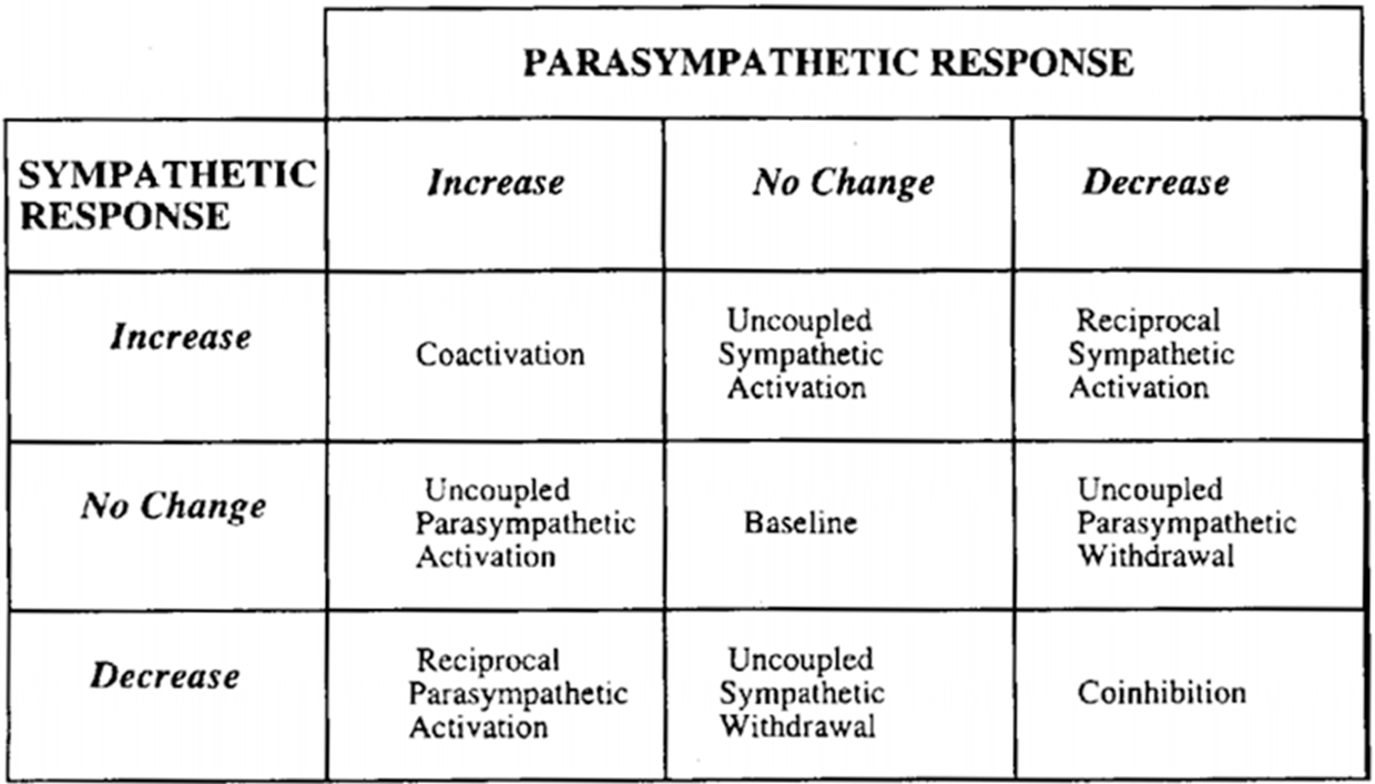
Sympatheitc Nervous System: Generally speaking it corresponds to arousal and energy generation. Primary function to stimulate the body’s fight-flight response. Prepares the organosm to reac and to respond optimally to threat (primes body for action and intense (muscular) effort). Mainly uses norepinephrine as a neurotransmitter at the postganglionic synapse
Parasympathtic Nervous System: Mostly associated with conserving body energy and maintaining homeostasis. Facilitates vegetative, non-emergency responses. Often describes as the “brake” for symp system. Postganglionix axons mostly release acetylcholine as a neurotransmitter
Cardiovascular system
Electrocardiogram
Systole - Ventricular contracton (blood pumped round body)
Diastole - Ventricular relaxation and refiling
Indices of Cariovascular activity
Heart Rate → number of heartbeats pm,
Heart Period → time (in ms) between adjacent hearbeats
Blood Pressure → the force exerted by the blood against the vessel walls and is generall measured in mmHG
Baroreflex → pressure-and-stretch sensors in the heart, carotid and other main arteries that that detect variations in blood pressure and signal to the brain the current state of cardiovascular arousal.
Heart Rate Variability (HRV)
Measuring Cardiovascular reactivity:
Initial heart deceleration: Orienting response. Sensory intake and perceptual processing (Parasympathetic mediated)
Subsequent heart acceleration: Cognitive processing and diminished sensory intake (Parasympatheitc withdrawal and sympatheitc dominance)
Electrodermal system
Electrodermal system (EDA) refers to variations in the electrical characterisitics of the skin.
Exosomatic: apply small electrical current across the skin to measure skin conductance levels
Endosomatic: Record skin potential without applying external electrical current
Eccrine sweat galnd’s primary function is thermoregulation. However, those on the palmar and plantar surfaces are more related to grasping behaviour than to cooling and are thought to be more responsive to psychological stimuli thna to thermal stimuli. Predominantly sympathetic cholinergic (achetilcolin) innervation
Measuring skin conductance: Typically the skin is not prepared, but some researchers ask participants to wash their hands. Conductive paste may be used. Ambient temperature and time of day are 2 enviro factors that should be controlled.
Tonic EDA → slow acting components and background components and background charactertics of the low signal (overall level, slow climbing, slow delinations over time)
Slow changes and DC components
Skin condcutance level (SCL) → changes in the SCL are thought to reflect general (slow) changes in autonomic arousal
Phasic SCRs → faster changing elements of the signal
Non-specific SCRs (NS-SCRs) → Phasic change in electrical conductivity of skin that occur in the absense of an indentifiable eliciting stimuli
Event-related SCRs (ER-SCRs) → phasic change in electrical conductivty of skin that can be attributed to specific eliciting stimuli
SCRs
Minimal amplitude threshold: historically: historically 0.05µS but now 0.01µS can also be seen in literature
Peak latency decision: typically between 1-4 seconds
Some type of normalization typically applied: e.g. square root transformation
General: Considerable inter-subject differences in responsivity, Reasonably fast habituation, Deep breaths and bodily movement generate responses
SCL: Deep breaths and bodily movement generate responses
NS-SCRs: Deep breaths and bodily movement generate responses
SCRs: Slow responses make it difficult to analyse response to stimuli close in time
Pupil dilation
Sphincter pupillae muscle: Parasympathetic
Dilator pupillae muscle: Sympatheitc
Pupil constricts as circular msucles of iris as the light gets brighter (p). Pupil dilates as radial msucles of iris contract (s)
Eye-trackers challenges:
Very sensitive to room and stimuli luminosity
You blink and you miss it!
Eye-tracker performance can depend: On the participant’s eye colour, Contact lenses or glasses, Eyelashes mascara is prohibited!
Sympathetic activation → constriction of iris dilator muscle
Paraysmpatheitc inhibition → relaxtion of the iris sphincter muscle
Facial Electromiography (EMG)
Electromiography (EMG) → electrical recording of muscle activity
Corrugator supercilli – draws the eyebrow downward and medially, producing the vertical wrinkles of the forehead
Zygomaticus major – Activity occurs when the cheek is drawn back or tightened
Orbicularis Oculi – Closes the eyelid
Skin preparation → clean the skin to remove the dirt, oil and dead skin (no makeup). Skin typically abraded gently.
Electrode placement → bipolard recording (2 electrodes), highle condiuctive gel is used between the electrode and the skin (reducing impedance, stabilising impedance, stablising skin hydration and conductivty) and electrodes attached to the skin with double-sided adhesive tape
Startle and defensive mechanisms → an abrupt sensory event prompt a startle response, a rapid chain of rapid extensor-flexor movements that cascade throughout the body (+eyeblink). Under threat or predication animals show exaggerated startle reflex and so do humans. Reflects defensive dispositions
Interesting discussion about startle reflex and the (cascade) of defensive dispositions in the book chapter (Bradley and Lang, 2007)
Week 16 - Psychopharmacology
Communication within a neuron
Nerve impulse - electrical message that is transmitted down the axon of a neuron. Transport is quick and without loss of energy. Electrical currents in the body refelct the flow of ions
Neutral Atom → Loss of electrons (cation) and Gain of electrons (Anion)
Resting potential → neuron refers to the state of the neuron prior ro the sending of a nerve impulse
Movement of ions (at rest) → 2 forces:
Electrical gradient: +ve charges attracted by -ve enviro, -ve charges attracted by +ve enviro
Concentration gradient: Ions move from high to low concentration.
Action potential - the resting potential remains stable until the neuron is stimulated
Hyperpolarisation = -80mV or -90mV
Depolarisation = -60mV or -55mV
Threshold of excitment = -55mV, opening sodium channels = the action potential
Returning to resting state - at a certain threshold (~+40mV), sodium channels are quickly closed and potassium channels open fully
Communication between neurons
Charles Shrrington → studied reflexes and temporal and spatial summation. Sppedof conduction <15 m/s vs 40m/s within single neuron
Sparkers vs Soupers → 2 camos that investigated this problem in the 1930s. No consensus could be reach until
Major neurotransmitters
Neurotransmitter | Role in the Body |
Acetylcholine | A neurotransmitter used by the spinal cord neurons to control muscles and by many neurons in the brain to regulate memory. In most instances, acetylcholine is excitatory. |
Dopamine | The neurotransmitter that produces feelings of pleasure when released by the brain reward system. Dopamine has multiple functions depending on where in the brain it acts. It is usually inhibitory. |
GABA (gamma-aminobutyric acid) | The major inhibitory neurotransmitter in the brain. |
Glutamate | The most common excitatory neurotransmitter in the brain. |
Glycine | A neurotransmitter used mainly by neurons in the spinal cord. It probably always acts as an inhibitory neurotransmitter. |
Norepinephrine | Norepinephrine acts as a neurotransmitter and a hormone. In the peripheral nervous system, it is part of the fight-or-flight response. In the brain, it acts as a neurotransmitter regulating normal brain processes. Norepinephrine is usually excitatory, but is inhibitory in a few brain areas. |
Serotonin | A neurotransmitter involved in many functions including mood, appetite, and sensory perception. In the spinal cord, serotonin is inhibitory in pain pathways. |
Steps in synpatic transmission
Synthesis of smaller neurotransmitters such as acetylcholine
Action potential causes calcium to enter, relleasing neurotransmitter
Neurotransmitter binds to receptor
Seperation from receptors
Reuptake of neurotransmitter by transporter protein
Postsynaptic cell releases retrograde transitters that slow further release from presynaptic cell
Negative feedback sites respond to retrigrade transmitter or to presynaptic cells own transmitter
Post-synaptic activation ~ will open or close ‘ion gates’. Depolarisation = excitation = excitatory PSP (EPSP). One EPSP will no create an AP → temporal summation, spatial summation
Biological basis of drug use and addiction
Drugs can either facilitate or inhibit synaptic transmission → Agonist or Antagonists
Nicotine, SSRI, MDMA, Sedative → gets released into the blood stream and floods the synaptic clef, and taking the place of acetylcholine
Bio basis → major target of recreational drugs: dopamine
Too much dopamine → Hallucinations & paranoia (schizophrenia), Uncontrolled speech and movement (Tourette’s), agitation & repetitive action (OCD), Overexcitement & euphoria (mania)
Too little dopamine → Tremor & inability to start movement (PD), Feelings of lethargy & misery (depression), Lack of attention & concentration (ADHD), Cravings & withdrawal
Addictive substance increase dopmaine activity in certain areas of the brain ‘reward pathway’
Drugs → Almost all abused drugs stimulate dopamine release in the nucleus accumbens. Substained bursts of dopamin in the nucleus accumbens inhibit cells that release the inhibitory neurtransmitter GABA
Alcohol addiction Alcohol has a number of diverse physiological effects including:
Inhibition of sodium (NA+) across the membrane
Expansion of the surface of membranes
Decreased serotonin activity
Enhanced response by the GABA receptor
Blockage of glutamate receptors
Increased dopamine activity
Various factors contribute to continued substance abuse: tolerance levelss, cravings in response to cues, brain reorganisation. Addiction and increased attentional biases: Attentional biases predicts heroin relapse (Marissen et al., 2006)
Medications used to combat addictions include → antabuse, methadone and many don’t respond to other treatments so meds have been used to reduce cravings
Week 17 - Visual perception
Historical roots of vision research
Structualism → debate where vision is innate (Descartes) or learned (Berkeley). ‘Sensory atoms’ sre coded on the retina. Direct correspondence between retina and coscious awareness. Theory based on introspection
Behaviour driven by enviro cues than internal, mental processes. Pereception doesn’t rely on complex cognitive processes or stored knowledge of the world; rather occurs through passive resonance with incoming info
Gibsin’s (1966) theory of direct perception: Perceptual invariants, 2 perceptual invariants:
Optic flow (can be used to judge speed and direction)
Texture (can be used to judge depth)
We don't perceive the world by breaking it down into individual parts. Instead, we see the world as a whole, with emergent properties that can't be understood from the individual components. According to Gibson, we directly perceive these properties through light entering our eyes, without the need for learning or internal representation.
Artificial intelligence and the cognitive revolution → The task of a psychologist trying to understand human cognition is analogous to that of a man trying to discover how a computer has been programmed
Behaviourist model = enviro = sensory perceptions, memories, beliefs, emotion, ideas or behaviour
Cognitive model = enviro = sensory perceptions, memories, beliefs, emotion, ideas → behaviour
Assumptions of the constructivist approach
Congition consists of an orderly series of stages of mental events that actively reonstruct the retinal input
That is, the detection of perceptual invariants such as optic flow and texture relies on complex, mental processes.
Given that mental processes take time to complete, they can be isolated and studied through experimental investigation.
Visual illusions → if perception were completely determined by the pattern of light hitting the eye, then the 2 lines would be perceived as the same length
Ambigious figures → if perception were completely determined by the pattern of light hitting the eye, then each pattern would trigger a single percept
Impossible figures → if our vision was an infallible reflection of the world, then how could impossible figures be percieved
Early visual processing
The process of forming a reconstruction of the visual world begins at the retina
Visbile light forms part of the spectrum of electromagnetic radiation, which also contains radio waves an xrays
Newton (1672) discovered that light can be split into many colours. Diff colours arise from diff parts of the electromagnetic spetrum. These differences are captured by 2 kinds of cone: red, green, blue
Human retina ~ some interesting observations:
7+ mill cones and 125 mill rods on the retina
Each cone and rod repsonds t o light that emanates from a diff part of the visual field
Point at which all nerve fibres converge to leave the retina is insensitive to light, producing a blind spot in our vision
Detailed structure of the human retina → made up from many diff kinds of cell, diff properites of light stinulate diff kinds of receptor
The eye in action → light particles (photons) enter the eye and travel towards the retina. Light is absorbed y retinal photoreceptors which, via a process known as isomerization, immediately discharge an action potnetial. The AP is transmitted to ganglion cells and thereon to the optic nerve and brain
Receptive fields → each photoreceptor is sensitive to a very small area of the visual field (receptive field). Adjacent photreceptors are sensitive to adjacent areas of the visual field. Every area of the visual field is coded y the retina and primary visual cortext (V1)
Some retinal ganglion cells, especially parvo-cellular cells, are very sensitive to changes in light intensity → Cell only fires when there is a change in light intensity across its outer and inner receptive fields. For only the inner field to be activated, some kind of edge or boundary must be present. This sensitivity to changes in light intensity may serve an important psychological function: edge detection
The combined output of retinal cells can be used to charcterise the size of an edge → the simple cell only fires when 4 feeder cells are activated, in this way it can encode length
The receptive fields of retinal ganglion cells are thought to play a role in these curious visual effects
Imposing order on the retinal output:
early vision is about detecting very basic features: edges, colour and motion. However, at this stage it’s not yet clear which edges and colours belong to the same object. In fact, there’s no sense of ‘objectness’.
Rather, all that exists is an alpabet soup of basic visual features - coloured blobs and lines made up from stimuli in neighbouring receptive fields that are static or moving, not organised into any coherent form. Somehow, these bits and pieces of info need to be assembled into recognisable forms
Texture
Texture also provides a means of organising the perceptual world into boundaries and surfaces. That is, texture provides a way bt which natural disconinuties can be used to infer perceptual boundaries.
Perception of texture is immediate and effortless, which suggests that it’s made available at a very early stage of processing
Textures - a variety of properties, including colour orientation, motion and depth
The gestalt psychologists → studies conducted suggest that the visual system uses a simple set of rules to decide what basic features probably fit together and belong to the same object. That is, to decide which feature are spatially correspondent. The gestaltists relied on the power of introspection to demonstrate this
Gestaltism proposes that the whole is more than the sum of the parts (i.e. the grouping of simple visual properties generates ‘higher-level’ emergent forms)
The process of grouping provides a way of combining visual elements into larger, more meaningful units
It also provides a nice example of how the visual system imposes its own laws on to the visual input. That is, it constructs its own reality of how the visual world is probably organized
Neural basis of similarity grouping - neural activation associated with high vs lower similarity
Effects of temporal-parietal damage on global/local perception: damage to the region of brain that is involved in perceptual grouping causes problems in global perception
Visual objects recognition
Recognition is not a unitart activity , and occurs at 2 general levels
Perceotual recognition ~ allows us to recognise an object across different viewingn angles and lighting nd when certain parts are occluded → semantic recognition ~ allows us to know the function of an object and to recall asscociations that we have made with it
APPERCEPTIVE AGNOSIA (Lissauer, 1890):
Patients are unable to properly assemble the individual attributes of objects. Perceptual recognition is therefore impaired, as often shown in figure copying performance
APPERCEPTIVE AGNOSIA (Lissauer, 1890):
Patients are unable to properly assemble the individual attributes of objects. Perceptual recognition is therefore impaired, as often shown in figure copying performance
APPERCEPTIVE AGNOSIA (Lissauer, 1890):
Patients are unable to properly assemble the individual attributes of objects. Perceptual recognition is therefore impaired, as often shown in figure copying performance
Perceptual recognition allows us to know that an object has a constant shape, despite the fact that it changes its appearance (and impression on the retina) as our viewing position changes. To obtain this knowledge, we need to compute ‘object constancy.
Template matching (Sutherland and Williams, 1969): Rotate and manipulate the shape until it fits with a template that is stored in memory. This method works well for very distinctive objects, but has a problem with visually similar objects
Critical features (Selfridge, 1959): Look for local features that are unique to that object (i.e. elephant’s trunk). Works well for distinctive objects, but has a problem when the critical feature is obscured, or the object is defined by the spatial arrangement of its parts (i.e. faces).
Structural descriptions (Marr and Nishihara, 1979):
Encode local features AND their relative positions relative to an internal frame of reference that can be seen across viewpoint. That is, pick out aspects of the object that are constant and unchanging
One property of an object that does not tend to change across viewpoint is its main axis. The way in which the components parts of objects relate to the main axis do not change across viewpoint
So axis information provides a means by which the parts of an object can be related to one another across viewing angle
Generalised cones: according to Marr and Nishihara, each object axis makes up a ‘generalised cone’. Classes of objects can be distinguised y the various lengths and arrangements of these cones. So once the various axes of an object are identified, it becoes possible to catergorise the object
Issues: In some instances, the main axis of a stimulus will be obscured, either by an overlapping object or from the viewing angle. Given that we can still recognise stimuli under these conditions, there must be more to recognition than deriving axis-based descriptions
Geon Theory (Biederman, 1987) - Biederman proposed that, in addition to axis information, there are other object properties that remain invariant across viewpoint. These properties consist of wedges, cylinders, spheres and other basic shapes, and are collectively described as ‘geons’.
Empirical support: Whenever geons intersect with one another, they form a concavity. Concavities therefore enable one geon to be distinguished from another
Week 18 - Cognitive neuroscience of LTM
Introduction
Varieties of Long-Term Memory: Explores the divisions in memory and the stages (encoding, storage, retrieval), with different methods/levels of analysis: biology, neuroscience, psychology, and cognitive neuroscience.
Focus Today: Highlights using cognitive neuroscience to study memory, focusing on research design, conceptual issues, and critical thinking.
Aims of Today's Lecture: Covers multiple memory systems and tests, cognitive neuroscience of memory, and advanced designs for encoding and retrieval.
Multiple Memory Systems and Tasks
Memory Systems Debate: Discusses whether there are distinct systems or just one system with multiple processes.
Direct vs. Indirect Retrieval Tests: Explains measures to study explicit (conscious) and implicit (unconscious) memory, with examples of different tests.
Amnesia Cases: Highlights damage to the medial temporal lobes leading to poor explicit memory but intact implicit performance in indirect tests.
Healthy People: Examines how encoding manipulations (e.g., deep vs. shallow processing) impact memory tests, suggesting separate systems but also noting limitations and confounds.
Cognitive Neuroscience of Memory: Basic Issues
Behavioural Measures: Critiques the "black box" nature of behavioural metrics and highlights neuroimaging methods like fMRI (spatial resolution) and EEG (temporal resolution) to study memory processes.
Research Questions: Probes how to isolate memory-related brain processes from general task demands using manipulation designs.
Advanced Cognitive Neuroscience Designs for Encoding
Task Manipulations: Highlights how differences in encoding tasks (e.g., shallow vs. deep) influence memory, with left prefrontal cortex activation implicated in deep encoding.
“Difference Due to Memory” (dM) Paradigm: Describes event-related designs to measure brain activity linked to memory success or failure, providing insights into encoding processes.
Advanced Cognitive Neuroscience Designs for Retrieval
Memory Test Sorting: Differentiates trials by memory type and subjective/objective experience, with studies showing brain activation patterns for familiarity and recollection.
Hippocampus Role: Explores its function in binding contextual details and supporting episodic memory, with distinctions in familiarity versus recollection using ERPs and fMRI.
Repetition Priming: Highlights reduced brain activity (repetition suppression) during repeated stimulus processing, suggesting procedural and episodic memory systems are distinct.
Final Summary
Core Takeaways: Long-term memory involves hippocampal recollection, distinct memory systems (e.g., procedural, episodic), and insights from advanced imaging methods.
Unanswered Questions: Emphasizes gaps in how different levels and perspectives (biological, psychological, clinical) interrelate.
Recommended Reading: Suggests textbooks and review papers for deeper understanding.
Term 2
Week 24 - Intro to spring term
The cognitive approach
Input → Mediational process → Output
Reaction time - time in ms between the presentation of a stimulus and the production of a motor response
Primarily used for
studying nature of mental processes and their underlying structures
Study the reaction process by manipulating the stimulus/response
Benefits:
Can probe the system when it’s working well
Sensitive measure of chnages in mental operations
Cogntive stage theory - cognition involves a series of component processes that are conducted sequentially, that each takes time. Sum of this time = RT
Can manipulate tasks, stimuli, etc and measure RTs to provide info about the diff stages
Stimuli → visual processing → conceptual processing → episodic memory → selecting response
Donders’ subtraction method (Donder 1868) → isolate the duration of critical process by comparing RTs across tasks presumed to differ the presence of a single stafe
How long does each component take? - can be used to time components underlying choice reaction time tasks. Task contains 3 components: Slide 62-65
Stimulus discrimination
Response selection
Response execution
Issues with subtraction method:
Strong assumptions about sequence of processes involved
Assumption of “pure insertion” → assumes components don’t interact, BUT components of a task may not be performed in the same way when new components are added (Sternberg, 1966)
Addictive factor logic (Sternberg 1969) → Sternberg manipulated the number of times a component occured across levels of variables instead of comparing tasks with different components. Determine time of component by comparing levels
Additive or interactive → by using multiple factors we can distinguish additive (independent) from interactive (non-independent) effects
Week 25 - Neural Network approach *
Intro
Machine learning: Input → Feature extraction → classification → output
Artificial intelligence: Input → feature extraction + classification → output
Artifical neural network (ANN)
Modelling
Core Concepts of ANNs
Node Layers:
Input Layer – Receives data (e.g., sensory input in cognition)
Hidden Layers – Processes information (akin to brain regions integrating signals)
Output Layer – Produces the final decision/prediction
Weights & Activation Functions:
Weights determine how much influence each input has.
Activation functions decide whether a neuron "fires" (e.g., similar to real neural firing).
Examples: Threshold-based activation (+1, 0, -1), Sigmoid or ReLU functions (common in deep learning)
Perceptron Model → Perception → single neuron with input(s) and output(s)
Single-layer perceptron: Mimics a simple neuron.
Multi-layer perceptron (MLP): Introduces hidden layers for complex processing., Similar to multi-synaptic pathways in the brain.
Training & Adaptation
Supervised Learning: Learns from labeled examples (e.g., training an ANN to recognize EEG patterns).
Unsupervised Learning: Finds hidden patterns without labels (e.g., clustering brain activity states).
Bias Values: In ANN, bias adjusts neuron activation. In cognition, electrical brain stimulation can modify excitability, similar to ANN tuning.
Machine learning
Supervised Learning
Uses labeled data to train ANNs.
Example: Recognizing brain states linked to emotions or decision-making. Studies (e.g., Zamani, Sadr & Javadi, 2022) show ANN applications in clinical neuroscience.
Unsupervised Learning
Finds hidden patterns in data (e.g., identifying new cognitive states from fMRI/EEG).
Used in studies to categorize different neural processing modes.
Reinforcement Learning
Learning through rewards and feedback (e.g., adaptive learning models in AI).
Example: AI playing Atari Breakout or simulating human decision-making in tasks.
Practical Applications in Neuroscience & Psychology
Training ANNs on neural data to predict behavior or diagnose cognitive disorders.
ANNs in cognitive modeling to simulate memory, learning, and perception.
Neural adaptation: Like brain plasticity, ANNs adjust weights over time to optimize performance
Week 26 - Face Perception
FRUs could just store lots of “instances” of a face
Face averages might be a good way of capturing within-person variability and implementing FRUs. Stable identity characteristics are extracted, meaningless variation (”noise”) is eliminated. Also provide simple account of face learning. But meaningful variation can also be extracted during averaging.
Introduction: Face Perception & Recognition
- Face Perception & Recognition:
- We recognize faces based on key features (e.g., expression, age, gender, race, attractiveness, trustworthiness, competence, intelligence).
- Face Recognition Units (FRUs) store facial information—either as multiple instances or as an average representation.
- Face Averages in Recognition:
- Face averages capture stable identity characteristics while filtering out noise (irrelevant variation).
- Example: MyHeritage FaceVACS system compares identity averages vs. individual photos to assess recognition accuracy.
- Variability in Face Recognition:
- Within-person variability: A single person looks different across photos (e.g., different lighting, expressions).
- Between-person similarity: Some people naturally resemble each other, making recognition difficult.
- Real-world challenge: Unfamiliar face identification is significantly harder due to within-person variability.
---
### Modelling: Face Recognition & Experimental Studies
#### 1. The Importance of Variability in Face Identification
- Study: Sorting photos into different identities (N = 20)
- Median: 7.5 identities
- Mode: 9 identities
- Range: 3-16 identities
- No participant arrived at the correct solution, highlighting the challenge of face perception.
#### 2. Face Recognition in Applied Contexts
- CCTV Surveillance & Passport Control ("The Passport Problem")
- Identity impostors exploit resemblance to travel illegally.
- Human accuracy in matching faces is only ~80%, even for professionals.
- Face Matching in the Laboratory vs. Real World
- Lab tests: Controlled, establish cause-and-effect, but use simplified displays.
- Field studies: Realistic but lack control and generalizability.
- Virtual Reality (VR) Studies:
- Offers immersive, realistic environments while maintaining experimental control.
- Tracks accuracy, reaction times, eye movements.
---
### Machine Learning & Cognitive Research on Face Recognition
#### 1. Avatar-Based Face Recognition Studies
- Experiment 1: Recognizing Avatars of Familiar People
- Participants viewed avatars vs. photos of familiar faces.
- Results:
- 92% accuracy for photos
- 88% accuracy for avatars
- Avatars successfully preserve identity.
- Experiment 2: Matching Avatars to Photographs
- Participants matched avatars to real photographs.
- Recognition rate: 43% of identity pairs.
- Experiment 3: Comparing "Face Space" of Avatars & Photos
- Used landmarking & PCA analysis to compare facial structures.
- Statistical results showed avatars and photos share structural similarities.
- Experiment 4: Avatar-to-Photo Matching in a VR Airport Simulation
- Participants (N = 60) tested face matching in a simulated airport.
- Findings: VR allows face-matching research in more realistic settings.
#### 2. Key Takeaways on Familiar vs. Unfamiliar Face Recognition
- Familiar Face Recognition is Robust:
- The brain extracts consistent identity patterns across different conditions.
- Unfamiliar Face Recognition is Difficult:
- High within-person variability makes matching harder.
- Psychologists Use Advanced Methods:
- AI, VR, and neural network models help study and improve face recognition accuracy.
Week 27 - Vestibular system
Fundamentals
Vestibular system - part of the inner ear, next to cochlear. 2 vestibular labyrinths (1 each side), each labyrith:
3 semicircular canals → detect angular acceleration of the head, constant velocity stimuli are adapted to → feeling of no rotation when actually spinning. After-effect when rotation stopped
Anterior/Superior = pitch, nodding
Posterior = roll, ear to shoulder
Horizontal/Lateral = yaw, shaking head
2 otolith organs → Detect linear acceleration of the head, 500 mill yrs old, oldest sensory system
Utricle = horizontal plane, walking forwards/backwards
Saccule = vertical plane, up/down movement, gravity
Vestibular projections → Vestibular nerve
Cerebellum
Vestibular nuclei in medulla and pons (spinal cords, eye muscles, cerebellum, thamalus: cortex and hippo)
Vestibular brain → unlike other senses, there’s no unimodal vestibular cortical region. Core vestibular brain regions in parietal lobe. Parieto-insular vestibular cortex (PIVC)
Defined in monkeys, analogues in humans
Parietal Operculum (OP2) in humans is key region
Vestibuloocular reflex → stabilises gaze in response to head movements
Stabilise body posture in response to head movements → maintain centre of gravity
Pathways from vestibular nuclei in brainstem in spinal cord → lateral tract
Extensor effects on the side towards the head tilt, flexor effects on the side away from the head tilt
Stabilises the head in response to unexpected movements → neurons involved in the VCR are suppressed during active head movements. Neck muscles activated when the head is moved unexpectedly
Researching the vestibular system
Vestibular system always on. Can't compared to conditions without vestibular signals as you could with other senses. Vestibular system primarily related to motion; Neuroimaging struggles with moving subjects, and allowing movement in experiments reduces control. Modulate vestibular activity:
Turntables/Motion Platforms/Rotating Chairs → rotate at a pre-determined on a given axis or plane, combine into multi axis rotations for more complex stimuli.
Passive vestivular stimulation: well-controlled, but not the same as many movements in everyday life
Investigate: responses to rotation, eye movements, effect of rotation on tasks, effect of rotation on tasks
Human or animal studies
Caloric Vestibular Stimulation: use of temp to modulate fluid flow in the semicircular canals
Temp anywhere between cold (0-30) or warm (44-55)
Multiple methods
Challenges stumulation times
Often used clincally to test brainstem functions
Galvanic Vestibular Stimulation → use of electrical currents to modulate vestibular nerve activity
Electrodes placed on the mastoids, 1 on eahc side (binaural-bipolar)
= Anode = hyperpolarizing = decreases activity in the nerve
- = Cathode = depolarizing = increases activity in the nerve
Participant feels a roll rotation towards the side of the cathode
Postural response (vestibulo-spinal reflex) towards the anode
Well-controlled parameters, with lots of flexibility
Individual responses to stimulation can vary widely, but tend to be stable over time
Higher-level functions
VS has huge amount of projections in the brain. Integrated with many other senses. These factors mean the VS is involved in a huge range of cognitive and perceptual functions: bodily self-consciousness, self-motion, gravity
Bodily self-consciousness
Multisensory signals can contribute to all of these aspects of bodily self-consciousness
Vestibular signals in particular can influence a number of aspects: Body schema, Body ownership, Self-location
Body Schema → size/shape of body segments, alteratins in body schema reported by patients. Vestibular stimulation can alter percieved size of the body. Clincal implications:
Patients with anorexia might have greater sensitivity to vestibular cues than control participants (only one study much more research necessary) (Brand-Gothelf 2016)
Vestibular stimulation can evoke phantom limb sensations in amputees, can reduce pain of phantom limbs (Andre 2001)
Body schema is constrained by gravity
Movements of the body take place in a gravitational field, which must be continually accounted for by the vestibular system
Alterations in vestibular processing modulate perception of gravity = altered body schema?
Body ownership → sense of “owning” the body (parts)
Rubber hand illusion
Feel your real hand is located where the fake hand is → visual capture
Vestibular stimulation can decrease ownership of the fake hand → ownership of your real hand increases (Ferrè 2015)
Full body illusion
Feel like you are embodying a mannequin/virtual person
Greater ownership of the mannequin when GVS matches visual cues vs visual cues alone (Preuss 2018)
Self-location → Where am “I” in space. Most fundamenta part of sense of self. We experience the self from within the body at one location in space
Out of Body Experience
Greater frequency with vestibular patients/dizziness (Lopez, 2018)
More often when lying down vs standing
Gravitational signals!
Overlap with vestibular regions – Temporoparietal Junction is key region of OOBEs (Blanke 2004)
Vestibular signals must be integrated with tactile, proprioceptive, and visual cues to perceive the body in a single location in space → Interruption of this integration can cause OOBEs
Self-motion perception → as we navigate the world, whole host of sensoy info (VS, Visual System, Proprioceptive system)
Information is integrated according to principles of optimal integration
More reliable senses given higher weighting
Multisensory percept more reliable than individual estimates
Visuo-vestibular integration vital for self-motion perception
Investigating visuo-vestibular self-motion integration requires three conditions: vestibular-only (platform movement), visual-only (optic flow), and combined visuo-vestibular. Vestibular cues are weighted more than optimal integration predicts (Fetsch et al., 2009). The MSTd region is crucial for integration (Gu et al., 2007). Early studies suggest reciprocal inhibition between visual and vestibular systems, but the interaction is likely more complex.
Self motion conflicts → During natural movement, visual and vestibular signals provide corroborating information = these two systems are almost always integrated. But, we can have visuo-vestibular conflicts in certain situations. This visuo-vestibular conflict can have serious consequences → motion sickness and after-effects
In VR → Cybersickness can be mitigated by preventing visuo-vestibular conflict, such as using artificial vestibular stimulation (e.g., GVS) to align visual and vestibular cues or pre-conditioning the brain to down-weight vestibular input before VR. Adaptation to VR through repeated exposure likely occurs due to this down-weighting, but it may have aftereffects, including coordination changes, altered vestibular reflexes, reduced vestibular sensitivity, and cognitive alterations (Stanney et al., 1999; Harm et al., 2008; Di Girolamo et al., 2001; Gallagher et al., 2019, 2020, 2023; Szpak et al., 2019).
Gravity is constant, unchanging, uniquitus = fundamental refernece for the brain. No specialised receptors for gravity – representation built from multisensory cues, including signals from the otoliths (saccule)
Influence of gravity on cognition
Verticality: Affected by vestibular signals → GVS, Aubert Effect
Proprioception – Joint position sense: Reduced “upwards bias” when the head is tilted (Gallagher, 2021)
Cognition – Behaviour control strategies: Reduced “exploration” when the body is supine (Arshad, 2019)
Huge overlap between areas of the brain which process gravity information (e.g., motion, verticality etc.) and vestibular cues
Microgravity → When in 0g, otoliths can no longer signal gravity. Space Adaption Syndrome → after a few days, vestivular system adjusts to 0g
Clinical factors
Peripheral Vestibular conditions
Affects the Peripheral VS (V labyrinths or nerves). Symptoms typically include:
Oscillopsia (unstable vision)
Instability/imbalance
Vertigo
BPPV, Ménière’s disease, Vestibular neuritis
Benign Paroxysmal Positional Vertigo
Otoconia escape and find their way into the semicircular canals. Head movements displace escaped otoconia, shifting fluid in the canals and triggering vertigo. Not serious, very common. Treament usually physical therapy:
Epley manoeuvre – helps to remove otoconia from canals. Effective in 90% of cases
Meniere’s diease → cause not know, could be linked to pressure in inner ear
Endolymphatic hydrops → excessive fluid in the inner ear which damages neurons. Vertigo, nausea/vomiting, hearing loss, balance problems, tinnitus → Unpredictable onset and duration, Can affect one or both ears
Treatments vary depending on symptoms: Medication, Vestibular rehabilitation, Hearing aids, Psychotherapy, Surgery
Vestibular Neuritis → Inflammation of the VN, usually caused by viral infection
Labyrinthitis is very similar, but also affects the cochlear/cochlear nerve
Vertigo, imbalance, nausea/vomiting → Hearing loss and tinnitus with labyrinthitis
Treatment is usually with medications → Antihistamines or motion sickness tablets. Vestibular rehabilitation if symptoms last a long time
Central Vestibular Condtions → affects CNS strcutures associated with vestibular processing: Vestibular nuclei, cerebellum, midbrain, cortex
Symptoms typically include: Vertigo, Spatial disorientation, Cognitive/perceptual disturbances?
Vestibular migraine, Stroke
Vestibular Migraine → mechanism unclear, but thought to involved links between the trigeminal nerve and brainstem vestibular networks, as well as the vestibulo-thalamo-cortical network
Migraine headache symptoms associated with vestibular symptoms
Photophobia/phonophobia, visual aura, and/or headache
Vertigo, unsteadiness, oscillopsia
Treatments varied and not standardised
Medications for migraine or anxiety might help
Neuromodulation trials are promising
Vagus nerve stimulation
Trigeminal nerve stimulation
Lifestyle changes to avoid triggers
Stroke: Vertebrobasilar Ischemic Stroke/Vertebrobasilar Insufficiency → A blockage in the vertebrobasilar system, which supplies blood to the inner ear, can cause vestibular symptoms.
Symptoms vary depending on which artery is blocked, but include: Vertigo, Nystagmus, Gait disturbance/imbalance, Facial paralysis
Treatment as for other types of ischemic stroke: Clot-busting drugs in first 4h, Thrombectomy
Vestibular Dysfunction = Any disruption to vestibular signalling can have profound impacts on the individual, beyond vestibular symptoms:
Anxiety
Depression
Memory impairments
Spatial memory particularly, Linked to atrophy of the hippocampus, Risk for dementia?
Depersonalisation/derealisation
Out of body experiences
Altered verticality/motion perception
Likely because this system is so interconnected throughout the brain
Week 28 - Interoception
Defining Interoception
Sight, Hearing, Smell, Touch, Tasting, Vestibular and Proprioception. BUT what about internal organs, the processing and awareness of internal body signals
Homeostasis: tendecy towards a relatively stable equilibrium between interdependent elements, especially maintained by physiological processes
Allostasis: efficent regulation required to prepare the body to satify its needs before they arise
Why are we bad at perceiving ineroceptive sensations
Mostly unconscious, i.e. below threshold for conscious detection.
Controlled voluntary by the autonomic nervous system and low-level brainstem reflexes
Reveal themselves typically in situations of: homeostatic imbalance, emotional states, sickness, etc…
Interoceptive sensation are often overshadowed, or inseparable from exteroceptive cues
Sensations from the internal organs are often referred, e.g. referred pain
Classic def → afferent processing of signals origanating within the body and which refer to the state of the body. Generated by mechanoreceptive organ-stretching, chemorecption. Transmitted by afferent pathway (neural, humoral)
Does not include: proprioception and sensing the enviro
Challenges: asses awareness of interoceptive sensation conveyed purely by introceptive pathways and other sensation are also important homeostasis & allostasis
Anatomical (functional def) - sense of physiologica condition of the body at any given time. Carry relevant homeostatic control info
Signals carries by spinal laminar 1 spinothalamic tract coverge to the posterior insula
Interoceptive hierachy: Model by Critchley and Garfinkel (2017)
Afferent signals ← preconscious impact ← interoceptive accuarcy → interoceptive sensibility → interoceptive awareness → execution/attribution
How to assess
Heartbeat counting task (aka mental tracking)
Heartbeat discrimination task
Interoceptive maniuplation (e.g. Bolus isoprotenol infusion
Participants should detect changes in airflow resistance, can also be achieved with changing the O2/CO2 concentration.
Detection accuracy: Real and Bogus changes in resistance at probe
Detection discrimination: Rate the resistive load of different loads
Other measures: minimum resistive load detection
Multidimensional Assessment of Interoceptive Awareness (MAIA; Mehling et al, 2012)
Noticing: Awareness of uncomfortable, comfortable, and neutral body sensations
Not-Distracting: Tendency not to ignore or distract oneself from sensations of pain or discomfort
Not-Worrying: Tendency not to worry or experience emotional distress with sensations of pain or discomfort
Attention Regulation: Ability to sustain and control attention to body sensations
Emotional Awareness: Awareness of the connection between body sensations and emotional states
Self-Regulation: Ability to regulate distress by attention to body sensations
Body Listening: Active listening to the body for insight
Trusting: Experience of one’s body as safe and trustworthy
Neural correlates of Interoception
The skin as a interoceptive organ
Touch
CT afferents -Specialized group of skin afferents that has been found mainly on the hairy skin of the body
In humans, CT afferents respond more vigorously to slow, caress-like touch, provided at a temperature typical of human skin:
Slow-conducting (they don’t respond very fast)
Don’t’ respond to pressure (gentle touch)
Fire mostly in velocities within the rage of 1-10 cm/s (2-3 cm/s for most people)
The more these fibres are activated on the skin the more pleasant is the touch
Anatomical (functional def) - shares more characterisitcs with the interoceptive system than with the exteroceptive system
Interoceptive accuracy has been shown to impact: Emotional Experience (e.g. increased reactivity to affective stimuli); Decision-Making (e.g. improved decision making); Social Cognition (e.g. improved ability to read other’s emotions); Self-awareness (e.g. improved self-awareness); Mental Health (e.g. Anxiety; Depression; Eating Disorders; Austim; Depersonalisation disorder; ….)
Introception and emotion
High interoceptive accuracy has been associated with inreased relationship between physiological and subjective responses to emotional stimuli
Design: Perception of emotional pictures
DVs: Heart responses and subjective ratings of arousal
Design: Classical conditioning of nonconsciously perceived pictures
DVs: Shock expectancy
Rubber hand illusion → modulaqting the experience of body ownership through cardio-visual synchrony. Individuals with high interoceptive accuracy reported strogner illusion. The experience of body ownership depends on the online integration of intero/extro-ceptive info
Introception and the self
Week 30 - Visual attention learning
How to explain selective attention
Broadbent’s filter theory of attention → early selection model (Broadbent, ‘58). Selection based on simple features/location. “Bottleneck” idea, filtering items to enter into STM
Evidence for → Psoner’s cue validity task
Evidence against → Cocktail party effect: in busy enviro, we can still listen to an individual (late election); Stroop test (1935): we get interference effects from irrelevant info even through we are attending to the relevant content
Late selection (Deutsch & Deutsch, ‘63) → al stimuli processed to “semantic” level, based on goal. Selection may be object-based
BUT attentional enhancement of e processing of vf
Yeshurun & Carrasco (1998) attention as providing signal enhancement of simple features. Consistent with a “spotlight” of attention.
Texture segregation task – optimum resolution
Varied target eccentricity. Resolution towards retinal periphery usually too low. Attention improves performance.
Mismatch between size of spatial filter and size of texture. Filter affected by attention.
Also, idea of distracter suppression
Evidence against: No interference from distracting stimuli
Bichotic listening task → message in unattended ear is largely uprocessed (Cherry, ‘53)
Lack of interference of vf → Yantis & Johnston (‘90) cued letter identification task. SOme trials reduncant letter displayed. Didn’t sign affect performance
The phenomena of change blindness and subitisation
Change blindness → failure to detect changes to unattended objects. Usually brightness changes or other low level cues signal that something is happening, but can be removed by adding a blank display during visual presentations or disrupting what is being observed
Simons and Levin (‘98) Door study: Only meaningful semantic changes NOT simple visual changes detected
Brain-imaging and visual attention
Evidene from fMRI and EEG studies
fMRI:
Beck et al (2001) investigated brain areas involved in visual selection and awareness using fMRI and dace and place stimuli
For detected relative to undetected changes, they found activation in fusiform gyrus, parietal lobe and dorsolateral prefrontal cortex
DLPFC is associated with executive function. It appears to work with partietal cortext in selecting stimuli
Replication of study using EEG
Replication of Beck study with Michael Wright at Brunel University (Wright & Lakha, 2006)
Prior study by Beck et al. (2001) looked at neural correlates of visual awareness of change in a change blindness task using fMRI
Similar face and place stimuli were used along with the letter task to increase the attentional demands
EEG gives better temporal resolution than fMRI
EEG methods
Experiment →
Findings →
DEcomping the VEP
P1, early visual processing
N1 & P2, visual discrimination → Luck, Woodman & Vogel (2000) activity in extrastriate areas
P3, discrimination, context updating → Yamaguchi & Knight (1991) activity in temporal-parietal areas / Cohen et al. (1997) frontal areas
Luck et al (1999) increased firing rate of cells in area V4 that correspond to feature and location
Luck, Woodman & Vogel (2000)
Stimulus presented randomly to left or right of fixation
Participants told to attend left or attend right
Compare ERP to attended and non-attended stimulus
What component of ERP affected
Early effects would suggest sensory coding
Late effects would suggest attention influences what enters visual memory / consciousness or response selection
Understanding the brain mechanisms through neuropsychology
Ventral stream - “What” system, (Ungerleider & Mishkin, 1982)
Object recognition. Detailed object representations based on sensitivity to object features
Primates only
Layers 2/3 of V1-V2-V4 to infero-temporal cortex
Based on parvo layers of LGN - high resolution, slow, colour selective
Dorsal stream - “Where" system, spatial processing, localising objects
Visuo-motor co-ord., spatial representation, visual attention
Layer 4B of V1-V2-V3-V5 to parietal cortex
Based on magno layers of LGN - low resolution, fast, no colour, respond transiently
Parietal lesions (dorsal route)
Spatial processing, visually guided action & attention
Balint (1909) - Balint’s syndrome: superior parietal damage, difficulty reaching & fixating on objects
V.K. (Jakobson et al., 1991) had poor visuo-motor skills and difficulty directing attention to specific objects. Good form & colour vision
Generally, problems with parallel processing of multiple objects (simultanagnosia), visual world fragmented as objects have no “where” about them
RM (Friedman-Hill et al, 1995) poor localisation, spatial representation. Miscombining of object features
Visual extinction
Fail to notice on contralesional side (Karnath, 1988)
Tends to result from right superior parietal lesions
Items to left not selected for conscious processing
Case of RM showed symptom
Items compete for attention, but right spatial bias in visual selection (Desimon, ‘88)
Occipital-temporal lesions (ventral route)
Difficulties with using vision to recognise objects
Suggested due to failure to combine visual features into objects
Oliver Sacks describes case of patient who looked at his wife and confused her with a hat
HJA could navigate his way using vision but failed to recognise objects
Week 31 - Cognitive control *
Cognitive processes and the PFC *
Important framework across most domains og psych. Schneider & Schiffrim (‘77)
Controlled processes: Limited capacity, require attention, can be used flexibly in changing circumstances.
Automatic processes: No capacity limitations, do not require attention, difficult to modify once learned.
Note: some debate regarding this dichotomy
Perceptual: Salient Stimuli can automatically attract attention
Memory/Language/Emotion: memory/concepts/feelings can be automatically activated
Motor actions: Many behaviours are automatic
Automaticity can be beneficial, can “free up” limited attentional capacity for other things. Highly skilled behaviour often relies on automaticity
Self control is important → Other times we need to overcome automatic impulses for more, goal directed behaviour. Bottom up VS Top down key distcinction
Humans need self-control to live cooperatively and healthily.
Failures -> obesity, addiction, poor financial decisions, sexual infidelity, etc.
Success -> improved relationships, job success and mental health…
Situations requiring congnitve control: Shallice and Burgess (‘91) → when automatic activation of behaviour wouldn’t be sufficient for optimal performance:
When an incorrect response has been or is liable to be produced by routine patterns
When no routine procedure is available to produce an appropriate response
Broad meaning of ‘response’, covert (internal cognitive) or overt (external action)
Basic/Specific tasks: Stroop, Action inhibition, WM tasks, etc
More complex: Tower of Hanoi puzzle, planning + reasoning tasks
Prefrontol Cortex (PFC) → cognitive control mechanisms have long been linked with PFC. PFC functioning also varies according to age:
one of the last brain regions to reach maturity during development
vulnerable to aging-related brain changes
PFC damage: due to location and possibility, it is often damaged as a result of traumatic brain injury
Patients with PFC damage show marked problems with cognitive control-dependent behaviours. Sometimes called “dysexecutive syndrome”. Collection of many symptoms, including:
Difficulty initiating, organising and carrying out activities
Poor concentration
Impulsivity and poor self control
Memory confabulation
But also social/emotional problems and more…
Early theories of cognitive control
Theoretical models: Working memory
Baddeley & Hitch (‘73) → extended by Baddeley (2000). Central executive (PFC) controls attention to info in slave systems (in posterior regions)
WM and Lateral PFC: Animal and human studies suggest role for the LPFC in executive control. Activity in LPFC is sustained during maintenance, increases with number of held items in WM. Suggests this region may somehow be involved in maintaing the content of WM, together with other stimulus-specific regions
Supervisory Attentional System: Norman & Shallice (‘80/6) → Perceptual inputs trigger schemas (learned sequences of behaviour), competing schemas mutually inhibit each other. If a situation requires flexible behaviours, SAS is invoked and biases schemas
Early models relate SAS linked with selective attention. Emphasises the role of control processes in top-down control over automatic processes. Alan Baddeley (‘93) - SAS roughly corresponds to CE in WMM
Many diff tasks recruit similar PFC regions (Duncan & Owen, 2000), suggests some PFC regions implement domain-general control
PFC and intelligence → several PFC (+partietal) regions are correlated with fluid intelligence (Abstract logical thinking and problem solving in novel situations, independent of acquired knowledge.)
Mysterious control “homunculus” → PFC linked with domain-general control system; however early models don’t specify details of how control system works
Fractionatng cognitive control
What is control → cogntive control is unlikely a unitary mechanism. Stuss & Alexander (2007) – tests that measure “dysexecutive syndrome” are poorly correlated
Amount of cognitive control processes stilll under debate. A number of diff way to divide cog control have been suggested:
Maintaining versus manipulating information
Detecting need for control
Execution of control (such as inhibiting automatic responses)
Updating and monitoring of working memory
And lots more…
Fractioning the PFC → very large area. Subregions, Several researchers (e.g. Petrides, 1996; D’Esposito et al., 1999) argue that:
VLPFC is involved in simple executive processes such as retrieving, selecting and maintaining information.
DLPFC is involved in manipulating and monitoring the content of working memory.
Cohen et al (2000) suggests 2 main components are needed for top-down control: detection of need for control, implementation
How does the PFC implement control?
Inhibition in memory control
Week 32 - Somatosensory system and motor control
Somatosensory System
Receptors: Mechanoreceptors, thermoreceptors, nociceptors, proprioceptors.
Pathways: Contralateral organization, processed in SI and SII areas.
Corpus Callosum: Facilitates interhemispheric transfer (CUD studies).
🔹 Sensory-Motor Integration
Tactile Repetition Suppression & Afferent Inhibition: Timing and location of touch affect motor responses.
Interactions are time- and space-sensitive, important for precise movements.
🔹 Motor Control
Ventral (What) vs Dorsal (How/Where) Streams: Object recognition vs action planning.
Hierarchical Planning:
Top: Intention and goal.
Middle: Object recognition & transformation.
Bottom: Movement execution.
🔹 Motor Expertise & Learning
Occlusion Paradigm: Experts use subtle visual cues (e.g., hand position in tennis).
Learning Stages: Fast vs slow. Structural/functional brain changes.
Expertise Studies: Musicians, athletes show distinct GM/WM differences (e.g., larger gray matter volumes).
Week 33 - Neurocognitive approaches to depression and psychosis
Psychological Abnormality: The 4 D's
Deviance, Distress, Dysfunction, Danger
🔹 Depression
Symptoms: Sadness, worthlessness, anhedonia, sleep issues.
Biology: Genetic risk (polygenic), GABA/glutamate genes, hormonal changes (postpartum, aging), and brain areas (amygdala, hippocampus).
Cognitive Models: Beck’s negative triad (self, world, future); attention and memory biases.
Therapies: CBT, neurofeedback, SSRIs, ECT.
🔹 Schizophrenia
Positive Symptoms: Delusions, hallucinations, disorganized behavior.
Negative Symptoms: Flat affect, social withdrawal.
Cognitive Models:
Predictive processing: Disrupted top-down predictions.
Source monitoring: Confusing internal thoughts for external voices.
Biological Basis: Enlarged ventricles, altered connectivity (e.g., fronto-temporal), dopamine dysfunction.
Theories:
Neurodevelopmental: Early-life disruptions.
Neurodegeneration: Progressive brain changes.
Two-Hit Model: Genetic + environmental “hits”
Week 34 - Language and Communication
🔹 Semantics vs Pragmatics
Semantics: Meaning of words and their relations (e.g., "coffee" as a drink).
Pragmatics: Meaning in context; how language is used in social interaction.
Example: “Would you like to come upstairs for coffee?” — implied meaning may vary by context.
🔹 Role of Context
Context is dynamic: includes co-text, physical setting, and speaker characteristics.
Examples include sarcasm, accent biases, and gesture-supported comprehension.
🔹 Interactive Alignment Model (Pickering & Garrod, 2004)
Dialogue is interactive; alignment at one level (syntax, semantics) triggers alignment at others.
Priming plays a key role; forward models help predict utterances.
Alignment builds over time and strengthens communication efficiency.
🔹 Presupposition & Implicature
Presupposition: Assumptions (e.g., “Your brother is outside” presupposes you have a brother).
Implicature: What’s implied, not said. Can be conventional (fixed) or conversational (contextual).
🔹 Grice’s Maxims
Quality: Be truthful.
Quantity: Be as informative as needed.
Relevance: Be relevant.
Manner: Be clear.
Flouting maxims creates implied meanings (e.g., sarcasm, understatement).
🔹 Social Motivation & Double Empathy
Older adults prioritize quality relationships (socioemotional selectivity theory).
Double empathy problem: Miscommunication arises from neurotype mismatch (e.g., autistic vs non-autistic individuals).
Autistic-autistic pairs often have better rapport than mixed pairs.
🔹 Counterfactual & Figurative Language
Counterfactuals: Hypothetical reasoning (e.g., “If I had studied, I would have passed.”)
Dual representations: factual vs imagined worlds.
Figurative Language: Includes metaphor, irony, idioms. Context + familiarity shape understanding.