Week 4 - Conduction Disorders - Chapter 22
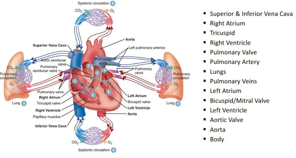
Patho review of how the blood flows^
Blood enters heart from:
superior vena cava & inferior vena cava
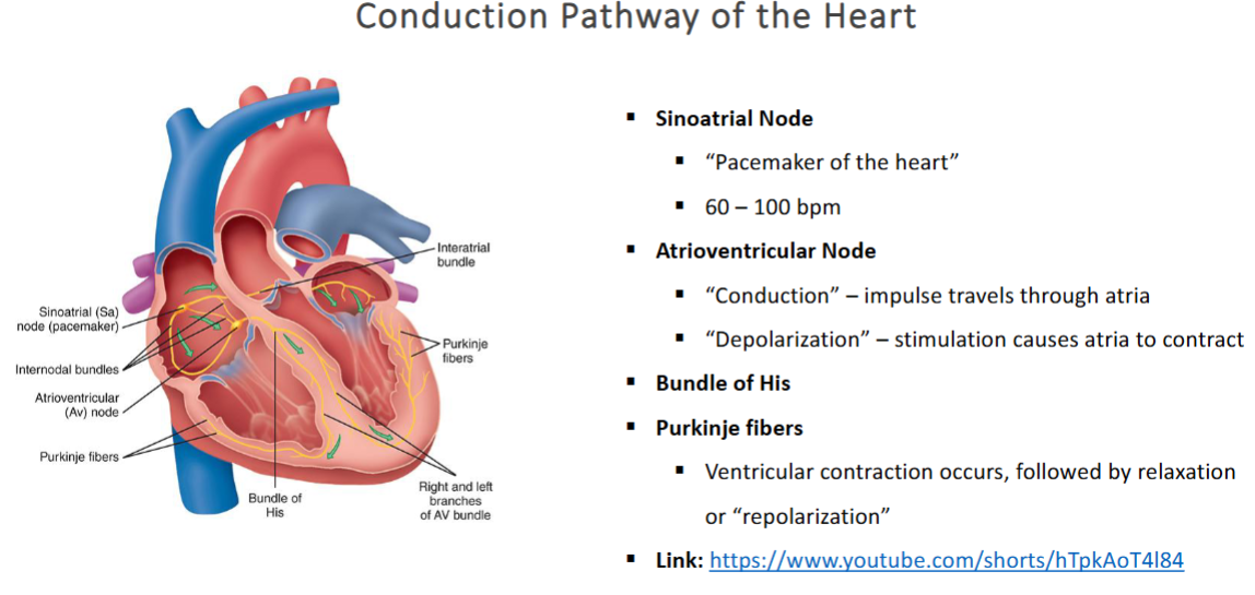
Electrical conduction of the heart^
Sinoatrial Node (SA node)
is the yellow bulb like gland on the left side of the picture above
when atrials contract they contract together
when ventricles contract, they contract together
depolarization = atrias have contracted (squeezed together) —> they are ejecting blood into ventricles
Bundle of his branches into Purkinje fibers in right & left ventricles, which ensures a synchronized contraction of the ventricles, allowing for effective pumping of blood to the lungs and the rest of the body.
SA node → AV node → bundle of his → Purkinje Fibers (in left & right ventricles) → ventricles contract (depolarization) → repolarization of ventricles
The Electrocardiogram:
Electrocardiography (ECG or EKG)
Records the electrical impulse that travels through the heart known as electrocardiogram (ECG)
putting leads/wires on patient chest to track electrical conductions of the heart
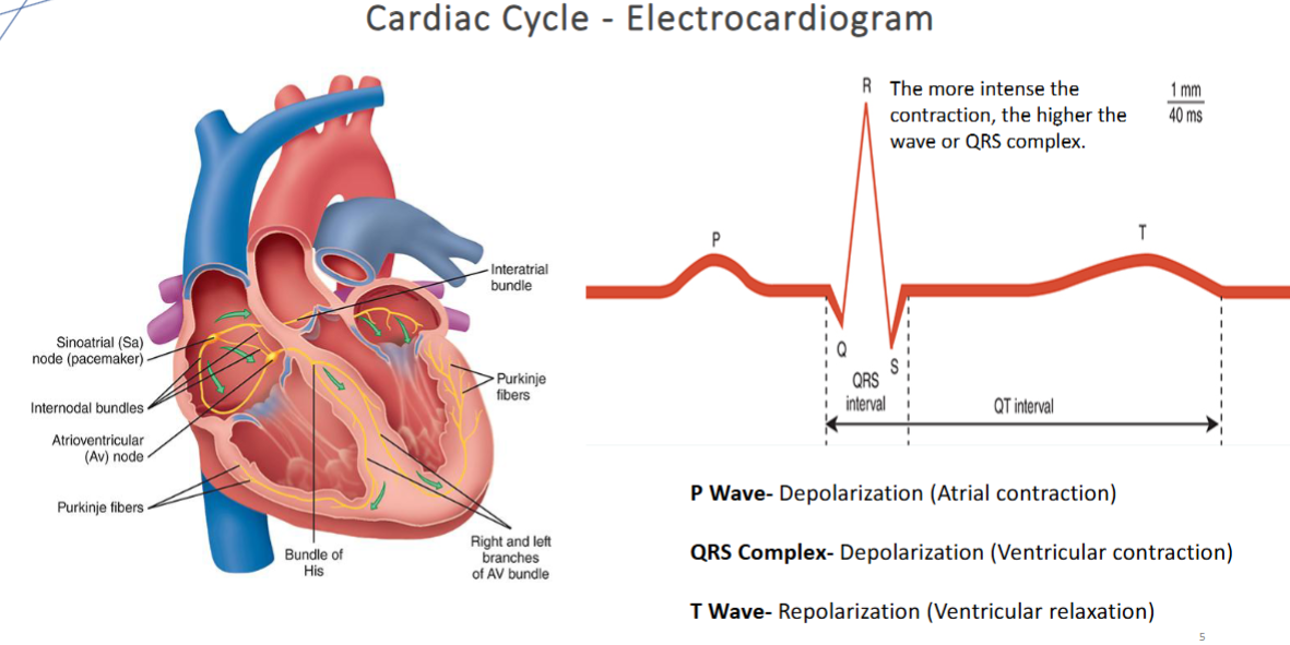
Each phase of the cardiac cycle is represented by specific waveforms
P wave
QRS Complex
T wave
These waveforms can be visualized via:
A cardiac monitor screen
A strip of ECG graph paper
ECG Monitoring Systems:
Electrodes are attached to cable wires connected to one of the following devices:
Standard 12-Lead ECG Machine
Placed at the patient’s bedside for an immediate recording
Hardwire Cardiac Monitor
Provides continuous monitoring at the bedside
Telemetry Monitor
Small portable transmitter, sends ECG data by radio waves to a central monitoring station
Continuous ECG Monitoring (Holter or Patch Monitor)
Small wearable device that records ECG continuously
Intermittent Monitoring (Loop Recorder)
Small implanted or external device that records ECG activity when the patient experiences symptoms
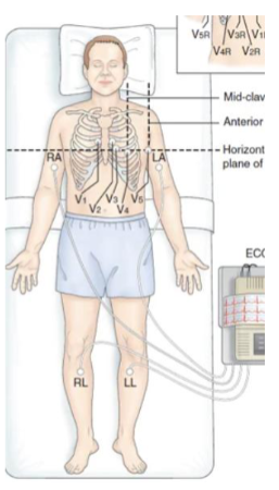
^This is the best patient position for the best ECG/EKG reading
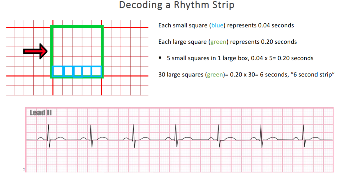
5 blue squares make 1 big green square (as shown in picture above)
you will receive 30 square strips (6 second long strips)
know how to count calculate P waves, QRS waves, T waves
know how to count the boxes
the amount of PQRST waves is the heart rate, which can be determined by analyzing the waveform patterns from the ECG strips.
WILL BE ON QUIZ^
Normal Sinus Rhythm (NSR):
Electrical conduction begins in the SA node, generating a sinus rhythm
Characteristics of NSR
Rate (Atrial & Ventricular): 60–100 bpm (adult)
IF NOT 60-100 IT IS NOT NORMAL SINUS RHYTHM (NSR)
Rhythm: Regular
QRS Complex: Usually normal shape and duration
P Wave: Normal, consistent shape; precedes each QRS
P wave MUST come before QRS or else = non sinus rhythm
P:QRS Ratio: 1:1
Clinical Significance
Indicates good cardiovascular health
Rise in resting heart rate ≥10 bpm increases risk of:
Sudden cardiac death
Atrial fibrillation
Heart failure
Coronary artery disease
Stroke
Cardiovascular disease
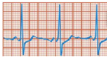
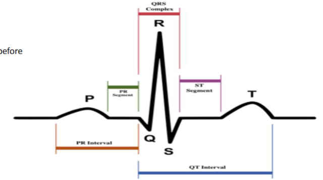
Electrocardiogram:
P wave
Electrical impulse from SA Node
Atrial depolarization
0.11 seconds or less
P-R interval
Time needed from SA node stimulation → period before ventricular depolarization (contraction)
0.12 – 0.2 seconds
QRS Complex
Ventricular depolarization
0.12 seconds
ST Segment
Ventricular repolarization
T wave
Ventricular repolarization
QT Interval
Measures total time for ventricular depolarization and repolarization
0.32 seconds – 0.40 seconds
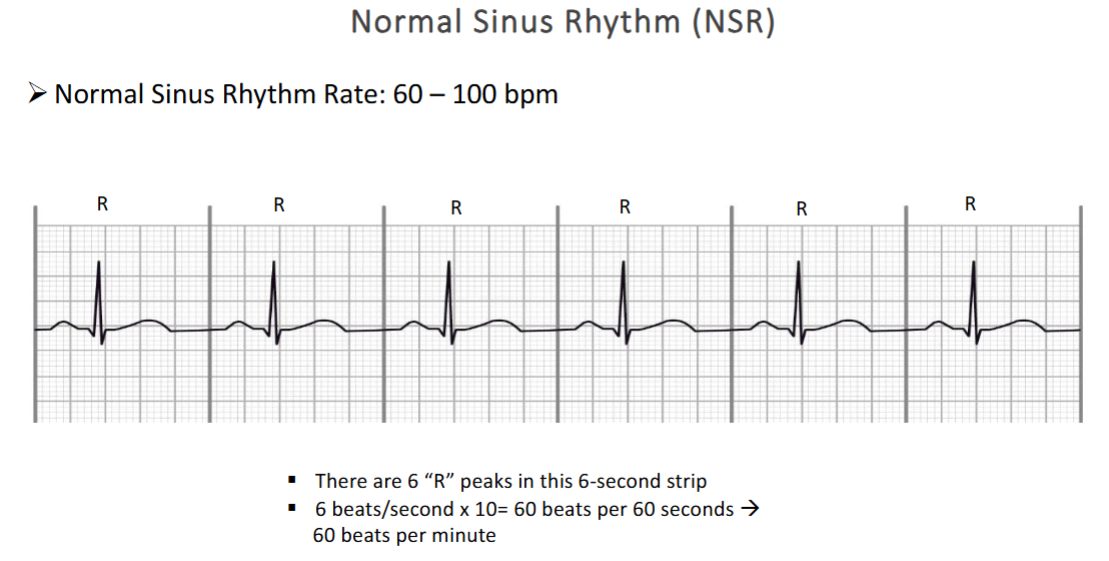
each peak is a cycle so you see 6 QRS complex (peaks) which means 6 cycles
6 beats within 6 seconds
6 seconds x 10 = 60 seconds
6 beats x 10 = 60 beats
MEANING: 60 beats per minute (60 bpm)
Sinus Bradycardia:
Bradycardia
Slowed heart rhythm < 60 bpm
Originates from the SA node
All other sinus rhythm features are normal
ECG Features:
Rate: < 60 bpm
Rhythm: Regular
P wave: Normal, before every QRS
PR interval: 0.12–0.20 sec
QRS: Usually normal
Causes:
Low metabolic needs: sleep, athletic training, hypothyroidism
Vagal stimulation: vomiting, suctioning, severe pain
Medications: beta-blockers, calcium channel blockers, amiodarone
Cardiac: coronary artery disease, inferior MI, sinus node dysfunction
Other: increased ICP, hypoxemia, acute heart failure, delirium
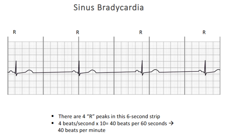
REGULAR PATTERN, same consistent pattern but going slow^
Management of Sinus Bradycardia:
Management:
Dependent on cause
Asymptomatic: monitor & treat underlying cause
Symptomatic/unstable:
Alteration in mental status, chest discomfort, hypotension
Treatment: Atropine, pacemaker, address underlying cause
Avoid vagal stimulation and medications that slow HR
Sinus Tachycardia:
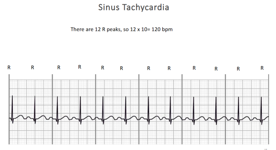
Sinus tachycardia occurs when the SA node fires faster than normal, leading to a heart rate >100 bpm in adults (usually <120 bpm).
It is essentially normal sinus rhythm but faster, and it is nonparoxysmal (does not start or stop suddenly)
Common Causes
Physiologic or psychological stress
Examples: blood loss, anemia, shock, heart failure, pain, fever, exercise, anxiety
Any condition increasing sympathetic activity or metabolic demand
Medications and stimulants
Sympathomimetic drugs: catecholamines, aminophylline, atropine
Stimulants: caffeine, nicotine
Illicit drugs: amphetamines, cocaine, ecstasy
Enhanced SA node automaticity or excessive sympathetic tone
Called inappropriate sinus tachycardia (heart rate high without clear physiologic need)
Autonomic dysfunction
Example: Postural Orthostatic Tachycardia Syndrome (POTS)
Characterized by: rapid heart rate without hypotension, palpitations, lightheadedness, weakness, blurred vision upon standing
Management of Sinus Tachycardia:
Goal: Identify and treat underlying cause; relieve symptoms.
1. Vagal Maneuvers / Non-Drug Interventions
Carotid sinus massage
massage someone carotid artery
IF NOT IN SINUS RHYTHM they will pass out
Gagging
Bearing down against a closed glottis (Valsalva maneuver)
Forceful, sustained coughing
Cold stimulus to the face (ice-cold wet towel)
Adenosine IV to interrupt tachycardia
2. Hemodynamically Unstable Patients
Signs: altered mental status, chest discomfort, hypotension
Synchronized cardioversion (electrical current timed with QRS)
if patient is unstable do cardioversion (the charge & clear defibrillators)
stable patients give some medications
Used if vagal maneuvers or adenosine fail
3. Hemodynamically Stable Patients
IV Beta-blockers (Class II antiarrhythmic)
IV Calcium channel blockers (Class IV antiarrhythmic)
Cardioversion if medications fail or are contraindicated
4. Persistent / Inappropriate Sinus Tachycardia
SA node catheter ablation if unresponsive to other treatments
means none of the medications or treatments are working so, ablation of SA node
5. POTS (Postural Orthostatic Tachycardia Syndrome)
Tachycardia without hypotension; presyncopal symptoms on standing
Treatment targets underlying cause:
Hypovolemia → increase fluids and sodium, or use salt tablets
Combination of approaches as needed
Summary: Stepwise management → Vagal maneuvers → Medications → Cardioversion → Ablation
Atrial Fibrillation:
Atrial Fibrillation (AFib)
Prevalence & Public Health Impact
2.7–6.1 million Americans have AFib (CDC, 2017)
Strongly associated with aging, making it a growing concern in older adults
Pathophysiology
Abnormal impulse formation in atrial tissue → rapid, disorganized, uncoordinated atrial twitching
Structural or electrophysiologic changes in atria trigger arrhythmia
structural issues or ions are off (electrophysiologic)
Central nervous system (brain & spinal cord) and Cardiac Autonomic Nervous System (CANS) both contribute
CANS: network of autonomic ganglia within epicardium, atrial myocardium, and pulmonary veins
Hyperactive ganglia → impulses from pulmonary veins → AV node
Ventricular response depends on:
AV node conduction
Accessory pathways
Medications
Atrial Fibrillation: Classification of AFib
Paroxysmal: sudden onset, terminates spontaneously
have patient have a heart monitor for 24-48 hrs; ask what symptoms they experienced
Persistent: sustained, requires intervention to terminate
will not resolve spontaneously; patient will need something done to fix this such as medication adjustment, electrical cardioversion, or in some cases, catheter ablation or repair the valve (like incase there is a leaky valve)
Permanent: continuous, resistant to intervention
not going anywhere, heart constantly in A-fib
heart may be resistance to treatments (pts live with chronic A-fib)
Nonvalvular: absence of moderate-severe mitral stenosis, absence of mechanical valve
Complications:
blood stasis:
ineffective atrial contraction causes blood to pool
stagnant blood eventually forms clots (thrombi)
risk of emboli
if a clot dislodges, it can travel through blood stream and cause a stroke such as pulmonary emboli, emboli in other organs
AFib is a common arrhythmia in adults, especially the elderly
Results in ineffective atrial contraction → risk of stroke, heart failure, and other complications
Ventricular rate and symptoms vary depending on AV conduction and treatment
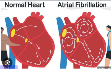
A-fib & Coagulation:
Atrial quivering:
Atria twitch or fibrillate instead of contracting efficiently
Blood stasis:
Ineffective atrial contraction causes blood to pool
Stagnant blood eventually forms clots (thrombi)
not good filling into heart so blood is stagnant and leading to possible clots
take blood thinners (aka anticoagulants) to combat blood clots (THIS IS THE MED YOU USE TO TREAT A-FIB)
LIFE-LONG TREATMENT
Risk of embolism:
If a clot dislodges, it can travel through the bloodstream →
Leading to stroke (most common), pulmonary emboli, emboli in other organs
Treatment: Patients often take anticoagulants (e.g., warfarin, apixaban) to prevent clot formation
Atrial Fibrillation – EKG Characteristics:
Rate
Atrial rate: 300–600 bpm
Ventricular rate: 120–200 bpm
Rhythm
Highly irregular
QRS Complex
Typically normal
May be abnormal if conduction changes are present
P Wave
Absent
Replaced by fibrillatory (f) waves
Irregular, varying in amplitude and shape
PR Interval
Cannot be measured
P:QRS Ratio
Many atrial impulses per ventricular response, not 1:1
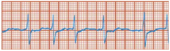
the 1st to 2nd box on strip is known as F wave, which represents the atrial activity and is a key indicator in diagnosing conduction disorders. It demonstrates the presence of multiple atrial impulses failing to conduct through the ventricular system.
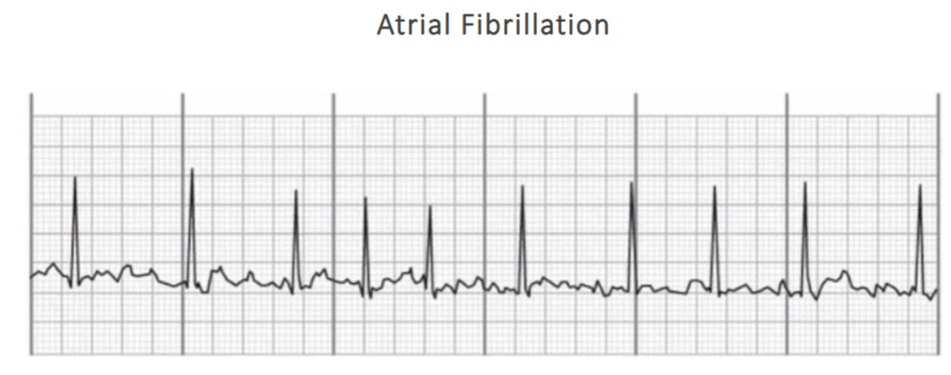
Fibrillatory “f wave” pattern
▪ “p waves” are indistinguishable, atrial rate is indeterminate
▪ There are 10 “R” peaks x 10 = 100 Ventricular Rate
Medical Management of Atrial Fibrillation (AFib):
Goal:
Prevent embolic events (e.g., stroke)
Control ventricular rate
Restore sinus rhythm when appropriate
1. Factors Influencing Treatment
Cause, pattern, and duration of AFib
Ventricular response rate
what’re the ventricles doing
Presence of structural or valvular heart disease
Coexisting cardiac conditions (e.g., coronary artery disease, heart failure)
Patient-provider shared decision-making
2. Management Approaches
Rate control: Use antiarrhythmic medications to control ventricular response
Rhythm control: Attempt to convert to sinus rhythm (cardioversion)
Observation: Some AFib converts spontaneously within 24–48 hours
Permanent AFib: When patient and provider decide not to attempt sinus rhythm restoration
3. Pharmacologic Therapy
A. Antithrombotic Medications
Reduce risk of stroke and embolic events
Include anticoagulants and antiplatelet drugs
difference is they work on different clotting factors
Indicated for most patients with nonvalvular AFib
Risk assessment: Use CHA₂DS₂-VASc score to guide therapy
B. Antiarrhythmic Agents
Control ventricular rate
Facilitate conversion to sinus rhythm if appropriate
can cardiovert a patient and be on antiarrhythmics
4. Catheter Ablation
destruction of specific cells that are causing the tachyarrhythmia (fibrillation)
5. Electrical Cardioversion
medical treatments that uses quick, low energy shocks to restore a regular heart rhythm
indications
hemodynamically unstable AF patients
ex: acute mental status changes, chest discomfort, hypotension
medication-refractory Afib
meds used to work for Afib but are no longer working
Antiarrhythmic drugs may be given before cardioversion to increase the success rate and maintain sinus rhythm
most common antiarrhythmics:
flecainide
propafenone
amiodarone
dofetilide
sotalol
______________________________________________________________
Electrical Cardioversion for Atrial Fibrillation:
Indications:
Hemodynamically unstable AF patients
Examples: acute mental status changes, chest discomfort, hypotension
Most effective when combined with antiarrhythmic therapy if appropriate
Medication-refractory Afib
Pre-treatment (Optional):
Antiarrhythmic drugs may be given before cardioversion to:
Increase the success rate
Maintain sinus rhythm
Common Antiarrhythmics Used:
Flecainide, Propafenone, Amiodarone, Dofetilide, Sotalol
Atrial Flutter:
Atrial Flutter
Caused by a conduction defect in the atrium
Rapid, regular atrial impulses at 250–400 bpm
AV node can blocks some impulses to prevent ventricular rates from reaching atrial rates
Protects against life-threatening ventricular fibrillation
Atrial Flutter – ECG Characteristics:
Rate
Atrial rate: 250–400 bpm
Ventricular rate: Usually 75–150 bpm
Rhythm
▪ Atrial rhythm: Regular
▪ Ventricular rhythm: Usually regular, may be irregular if AV conduction varies
QRS Complex
Usually normal
May be abnormal or absent
P Wave
Saw-toothed shape, called F waves PR Interval
Difficult to measure due to multiple F waves
P:QRS Ratio
Commonly 2:1, 3:1, or 4:1
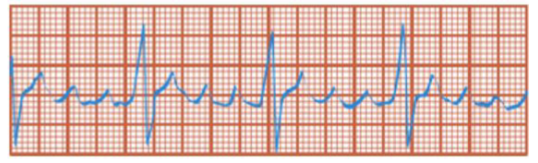
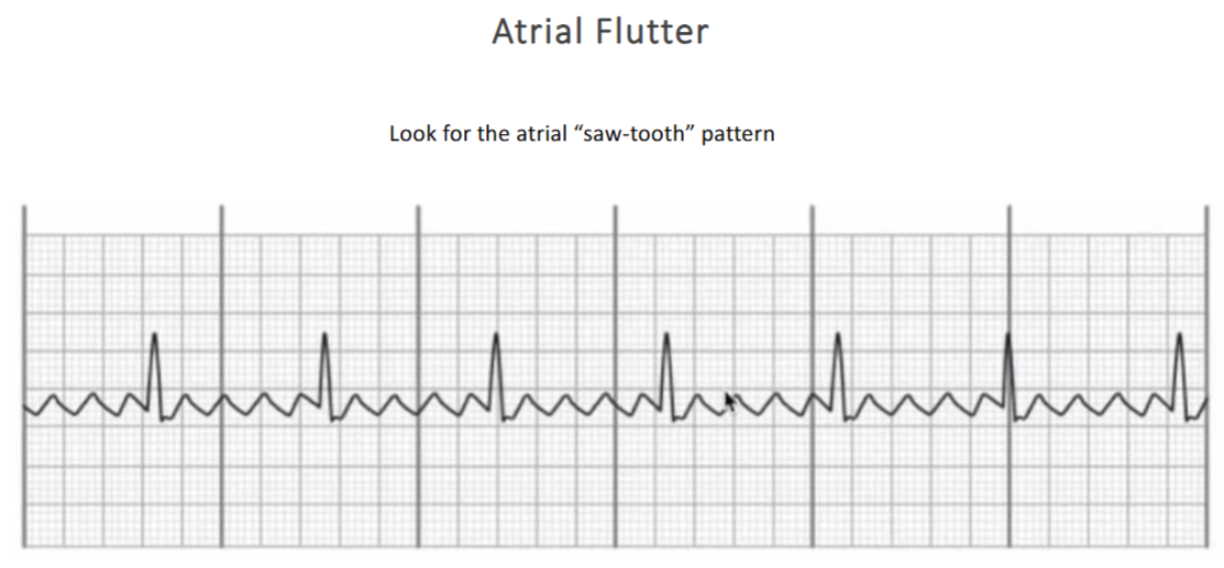
Look for the atrial “saw-tooth” pattern
▪ There are 27 “p waves” x 10 = 270 Atrial Rate
▪ There are 7 “R” peaks x 10 = 70 Ventricular Rate
Medical Management of Atrial Flutter:
Symptoms:
Chest pain
Shortness of breath
Hypotension
Acute Management:
Vagal maneuvers
increase parasympathetic activity to slow AV node conduction
massage the patients carotid or have patient bear down
Adenosine (IV):
Slows AV node conduction → may terminate tachycardia or reveal flutter waves for diagnosis
Rapid IV push followed by 20 mL saline flush
Elevate arm to promote rapid circulation
Ongoing Management:
Antithrombotic therapy
Rate control - beta-blockers, calcium channel blockers
Rhythm control - antiarrhythmics
Electrical Cardioversion:
Often successful in converting atrial flutter to sinus rhythm
Vagal maneuvers:
Physical actions that stimulate the vagus nerve
Goal: temporarily slows conduction through the atrioventricular (AV) node in conditions such sinus tachycardia, atrial flutter, or AV nodal reentrant tachycardia (AVNRT)
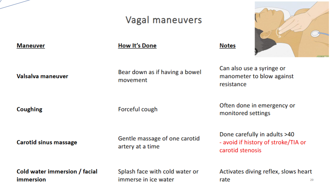
carotid massage is only on one side to prevent potential complications such as carotid artery dissection or ischemic stroke.