4BBY1020 Chemistry for the Biosciences Lecture 11: Protein Folding
Thermodynamics of Protein Folding
Stabilising Interactions: Folded proteins are stabilised by a mixture of non-covalent and covalent interactions, as well as entropic effects.
Hydrogen bonds
Electrostatic interactions (Charged side chains and dipole interactions)
London forces (dispersion interactions)
Hydrophobic effect
Disulphide bonds
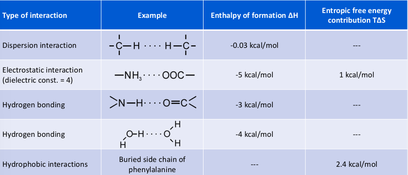
Typical Energetic Contributions (Theoretical):
Solvation Effects and Water Dynamics
Structure and Dynamics of Water ():
Forms hydrogen bonds with itself.
Each molecule has two hydrogen atoms and two lone pairs: acts as two hydrogen bond donors and two acceptors.
Water molecules are highly dynamic, exhibiting constant rotational and translational motions and rearrangements in hydrogen bonding patterns.
Water as a Solvent:
Polar substances dissolve readily due to water's polar nature and H-bonding ability.
Non-polar substances interrupt hydrogen bonding, leading to phase separation to reduce the water interface.
The Hydrophobic Effect:
Water surrounding non-polar molecules forms an "ice-like" cage.
the ability to move around is severely limited for all water molecules forming this cage → significant loss of entropy
Effect 1 (Hydrophobic Interactions): Removing hydrophobic residues from the water-protein interface to form a hydrophobic core is entropically favourable because water has higher entropy when not forming a "cage."
Effect 2 (Hydrophilic Interactions): Interactions between water and hydrophilic residues (charged/polar side chains) are enthalpically favourable due to hydrogen bonding and charge interactions. Though mobility is still limited, there is a significant energy benefit of forming the interactions
Protein Shape and Solvation:
A globular shape with hydrophobic residues are in the core and hydrophilic residues are on the outside is favourable
Hydrophobic collapse is considered a key step in folding. the globular shape of proteins is a combination of hydrophobic and hydrophilic interaction
Hydrophobic surfaces are frequently involved in protein-protein interactions.
Membrane Proteins:
Membrane proteins need to be anchored inside the membrane but may have other protein regions sitting on the membrane
the difference in hydrophobicity between membrane and non-membrane must be accounted for during folding
Examples: E. Coli BAM complex (beta-barrel-assembly machinery, PDB id: 6LYR) and E. Coli Multidrug exporter protein.
Stability and Entropy Calculations
Relative Contributions in Context:
Hydrogen bonds: While the theoretical contribution is , in an unfolded state, and groups already form H-bonds with water (weaker, but lost when folding). net contribution is only about .
Electrostatics: The theoretical enthalpic contribution is , when unfolded charged side chains are fully solvated in water (weakens enthalpic contribution). entropic contribution remains as the water in the solvation shell around the charge is released
Mathematical Representation of Entropy:
Entropy is related to the number of states () or degrees of freedom a molecule can adopt:
Where is Boltzmann's constant and W is the number of states (for a molecule, W = number of different conformations). For one mole of molecules, the gas constant is used:
Na is avogadro’s number and R is the gas constant
Entropy of Folding ():
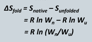
Because there is only one native state, .
For a protein of 100 residues with 10 conformations each, .
At , the free energy contribution is approximately .
Net Free Energy Calculation for a 100-Residue Protein:
Hydrophobic: 40 buried residues ( each) = .
Hydrogen bonds: 40 bonds ( each) = .
Electrostatic: 3 salt bridges ( each) = .
Dispersion: 100 interactions ( each) = .
Total Enthalpic/Hydrophobic Factors: .
Entropy of Folding Contribution: .
Net for folding: .
Marginal Stability:
free energy of folding has a large entropic and enthalpic contribution
enthalpically: non-covalent interactions favour folding
entropically: the separation of hydrophobic residues and solvent is favourable but the folding is unfavourable.
The net free energy () indicates that proteins are only marginally stable and can be easily destabilised by mutations, or changes of the environment (pH, temperature, or salt changes).
Protein Folding Kinetics and Pathways
Experimental Findings:
Folding is spontaneous.
Folding is fast (milliseconds to seconds).
Folding is cooperative (not a slow step-by-step process).
thermodynamics suggests a free energy for folding of around -10kcal/mol
Levinthal’s Paradox:
A protein with 100 amino acids and 10 conformations per residue has total possible conformations.
assume each conformation can be sample via internal rotations
If rotations occur at per second, sampling all conformations would take seconds ( years).
Since the universe is only years old and folding takes <1\,s, random "trial and error" sampling is impossible.
Consequence: folding times in experiment of up to 1s: there is more folding than just the sequence-structure relationship
folding must be biased towards native structures
proteins follow some pathways rather than randomly sampling
the amino acid sequence encodes protein structure and the different folding pathways
Anfinsen's Experiment (1961):
Studied Ribonuclease A folding in bovine pancreatic ribonuclease
The protein was reduced with mercaptoethanol in urea to yield a randomly coiled polypeptide chain.
Under optimal conditions of polypeptide pH and concentration, the protein reformed disulfide bonds of the native enzyme which occurs in the presence of molecular oxygen
Disulfide Bond Formation (Bovine Pancreatic Trypsin Inhibitor - BPTI):
58-residue protein with three native disulfide bonds: 5-55, 30-51, and 14-38.
Experimental folding shows multiple intermediate species (e.g., 5-51, 5-55, 30-51, 5-30, etc.).
Folding is complex and can involve non-native intermediates.
Folding Pathway of Lysozyme:
multiple intermediates and multiple pathways observed
alpha-helices form first.
beta-sheets form later.
The Role of Chaperones
Functions:
Actively support protein folding directly.
Stabilise folding intermediates to prevent protein aggregation (e.g., Hsp33).
Assist in protein transport across membranes.
GroEL/GroES Complex:
Dimensions: Height of 184 Å with GroES; GroEL width varies from 140 Å to 80 Å. Cavity heights and widths: 33 Å, 10 Å, 71 Å.
Mechanism:
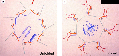
The "Closed state" exposes hydrophobic surfaces to attract the unfolded protein.
The "Open state" (hydrophilic residues exposed) hides hydrophobic surfaces, allowing the protein to fold in a protected environment.
Protein Misfolding and disease
Sickle Cell Anaemia:
Caused by a single mutation of Glutamate (Glu) to Valine (Val).
The Valine in the beta-chain fits into a hydrophobic pocket formed by Phenylalanine (Phe) and Isoleucine (Ile).
This causes haemoglobin molecules to link up and form long fibres that rupture red blood cells.
Alzheimer's Disease:
Described by Alois Alzheimer (1906) as striking changes in neurofibrils.
Proteins aggregate into thick bundles or tangles of fibrils, leading to neuron disintegration.
Associated with Amyloid-beta 42 fibrils.
Prion Diseases (e.g., Creutzfeld-Jacob Disease):
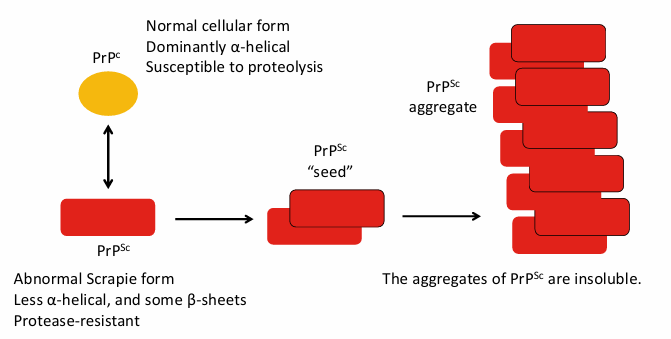
PrP protein exists in two inconvertible forms (causative agent):
PrPc (Normal cellular form): Dominantly alpha-helical, susceptible to proteolysis.
PrPSc (Abnormal Scrapie form): Higher beta-sheet, less a-helical content, protease-resistant, aggregates of PrPsc are insoluble.
The PrP^{Sc} form acts as a "seed" to aggregate normal PrP^c.
CryoEM structures (PDB: 6AXZ, 6UUR) show tight, regular packing of beta-sheets in a hydrophobic core.
General Pathological Misfolding:
Aggregation via beta-sheet formation is common.
Examples including Parkinson's disease, Huntington's disease, Type II diabetes, and cataracts.