Immunotechnology
LOs:
To become more familiar with some of the primary techniques used in the laboratory with regards to immunology:
hybridoma and mAb generation
flow cytometry/FACS
ELISA/ELISPOT
Definitions:
flow cytometry = study of cells as they move in fluid suspension, allowing multiple measurements to be made per cell
Notes:
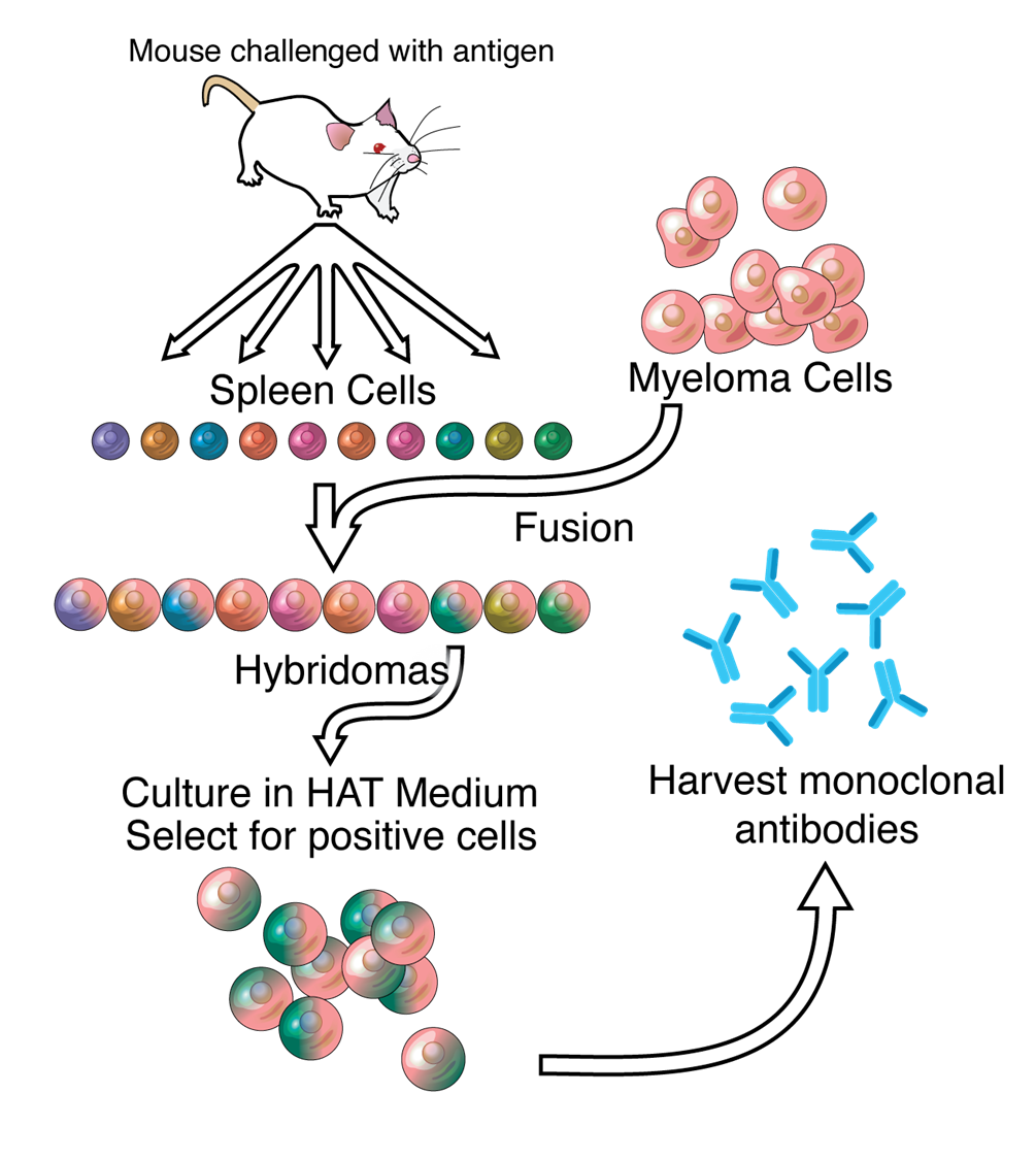
3: Monoclonal antibodies are produced in vitro by hybridoma technology, based on fusing myeloma cells with spleen lymphocytes. Monoclonal antibodies used frequently in the lab, preferential to polyclonal antibodies as they only bind one antigen/protein:
mice immunised with antigen
spleen cells isolated then mixed with myeloma cells (cancer, divide rapidly and uncontrollably, unlikely to die) and polyethylene glycol - supports growth and fusion
hybridomas cultured in HAT medium (hypoxanthine, aminopterin, thymidine). HAT is a selective media which selects for cells presenting the antibody
Hybridoma technology doesn’t always work and often results in wrong antibody. Need to grow hybridomas for a long time to collect enough antibody. May need to undergo thousands of different fusions to get right Ab with right specificity
4: Isolating cells/purifying cell populations allows scientists to confidently answer specific research questions while minimising interference from other cell types in the sample. Many downstream applications are made possible following purification.
The defined cell type is usually isolated based on a particular protein or cell marker which is expressed by the cell and absent in unwanted cells. Selection is usually performed using monoclonal antibodies, or a reporter surrogate marker in some cases.
FACS and flow cytometry different but same technology can be used for both. FC counts cell types and is the overall technology then used for FACS
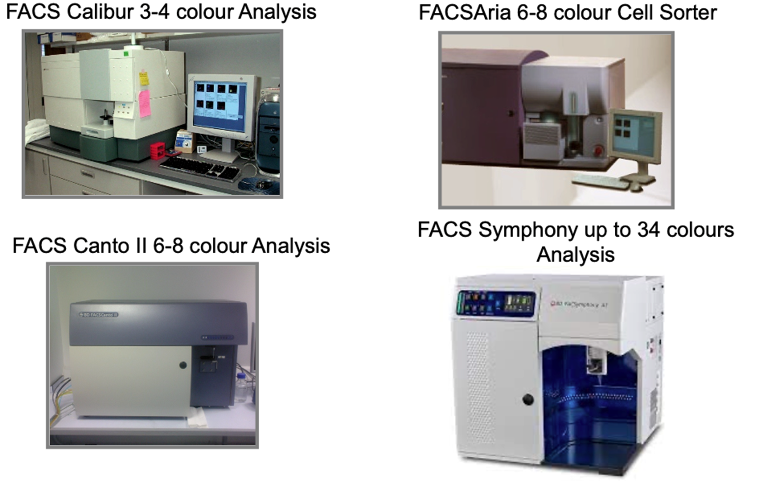
5: FACSTM = Fluorescence-Activated Cell Sorting. Commercially available since 70s but there has been major progression in the technology.
FACS = Mixed culture of tagged cells to sort exact type
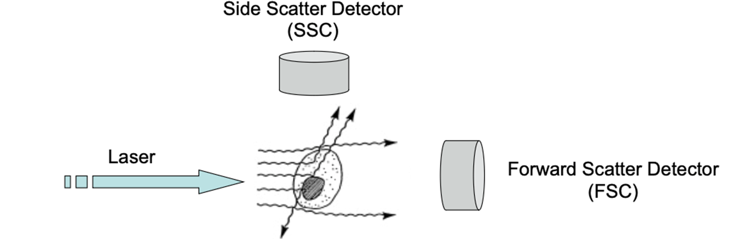
Forward light scatter (FSC) proportional to cell size, side light scatter (SSC) proportional to cell granularity - FSC and SSC most basic properties, rest of information comes from the colours shown
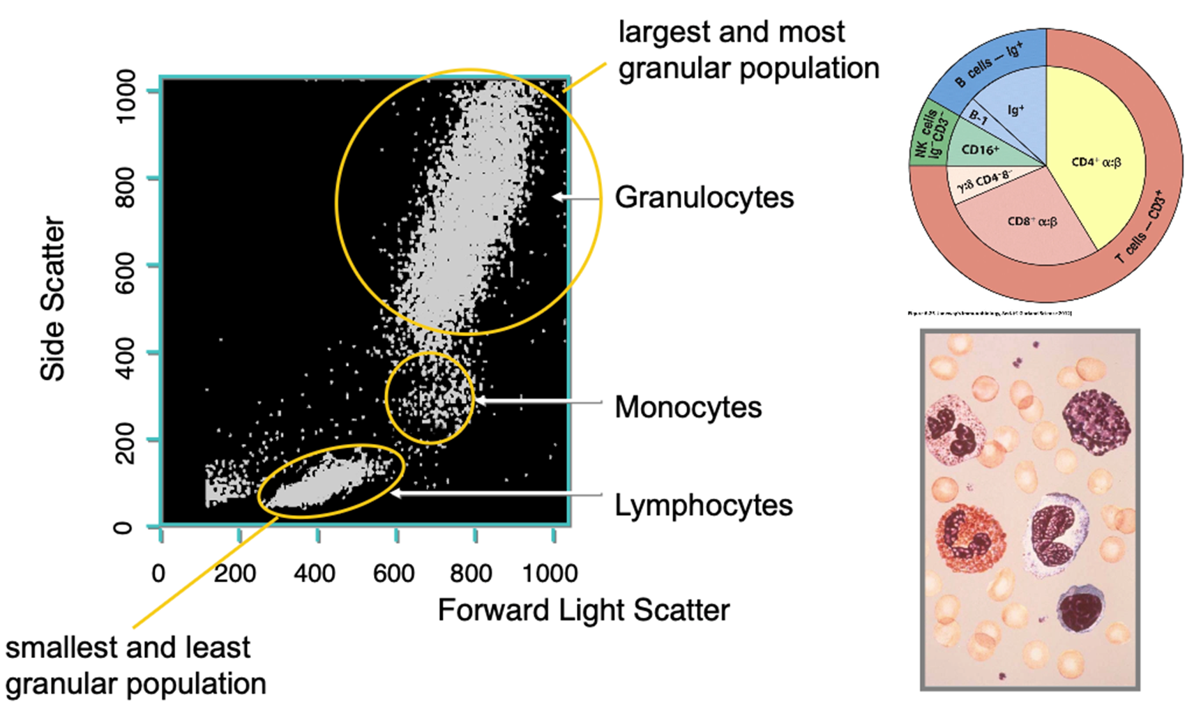
Neutrophils = large, granular - very easy to detect in flow cytometry
Fluorescence comes about via:
binding of fluorescent-labelled antibodies
calcium sensitive dyes within cells
fluorescent proteins expressed by cells
binding DNA dyes for cell cycle analysis
6: 6-8 colour most commonly found, FACS symphony vv expensive at around 1.5 million pounds with each antibody for it costing around 500
7:
Cells come through in sample stream with fluid running alongside it in sheath stream to keep cells cool. Quite fast depending on machine used however high speed isn’t always desired as errors may occur eg counted twice, things not picked up
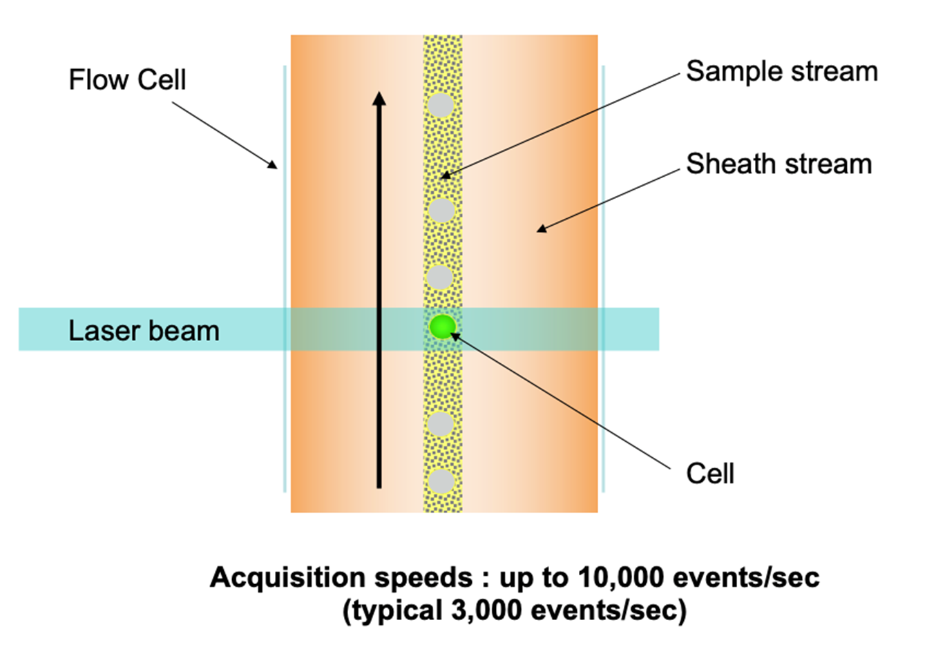
8: Forward scatter is proportional to cell size, meaning the bigger the cell, the more light is scattered and the higher the detected signal.
Side scatter is proportional to cell complexity/granularity so the more organelles/granules etc in the cytoplasm, the more light scatter and higher the detected signal. Measuring how often side or forward detectors are hit by refracted laser
When the laser beam strikes the stream, majority of light passes through unobstructed. Some of the photons diverge slightly, primarily via light diffraction, from their path as they contact membranes of passing cells.
9: Forward light scatter of immune cells of different sizes and granularities in the blood:
bottom left cut out = RBCs
10:
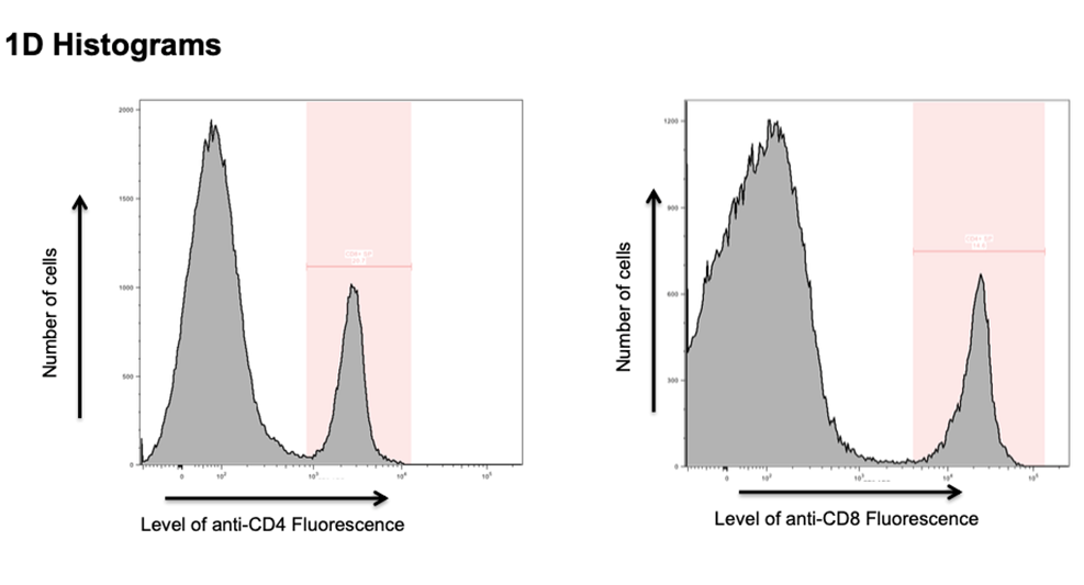
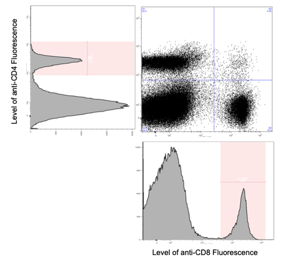
1D histograms only provide measure of 1 aspect eg in this case CD4 or CD8 - cells separated on size and granularity to narrow down spectrum of what’s being looked at then Igs used to select for properties - big peak = CD4/CD8 negative, smaller peak = CD4/CD8 positive
2nd shows what it looks like on a scatter plot, bottom left = negative for both, further up left = CD4+, further right = CD8+
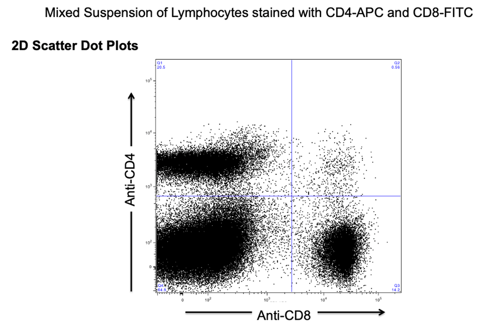
11: shows how peaks are constructed - both slides 10 and 11 generally wouldn’t be presented in a scientific paper
12:

13: There are 4 different types of enzyme linked immunosorbent assay (ELISA):
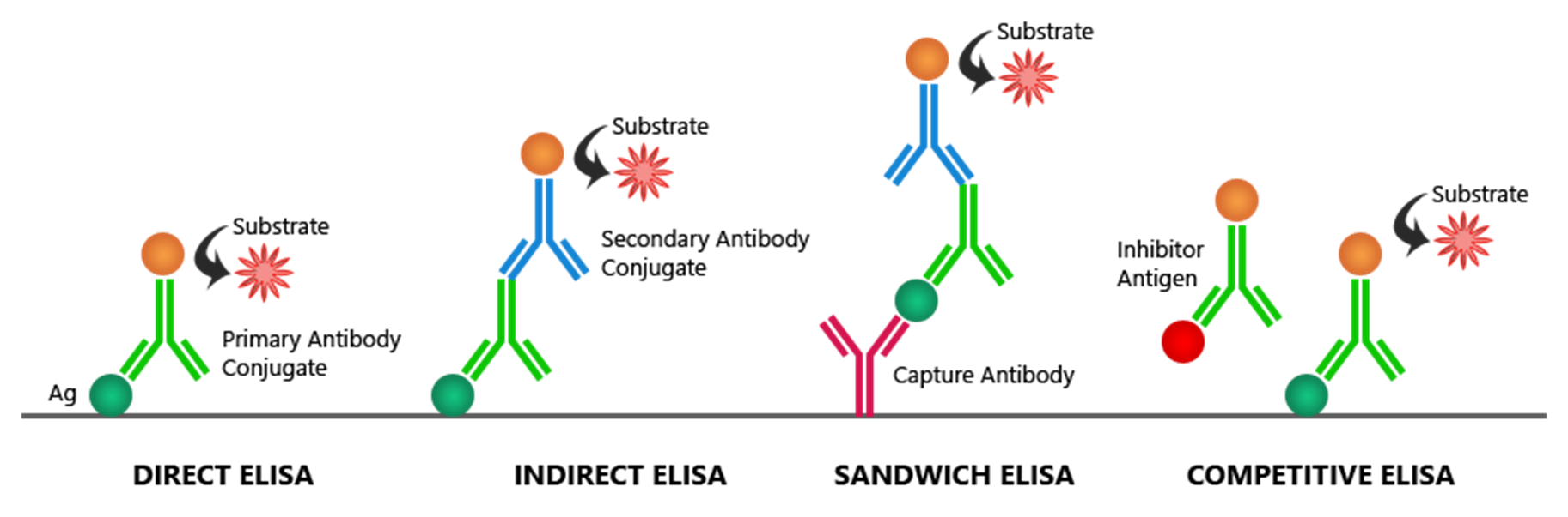
Differences include number and types of antibodies used
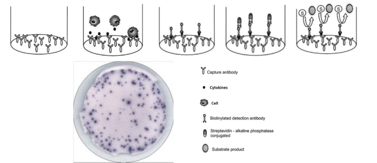
14: Enzyme linked immunosorbent spot assay (ELISPOT) is based on sandwich ELISA. It allows secretory activity of individual activated/responding cells to be measured (eg antibody or cytokine secretion). Cellular secretions are captured and shown as coloured spots, each of which represents a single reactive cell.
ELISPOTs pain in ass to get reliable data from, generally only used when no other option/low lab budget