Neuro Dev pt3
🧠 DETAILED MULTI-PARAGRAPH SUMMARY
Neurogenesis in the developing nervous system begins with neuroepithelial cells, which serve as the earliest stem cells of the neural tube. These cells initially divide symmetrically to expand the stem cell population, and later asymmetrically to produce differentiated progeny such as neurons and glial cells. As development progresses, neuroepithelial cells transition into radial glial cells, which act both as neural stem cells and as scaffolding structures that guide migrating neurons. These radial glial cells extend long processes across the thickness of the neural tube, providing a physical pathway for neuronal migration.
In the developing cerebral cortex, neurons are generated at the ventricular zone (inner surface of the neural tube) and migrate outward toward the pial surface. This migration occurs primarily along radial glial fibres and results in the formation of the characteristic six-layered cortical structure. A key feature of cortical development is its “inside-out” pattern: early-born neurons settle in deeper layers, while later-born neurons migrate past them to form more superficial layers. This precise layering depends on tightly regulated migration mechanisms and signalling cues.
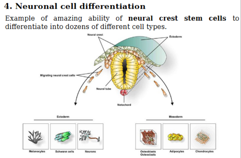
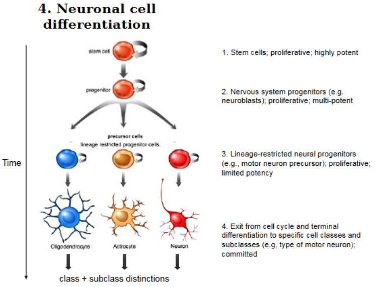
Cell differentiation during neurogenesis follows a progressive restriction model. Early stem cells are highly potent and can give rise to multiple cell types. However, as progenitor cells divide and differentiate, their potential becomes increasingly limited. External signalling molecules—such as growth factors and morphogens—play a crucial role in directing cell fate by altering gene expression. A striking example of cellular plasticity is seen in neural crest cells, which originate from the dorsal neural tube and migrate extensively to form a wide variety of cell types, including neurons, glia, melanocytes, and even skeletal elements.
Neuronal migration occurs via multiple mechanisms. Radial migration is relatively short-range and involves movement along radial glial scaffolds, while tangential migration involves long-distance movement across the neural tube, such as interneurons migrating from the ganglionic eminence to the cortex. These processes are guided by molecular cues, including the protein Reelin, which is secreted by cells in the outer cortical layer. Reelin ensures proper cortical layering by regulating both neuronal movement and radial glial organisation. Disruption of Reelin signalling leads to severe developmental abnormalities, including inverted cortical layers and neurological disorders such as lissencephaly(smooth brain).
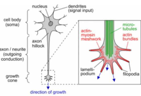
Axon and dendrite formation relies on the dynamic behaviour of the growth cone, a specialised structure at the tip of extending processes. The growth cone contains actin-rich filopodia and lamellipodia that explore the environment, while microtubules provide structural support. Growth cones respond to attractive (Netrins and WNT)and repulsive cues (Semaphorin and reelin)—either diffusible signals or contact-based interactions—which guide them toward their appropriate targets. Cytoskeletal remodelling, driven by actin and microtubule dynamics, is essential for directional movement and extension.
Finally, once axons reach their target regions, they undergo target selection and synapse formation. Axons may leave bundles (defasciculation), enter target regions, and refine their position through branching and signalling interactions. Synapses form through a stepwise process involving membrane contact, accumulation of synaptic vesicles, and development of the postsynaptic density. Over time, synaptic connections are refined through synaptic pruning, where excess connections are eliminated, and functional networks are strengthened, forming the basis of learning and memory.
📌 BULLET POINT SUMMARY
Neurogenesis Basics
Neuroepithelial cells = earliest neural stem cells
Symmetric division → more stem cells
Asymmetric division → neurons + glia
Transition into radial glial cells
Note: First step is that a patch of ectoderm is induced to become neuro-ectoderm. Requires
BMP inhibition
Radial Glial Cells
Act as stem cells + scaffolds
Guide neuronal migration
Eventually differentiate into glia
Cortex Formation
6-layered structure in the cerebral cortex
Neurons born at ventricular zone
Migrate outward along radial glia
Inside-out layering pattern
Differentiation
Early cells = high potency
Later cells = restricted fate
Controlled by signalling molecules (GF and Morphogens)
Neural crest cells → highly multipotent
Neural Crest Cells
Originates from the dorsal neural tube
Migrate widely
Form diverse cell types (neurons, glia, skin, bone)
Migration Types
Radial migration → short distance
Tangential migration → long distance
Guided by signals like Reelin
Migration Disorders
Reelin defects → inverted cortex
Leads to conditions like lissencephaly
Axon Growth
Growth cone = dynamic tip
Actin (movement) + microtubules (support)
Guided by:
Attractive signals (e.g., netrin)
Repulsive signals (e.g., semaphorin)
Target Selection
Axons leave bundles (defasciculation)
Enter target region
Branch and refine position
Synapse Formation
Pre- and postsynaptic membranes align
Vesicles accumulate
Postsynaptic density forms
Synapses mature over time
Synaptic Pruning
Excess synapses removed
Remaining connections strengthened
Essential for learning
📝 FILL-IN-THE-BLANK (WITH ANSWERS BELOW)
Section A
The earliest stem cells of the neural tube are called __neuroepithelial______ cells.
These cells later differentiate into _radial___ glial cells.
Neurons are generated at the _ventricular_ zone of the neural tube.
Cortical layers form in an ___inside_______-out pattern.
Migration along scaffolds is called __radial____ migration.
Section B
_neuronal crest__ cells originate from the dorsal neural tube and migrate widely.
The protein _Reelin__ is essential for proper cortical layering.
Loss of this protein can cause __lisscephaly__, a smooth brain condition.
Long-distance neuron movement is called _tangential___ migration.
Growth cones contain __actin___ filaments at their periphery.
Section C
Microtubules are made from __alpha__ and __beta__ tubulin.
Growth cones respond to _attractive____ and __repulsive________ signals.
Axons leaving bundles undergo __fassiculation______.
Synapses contain ___synaptic____ vesicles in the presynaptic terminal.
Removal of excess synapses is called __synaptic pruning________.
✅ ANSWERS
Neuroepithelial
Radial
Ventricular
Inside
Radial
Neural crest
Reelin
Lissencephaly
Tangential
Actin
Alpha and beta
Attractive and repulsive
Defasciculation
Synaptic
Synaptic pruning
🎓 HARD EXAM-STYLE MCQs (40)
Questions
Which cells are the earliest neural stem cells?
A. Astrocytes
B. Neuroepithelial cells
C. Neurons
D. OligodendrocytesRadial glial cells primarily function as:
A. Immune cells
B. Synaptic vesicles
C. Scaffolds and stem cells
D. Myelin producersCortical neurons originate from:
A. Pial surface
B. Ventricular zone
C. White matter
D. Neural crestCortical layering follows which pattern?
A. Outside-in
B. Inside-out
C. Random
D. BilateralNeural crest cells arise from:
A. Ventral tube
B. Dorsal neural tube
C. Cortex
D. Spinal cord onlyWhich molecule guides cortical migration?
A. Dopamine
B. Reelin
C. Serotonin
D. InsulinReelin is secreted from:
A. Ventricular zone
B. Marginal zone
C. White matter
D. AxonsLoss of Reelin causes:
A. Hydrocephalus
B. Lissencephaly
C. Parkinson’s
D. MSRadial migration occurs:
A. Along glial scaffolds
B. Randomly
C. Through blood
D. Only in adultsTangential migration is:
A. Short distance
B. Long distance
C. Static
D. PassiveGrowth cones are located at:
A. Soma
B. Axon tip
C. Nucleus
D. SynapseFilopodia are rich in:
A. DNA
B. Actin
C. Lipids
D. CalciumMicrotubules are composed of:
A. Actin
B. Tubulin
C. Keratin
D. MyosinGrowth cone movement depends on:
A. DNA replication
B. Cytoskeleton remodeling
C. RNA transcription only
D. ATP absenceAttractive signals cause growth cones to:
A. Stop
B. Move away
C. Move toward
D. DivideRepulsive signals cause:
A. Attraction
B. Growth
C. Turning away
D. FusionNetrin is:
A. Repulsive
B. Attractive
C. Neutral
D. ToxicSemaphorin is typically:
A. Attractive
B. Repulsive
C. Structural
D. EnzymaticDefasciculation refers to:
A. Axon bundling
B. Axon leaving bundle
C. Synapse formation
D. Cell deathSynaptic vesicles are found in:
A. Dendrites
B. Axons
C. Soma
D. NucleusPostsynaptic density is:
A. Lipid layer
B. Protein-rich region
C. DNA
D. RNANeural crest cells can form:
A. Only neurons
B. Only glia
C. Multiple cell types
D. Only muscleDifferentiation leads to:
A. Increased potency
B. Reduced potency
C. No change
D. Random fateEarly progenitors are:
A. Restricted
B. Multipotent
C. Dead
D. StaticLate progenitors are:
A. Multipotent
B. Restricted
C. Totipotent
D. ImmortalSynapse formation begins with:
A. Vesicle release
B. Membrane contact
C. DNA replication
D. Axon deathMature synapses have:
A. Few vesicles
B. Many vesicles
C. No receptors
D. No densitySynaptic pruning occurs:
A. Before birth only
B. During development
C. Never
D. Only agingCytoskeleton includes:
A. DNA
B. Actin & microtubules
C. Lipids
D. SugarsGrowth cone turning depends on:
A. Symmetry
B. Asymmetry
C. DNA
D. RibosomesVentricular zone is:
A. Outer surface
B. Inner surface
C. Middle
D. AbsentCortex has how many layers?
A. 3
B. 4
C. 6
D. 8First neurons form:
A. Outer layer
B. Inner layer
C. Random
D. White matterLater neurons migrate:
A. Below earlier
B. Past earlier
C. Nowhere
D. BackwardsNeural crest cells migrate:
A. Locally only
B. Widely
C. Not at all
D. Only ventrallyRadial glia later become:
A. Neurons
B. Glial cells
C. Bone
D. MuscleLissencephaly results in:
A. Folded brain
B. Smooth brain
C. Large brain
D. No brainAxon extension occurs at:
A. Soma
B. Growth cone
C. Nucleus
D. SynapseSignals guiding axons can be:
A. Only chemical
B. Only physical
C. Both chemical and contact
D. NoneSynaptic refinement leads to:
A. More random connections
B. Stronger networks
C. No change
D. Cell death
✅ ANSWERS
B
C
B
B
B
B
B
B
A
B
B
B
B
B
C
C
B
B
B
B
B
C
B
B
B
B
B
B
B
B
B
C
B
B
B
B
B
B
C
B