Cell Adhesion and Junctional Complexes Study Guide
Learning Outcomes for Cell Adhesion and Junctional Complexes
The primary goals of the lecture are to explain:
- How cells adhere to one another via cell-cell adhesion mechanisms.
- How cells adhere to the extracellular matrix (ECM) through cell-matrix adhesion.
- The specific composition of the extracellular matrix.
Classification and Characterization of the Extracellular Matrix (ECM)
The extracellular matrix is broadly categorized into two types based on cell density and organization:
- Epithelium: Characterized by tightly packed cells that bind directly to one another.
- Mesenchyme: Characterized by sparse cell distribution within the matrix.Two distinct forms of ECM organization:
Basement Membrane (Basal Lamina): A sheet on which epithelial cells reside, secreted by epithelial cells. It underlies epithelia and surrounds certain non-epithelial cells.
Composed of laminin, collagen IV, nidogen and perlecan
Fibrillar Matrix: A matrix composed of various fibers. Cells such as fibroblasts are buried within this matrix. It is deposited and constantly re-modelled by its embedded cells.
composed of collagen I, Fibronectin, elastin, proteoglycans
Overview of Junctional Complexes
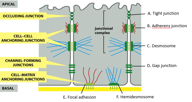
There are six primary types of junctional complexes used in cell-cell and cell-matrix interactions
A. Tight Junction: Forms a barrier close to the apical surface.
B. Adherens Junction: Linked to the actin cytoskeleton.
C. Desmosome: Cell-cell junction linked to intermediate filaments.
D. Gap Junction: Pores for chemical and electrical coupling.
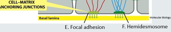
E. Focal Adhesion: Cell-matrix junction linked to actin.
F. Hemidesmosome: Cell-matrix junction linked to intermediate filaments.
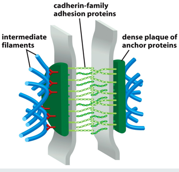
Desmosomes and Hemidesmosomes
Terminology:
Desmosome: Derived from the Greek desmos meaning "bond," "fastening," or "chain," and soma meaning "body."
maintain cell to cell adhesions very strongly via desmosomal cadherins (desmoglein and desmocollin)
cadherins are bound to cytoskeletal proteins and intermediate filaments via desmoplakin
connecting intermediate filaments in neighboring cells
Hemidesmosome: Derived from the Greek hēmi- meaning "half."
found in epithelial cells
major component is alpha-6 beta-4 integrin
transduces signals from ECM to the interior of the cell
intracellular domain of integrins are linked to keratin IF via plectins (anchor proteins) which are anchored to the basal lamina
Morphology and Structure:
- Desmosomes appear under an electron microscope as pairs of dark, disk-like or button-like structures at cell-cell contacts.
- Hemidesmosomes appear as "half-desmosomes" located at the cell-ECM interface.Scale and Dimensions:
- A single hemidesmosome is approximately wide.
- A desmosome is approximately high.
- An average cell is approximately wide.Intracellular Connections:
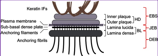
- Both are attached intracellularly to intermediate filaments (IFs), specifically Keratin IFs in certain cells.Detailed Anatomy of the Hemidesmosome and Associated Structures:
- Keratin IFs
- Inner plaque and Outer plaque
- Plasma membrane
- Sub-basal dense plate
- Anchoring filaments
- Anchoring fibrils
- Layers: Lamina lucida (LL) and Lamina densa (LD).
- EBS (Epidermolysis Bullosa Simplex), JEB (Junctional Epidermolysis Bullosa), and DEB (Dystrophic Epidermolysis Bullosa) are associated with different structural defects in these layers.
Clinical Implications of Mechanical Support and Adhesion
Intermediate filaments provide essential support against mechanical stress. Failure in these systems leads to severe pathologies:
- Epidermolysis Bullosa: Caused by mutations in intermediate filaments or desmosomal factors, leading to fragile skin and blistering.
- Pemphigus: An autoimmune condition where the body produces autoantibodies against desmosomes, causing loss of cell-cell adhesion.
Tight Junctions (TJs)
Localization: Found close to the apical surface of epithelial cells, forming a continuous ring around the cell.
Function: They turn epithelia into barriers by preventing the passage of molecules between cells.
important in cell polarity, because they act as permeability barriers and restrict macromolecular transport between cells
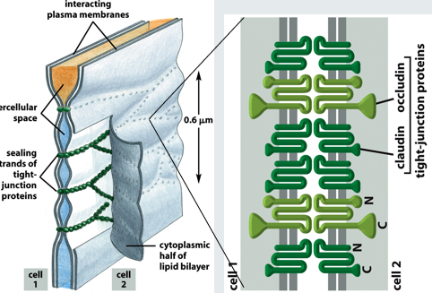
Molecular Components: Primarily formed by the proteins claudin and occludin.
Experimental Evidence (Claudin knockout in mice):
- In control mice, dye injected under the skin is contained.
- In Claudin knockout mice, purple dye spreads into upper layers of the skin.
- Claudin knockout pups experience rapid weight loss and death due to evaporation of water through the skin.Reference: Furuse et al. 2002, J Cell Biol. 156:1099-111.
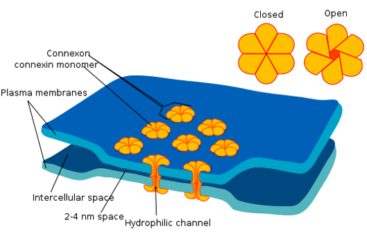
Gap Junctions (GJs)
Function: They couple cells via permeable pores known as "fenestrations."
Capabilities:
- Allow small molecules and ions to pass directly between cells.
- Facilitate chemical and electrical coupling.
- Ions which maintain the membrane potential of neurons can pass through gap junctionsLocalization: Dispersed in the lateral membrane between the apical and basal surfaces.
Components: connexons - which connect to form channels and can open and close, important in heart muscle (contraction signal) and neuronal synapses
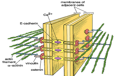
Adherens Junctions (AJs)
Linkage: Found in all epithelial and endothelial cells but also maintains cell-to-cell adhesions via cadherins and connects actin networks of neighboring cells.
Cadherins are a family of transmembrane proteins which form calcium-dependent homodimers with other cadherins. Link actin via anchor proteins (catenins, actinin and vinculin)
Structure: Often referred to as a "double headband" arrangement below the tight junctions.
Function: The actin-myosin complex associated with AJs can mediate contractile forces, which facilitates the bending of epithelia.
Binding Mechanism: Mediated by homophilic binding of E-cadherins, which is strictly dependent on calcium ions ().
Focal Adhesions
Function: Anchor the cell to the ECM with multi-molecular assembly.
Mediator: Uses integrins to bridge the intracellular actin to the basal lamina/ECM fibers. Main receptor family, heterodimeric transmembrane receptor family which recognizes motifs in ECM ligands
Dynamics: Focal adhesions are transient; they form and disassemble rapidly during cell movement (cell migration).
Cadherins and the Discovery of Cell Adhesion Molecules (CAMs)
Masatoshi Takeichi: Pioneered the discovery of cadherins (Kyoto University/Carnegie Institution) by re-investigating the dissociation of amphibian embryos (originally studied by Johannes Holtfreter).
Chemical Mechanism: EDTA is used to sequester calcium (). Without calcium, cadherins cannot maintain adhesion.
Calcium Dependency Thresholds:
- Adhesion occurs at concentration > 1\,mM\,Ca^{2+} .
- Dissociation occurs at concentration < 0.05\,mM\,Ca^{2+} .Classical Cadherins and their Locations:
- E-cadherin: Many epithelia.
- N-cadherin: Neurons, heart, skeletal muscle, lens, and fibroblasts.
- P-cadherin: Placenta, epidermis, breast epithelium.
- VE-cadherin: Endothelial cells.
Additional Adhesion Systems and Disease Links
Other Junctions/Mechanisms:
- Selectin-based adhesion: Responsible for "leukocyte rolling" of white blood cells in vessels.
- Neuronal synapse: Specialized adhesion for signal transmission.
- Plasmodesma: The plant equivalent of gap junctions.
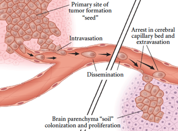
- Septate junctions: The invertebrate equivalent of tight junctions.Cancer Metastasis:
- Metastasis is intricately linked to changes in cell adhesion.
- Process: Primary tumor formation ("seed") → Intravasation (entering vessels) → Dissemination → Arrest in cerebral capillary bed → Extravasation (exiting vessels) → Colonization and proliferation in target organs (e.g., Brain parenchyma/"soil").