4.1 Thorax and Thoracic Skeleton
THORAX COMPONENTS
THORACIC WALL
Components:
Breast
Skeleton/cage
Muscles
Vessels & Nerves
Functions:
Milk production
Protecting thoracic contents
Respiration
Muscle attachment
THORACIC CAVITY
Components:
Pleural cavity - space between the lungs and the chest wall
Mediastinum - contains pericardial cavity, heart, esophagus, trachea, blood vessels, nerves, etc.
Functions:
Gas exchange
Pumping blood
Transporting food & air
THORACIC SKELETON
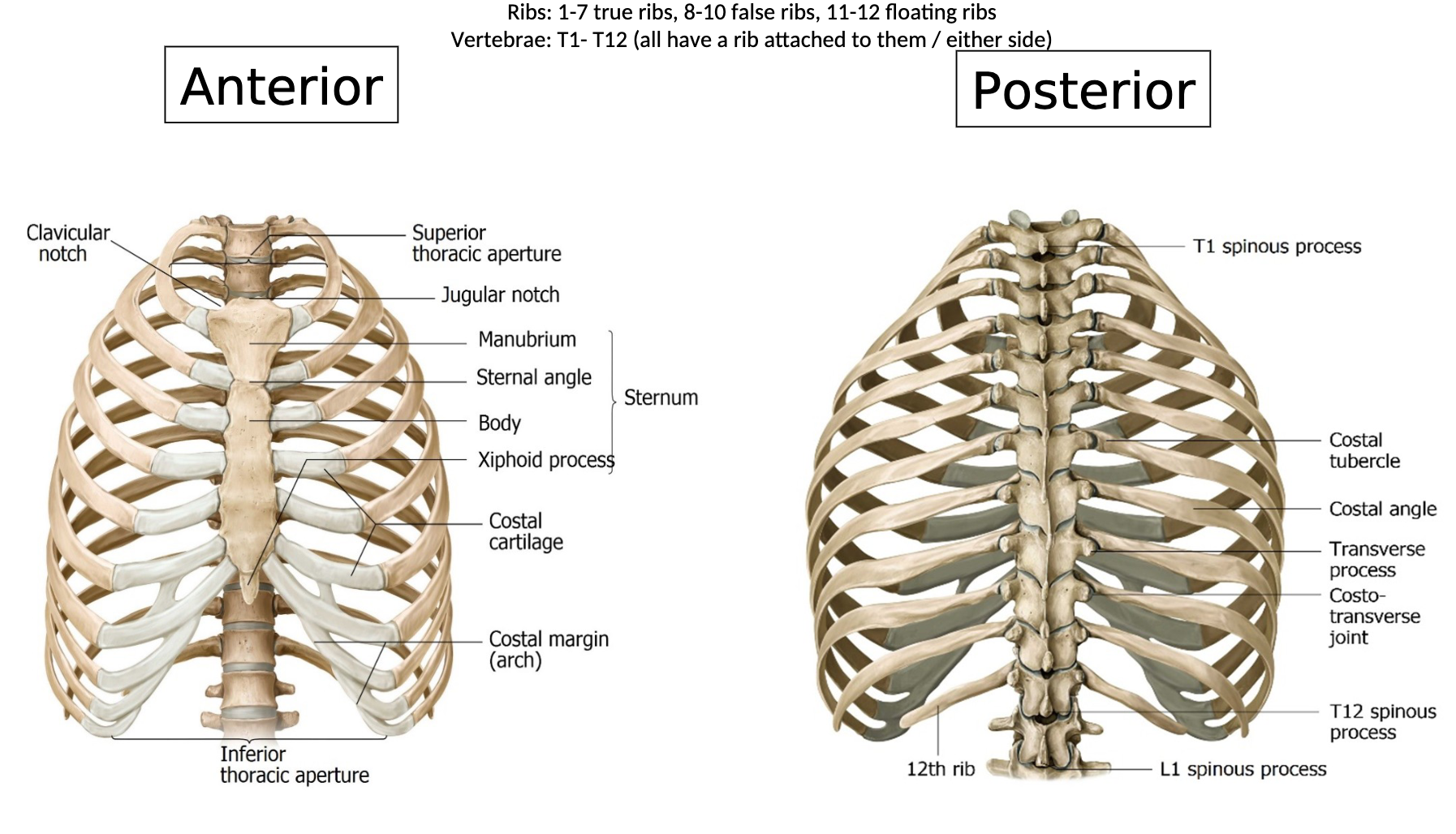
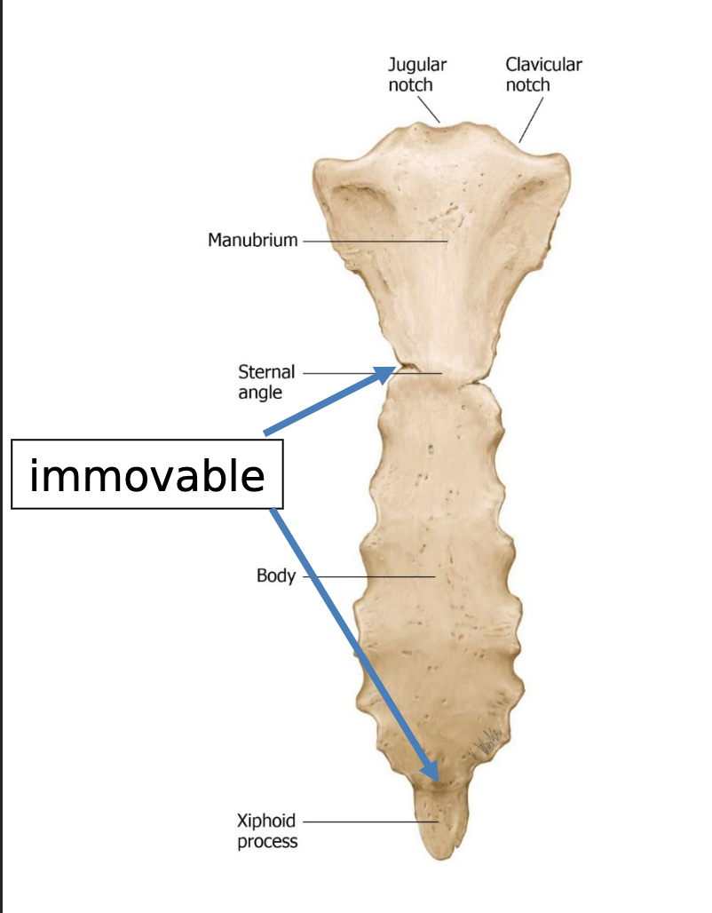
Sternum: Consists of 3 fused bones (manubrium, body, xiphoid process) linking to ribs 1-7. Classified as synchondrosis (type of joint in which bones are connected by hyaline cartilage) same as ribs, non movable
Ribs:
True Ribs (1-7): Directly connected to sternum
False Ribs (8-10): Attach to costal cartilage
Floating Ribs (11-12): No anterior articulation, no articulation with sternum
Vertebrae: T1-T12 have ribs attached on either side.
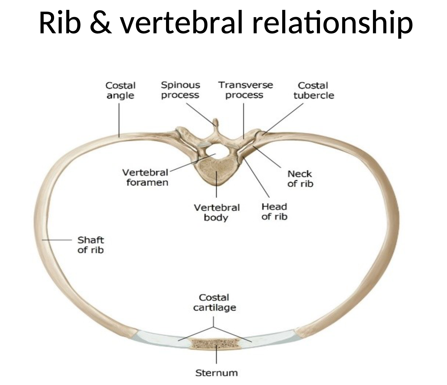
Thoracic vertebrae:
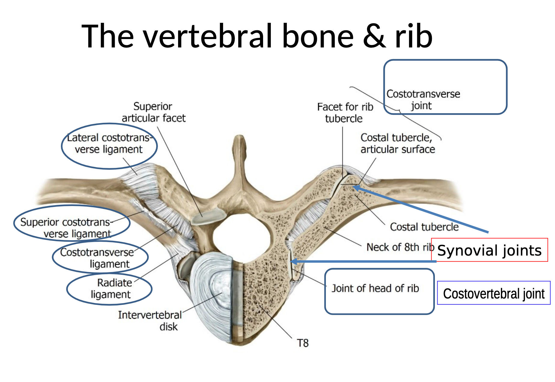
costal facets on transverse process (for costotransverse joint)
2 facets on body (for costovertebral joints)
RIB ARTICULATION
Key points about rib articulation:
Costal facets on transverse processes & bodies of thoracic vertebrae.
Costovertebral joints are synovial joints with gliding movement, enabling rib movement during respiration.
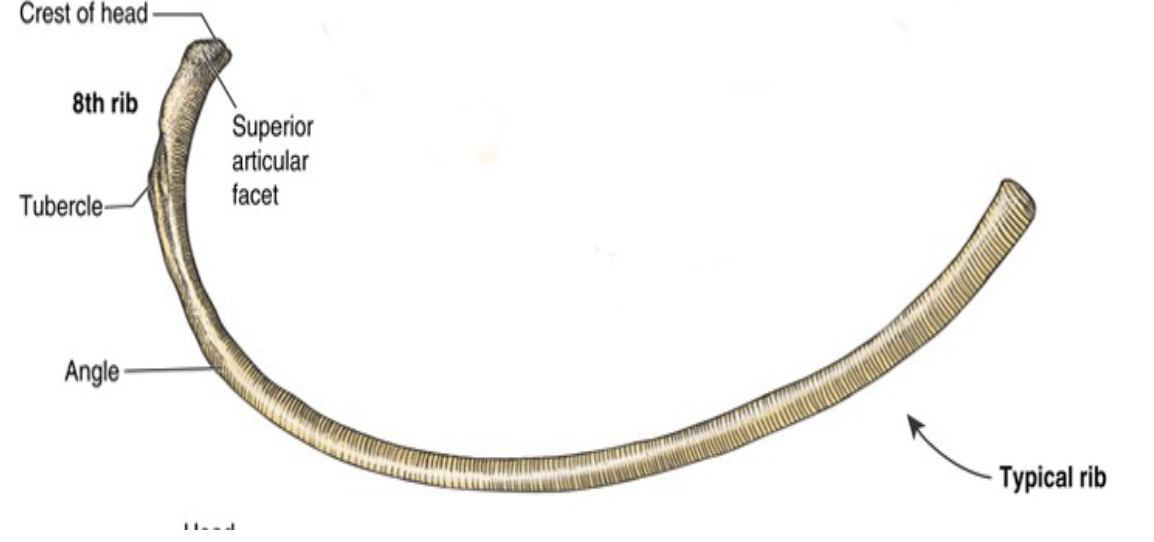
TYPICAL AND ATYPICAL RIBS
Typical Ribs (3-9):
Head with 2 facets, neck, tubercle located at the neck, and a flattened body.
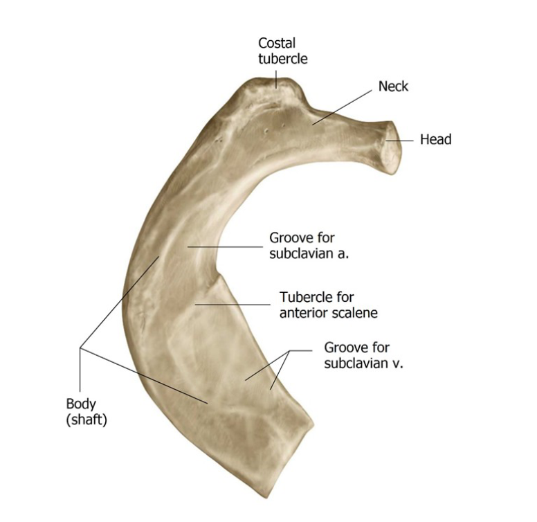
Atypical Ribs (1, 2, 10-12):
Rib 1: Broad, short, one facet.
Rib 2: More typical in structure with rough upper surface.
Ribs 10-12: One facet, structures of 11-12 lack neck and tubercle.
Rib 11-12: no neck or tubercle
RESPIRATION MECHANICS
Inhalation: Muscles contract, causing superior and anterior movement of the thoracic cage (handle on a backet)
Exhalation: Passive process; muscles relax, leading to downward movement of the thoracic cage.
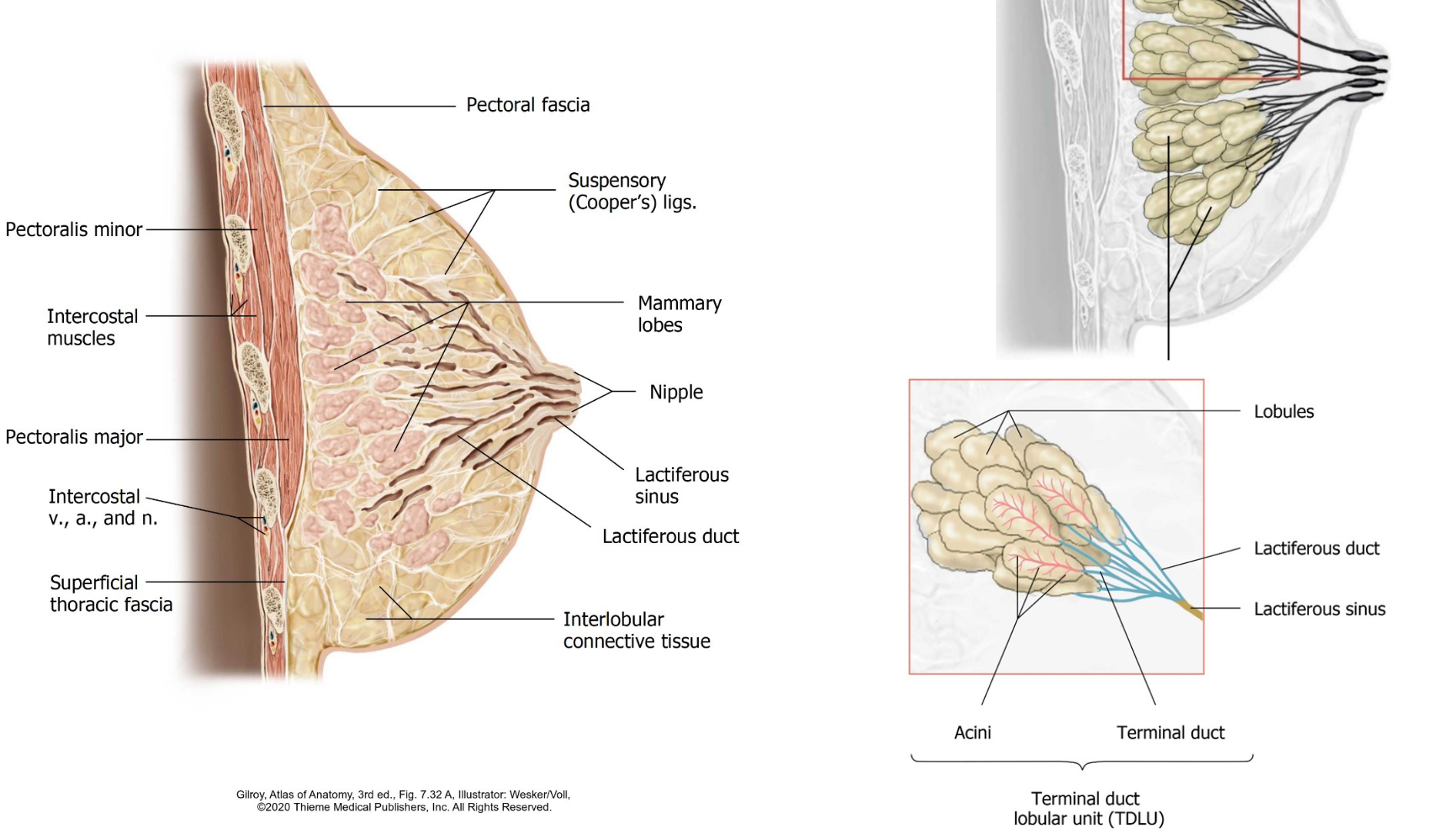
BREAST ANATOMY
Components:
Areola, nipple, adipose tissue, lobules (15-20), lactiferous duct.
Changes noted during puberty, pregnancy, and lactation.
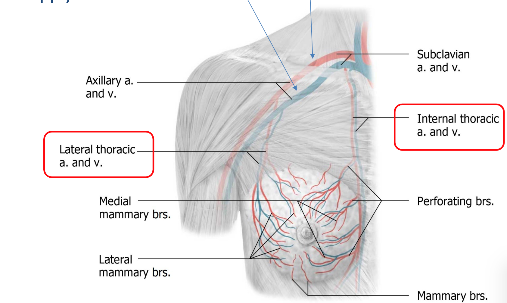
BLOOD AND NERVE SUPPLY TO BREAST
Arterial Supply:
Axillary artery, subclavian artery, aorta.
Venous Drainage:
Mainly through axillary vein.
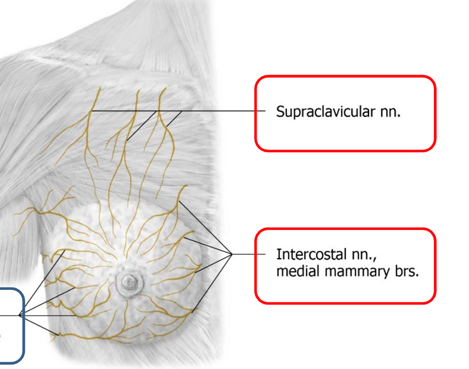
Nerve Supply:
Intercostal nerves.
LYMPHATIC DRAINAGE OF THE BREAST
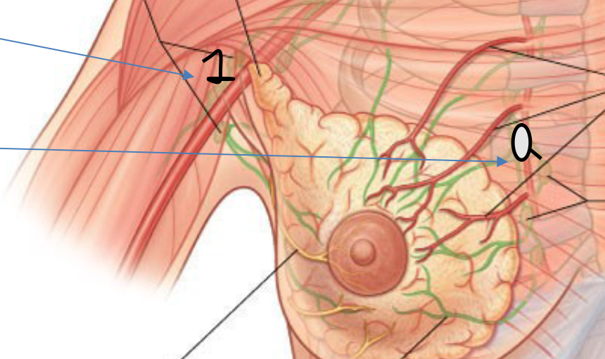
>75% lymph drained via axillary nodes (lateral breast). (1)
Medial breast drained via sternal nodes. (2)
Important for understanding cancer spread.
THORACIC STRUCTURE AND FUNCTION
Thoracic cavity divided into:
Mediastinum (contains heart and great vessels).
Pleural cavities (houses lungs).
Thoracic cage protects thoracic structures and serves as a muscle attachment site.
BLOOD SUPPLY AND NERVOUS INNERVATION
Blood Supply:
Intercostal arteries from aorta and internal thoracic artery.
Nervous Supply:
Intercostal nerves serving dermatomes(1 nerve root) and myotomes (<1 nerve root).
MUSCLES OF THE THORACIC WALL
Intercostal Muscles:
External: Most superficial, aids in inhalation.
Internal: Intermediate layer, aids in forced exhalation.
Innermost: Deepest layer, lies close to the pleura (membrane).
JOINTS AND LIGAMENTS
Costochondral Joint:
Cartilaginous joint (synchondrosis) connecting ribs and cartilage.
Costovertebral and Costotransverse Joints:
Synovial joints allowing rib movement and stability.
CLINICAL IMPORTANCE
Knowledge of thoracic anatomy is crucial for procedures like sternotomy, pericardiocentesis, and thoracentesis.
Boundaries of thoracic wall serve as anatomical landmarks for various clinical interventions.