Comprehensive Study Guide to Chromatin Architecture, Epigenetic Modifications, and Transcriptional Regulation
Genome Organization and the Physical Nature of Chromatin
Fundamental Problems in Genome Regulation
Development and Multicellularity: Generating stable phenotypic diversity from a single genome ( ). Epigenetic memory establishes and maintains distinct cellular identities across cell divisions despite an identical DNA sequence.
Adaptive Responses to Environment: Generating transient phenotypic diversity to maintain homeostasis (). Dynamic transcriptional switches modulate gene expression programs in response to internal metabolic shifts, hormonal cues, and external environmental stresses.
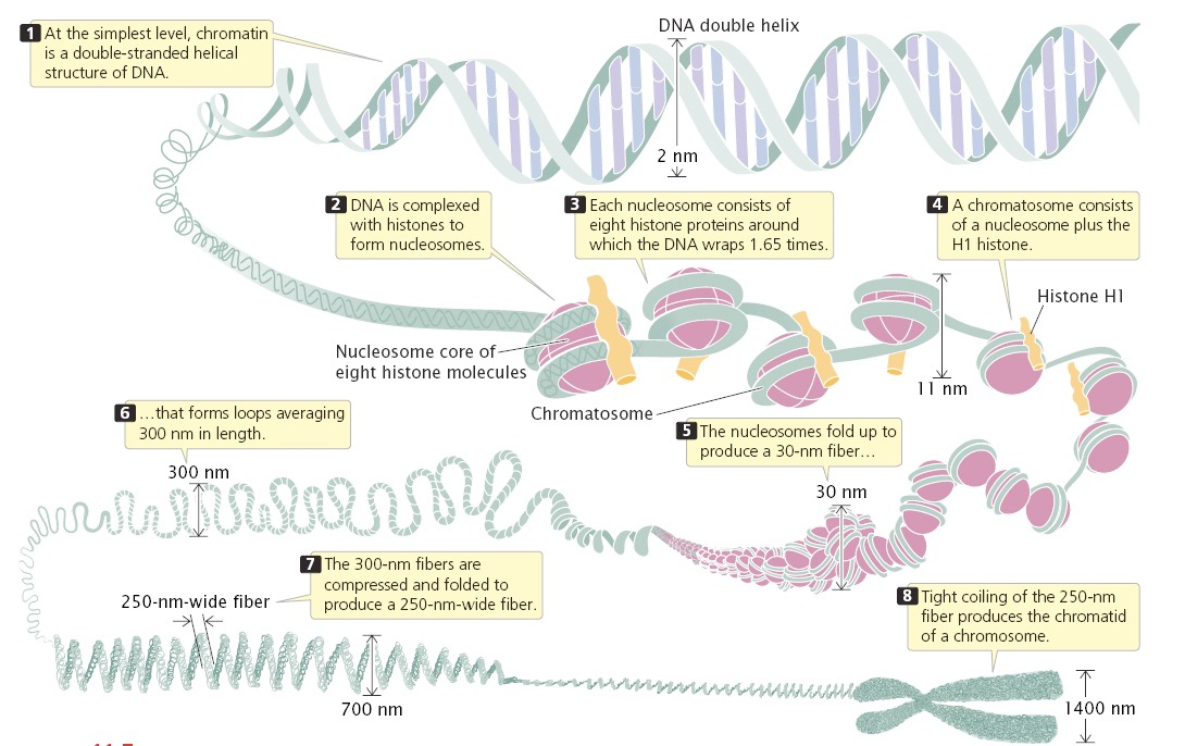
Physical Packing Challenge: Approximately of linear double-stranded DNA must be packaged inside a eukaryotic nucleus typically in diameter. This architecture resolves two conflicting imperatives:
Structural Compaction: Compacting the genome by a factor greater than to fit into the nuclear volume.
Functional Accessibility: Providing selective, dynamic, and temporal access to regulatory sequences for DNA replication, repair, recombination, and transcription.

Genomic Complexity, Gene Density, and the Non-Coding Landscape
Chromosome Dynamics Across the Cell Cycle: Mitotic chromosomes are highly condensed, transcriptionally inactive, and visually distinct, whereas interphase chromosomes decondense into less distinct, functional chromosomal territories within the nucleus.

Lack of Correlation Between Chromosome Number, Size, and Organismal Complexity: Chromosome counts and total genome size do not correlate with phylogenetic complexity (the C-value paradox). For example, the Chinese muntjac (Muntiacus reevesi) possesses chromosomes, whereas the Indian muntjac (Muntiacus muntjak) possesses (\female) or (\male) chromosomes, despite having nearly identical genome sizes and organismal complexity.
Epigenetic Regulation of Chromatin: Epigenetic modifications such as DNA methylation and histone acetylation play crucial roles in defining chromatin structure and function, influencing gene expression patterns and cellular identity. Furthermore, these modifications can create a cellular memory that persists through cell division, affecting how genes are accessed and expressed in response to environmental stimuli, which is essential for processes like development and differentiation.
Cross-Species Genome Variations:

Composition of the Human Genome ( Total):
Intergenic Regions (): Genome-wide repeats account for (LINEs , SINEs , retroviral-like LTR retrotransposons , DNA transposon fossils ); microsatellites and simple sequence repeats account for ; other unique intergenic regions account for .
Genes and Gene-Related Sequences (): Protein-coding exons constitute only ( of the genome). Related sequences comprise , containing introns, 5' and 3' untranslated regions (UTRs), pseudogenes, gene fragments, non-coding RNAs, and unique regulatory regions (promoters, enhancers).


B-DNA Structure and Physical Parameters
Composed of four deoxynucleotide building blocks: deoxyadenosine (), deoxythymidine (), deoxycytidine (), and deoxyguanosine ().
Monomers are joined by covalent phosphodiester linkages forming a polar, negatively charged sugar-phosphate backbone.
Double-stranded B-DNA consists of two antiparallel, right-handed helical chains held together by complementary base pairing:
base pairs share hydrogen bonds.
base pairs share hydrogen bonds.
The double helix has an outer diameter of , a helical rise of per base pair, and roughly per turn ( pitch).
B-DNA possesses a wide, deep major groove and a narrow, shallow minor groove. The major groove displays unique chemical recognition codes (hydrogen bond donors, acceptors, nonpolar hydrogens, and methyl groups) that allow sequence-specific binding proteins to read base sequences without unwinding the helix.

The Nucleosome: Structural Unit of Chromatin
Nucleosome Core Particle Architecture
The nucleosome is the primary repeating packaging unit of eukaryotic chromatin, creating the "beads-on-a-string" () fiber.
Core Particle Composition: An octamer of core histone proteins containing two copies each of H2A, H2B, H3, and H4, around which of DNA is wrapped in left-handed superhelical turns.
Symmetry: The nucleosome core particle exhibits pseudo-twofold dyad symmetry. A twofold dyad axis passes through the center of the wrapped DNA, dividing it into two symmetrical halves.


Core Histones and Evolutionary Conservation
Core histones are small, highly basic proteins enriched in positively charged lysine () and arginine () residues that electrostatically stabilize the negatively charged phosphodiester backbone of DNA.
Core and Linker Histone Specifications:
H1 (Linker histone): Lysine-rich, molecular weight , molar ratio .
H2A: Slightly lysine-rich, molecular weight , molar ratio .
H2B: Slightly lysine-rich, molecular weight , molar ratio .
H3: Arginine-rich, molecular weight , molar ratio .
H4: Arginine-rich, molecular weight , molar ratio .
Extreme Sequence Conservation: Core histones (particularly H3 and H4) are among the most evolutionarily conserved proteins known. Calf thymus H4 and garden pea H4 differ by only two conservative amino acid substitutions out of residues across more than a billion years of evolutionary divergence.
The Histone Fold and Assembly Pathway
Each core histone shares a conserved structural domain termed the histone fold, consisting of three -helices (, , ) separated by two short loop segments (, ).
Histone folding occurs via a reciprocal "handshake" motif:
H3 pairs with H4 to form stable heterodimers, which subsequently homodimerize into a central tetramer.
H2A pairs with H2B to form two independent heterodimers.
Stepwise Assembly Pathway:
The tetramer binds genomic DNA first, forming an unstable intermediate called the tetrasome.
Two heterodimers bind sequentially to the flanks of the tetrasome, forming a hexasome and then the completed octameric nucleosome.
Linker histone H1 binds the outside of the nucleosomal disc at the DNA entry and exit sites, sealing the turn and organizing linker DNA.

DNA-Histone Contacts and Sequence Positioning
The histone core makes approximately distinct contacts with DNA, almost exclusively with the phosphodiester backbone and minor groove, utilizing main-chain amides, basic side chains, and water molecules.
Sequence Preferences for Positioning:
Bending DNA around the octamer compresses the inner minor groove while expanding the outer minor groove.
, , and dinucleotides bend easily toward the minor groove; they show a periodicity localized where the minor groove faces inward toward the histone core.
dinucleotides prefer conformations where the minor groove faces outward toward solvent.
Active gene promoters and terminators exhibit Nucleosome-Depleted Regions (NDRs) flanked by strongly positioned and nucleosomes.
Histone Chaperones
Unchaperoned histones aggregate nonspecifically with DNA via charge interactions. Histone chaperones shield basic histone charges, preventing aberrant precipitation and directing stepwise chromatin assembly and disassembly:
NPM1/Numatrin/Nucleophosmin, NPM2, NPM3, Nucleoplasmin, NLP: Bind H2A/H2B; participate in oocyte storage, sperm chromatin decondensation, and ribosome biogenesis.
NAP1 (NAP1L1–NAP1L4): Binds H2A/H2B; mediates histone shuttling, nucleosome assembly, and variant exchange.
N1/N2, NASP, Hif1: Bind H3/H4; maintain soluble H3/H4 pools and support nuclear transport.
CAF1 (Chromatin Assembly Factor 1; subunits p150, p60, p48/cac1): Coordinates replication-coupled deposition of canonical H3.1/H3.2 and H4 behind DNA replication forks.
Biochemical Characterization by Micrococcal Nuclease (MNase)
MNase acts as both an endonuclease and an exonuclease, selectively cleaving unprotected linker DNA while sparing nucleosome-bound DNA.
Digestion Profile:
Partial MNase digestion: Generates an ordered nucleosomal ladder visible on an agarose gel at integer multiples of ( mono-, di-, tri-, tetra-nucleosomes).
Extensive/Complete MNase digestion: Trims linker DNA down to the core particle limit of .
Ultracentrifugation on a sucrose gradient resolves distinct sedimentation peaks (, , , ) corresponding to mono-, di-, tri-, and oligonucleosomal particles.

Dynamic Nucleosome Remodeling by ATP-Dependent Complexes
Nucleosome Breathing: Nucleosomal DNA spontaneously and transiently unwraps from the histone core at an equilibrium frequency of of the time. This transient exposure allows DNA-binding factors brief access.
ATP-Dependent Remodeling Complexes: Multi-subunit molecular machines that utilize ATP hydrolysis to alter histone-DNA contacts, classified into four major families:
SWI/SNF Family (yeast SWI/SNF, Drosophila BAP/PBAP, human BAF/PBAF): Contains catalytic ATPase subunits Swi2/Snf2, BRG1 (SMARCA4), or hBRM (SMARCA2). Drives nucleosome sliding, octamer displacement, and nucleosome ejection to expose promoter and enhancer elements.
ISWI Family: Catalyzes regular nucleosome spacing, optimizing chromatin compaction and transcriptional repression.
NuRD (Nucleosome Remodeling and Deacetylase) Family: Combines ATP-dependent remodeling with histone deacetylase subunits (HDAC1/2) for transcriptional silencing.
INO80/SWR1 Family: Catalyzes exchange of canonical core histones for histone variants (e.g., depositing H2A.Z).
Mechanism: Complexes bind both the histone octamer and nucleosomal DNA, forming a loop or "bulge" of unspooled DNA that propagates around the octamer in an ATP-dependent wave, resulting in nucleosome sliding or dissociation.
Epigenetic Mechanisms: Post-Translational Histone Modifications
Covalent Histone Tails and the Diversity of PTMs
The amino-terminal (-terminal) and carboxy-terminal (-terminal) tails of histones protrude outward through the DNA gyres. These tails are intrinsically disordered and undergo extensive, reversible enzymatic modifications.
Primary Classes of Post-Translational Modifications (PTMs):
Acetylation (Ac): Targets Lysine () residues; neutralizes the positive charge on the -amino group, weakening histone-DNA and internucleosomal contacts.
Methylation (Me): Targets Lysine () (mono-, di-, or trimethylation: , , ) and Arginine () (monomethylation, symmetric dimethylation , asymmetric dimethylation ). Retains the positive charge.
Phosphorylation (Ph): Targets Serine (), Threonine (), and Tyrosine (); adds a negative charge ().
Ubiquitination (Ub): Covalent attachment of a ubiquitin moiety to Lysine residues (mono-ubiquitination of H2AK119 or H2BK120).
SUMOylation (SUMO): Attachment of Small Ubiquitin-like Modifier to Lysines; often antagonizes acetylation and mediates transcriptional repression.
Short-Chain Acylations: Propionylation (), butyrylation (), crotonylation (), 2-hydroxyisobutyrylation (), malonylation (), succinylation (), and formylation ().
Other PTMs: Citrullination (conversion of arginine to citrulline by PAD4), ADP-ribosylation, and O-GlcNAcylation.

Histone Acetylation, HATs, and HDACs
Mechanism: Acetyltransferases transfer an acetyl group from Acetyl-Coenzyme A (Acetyl-CoA) to the -amino group of lysine, transforming a positively charged amine into an uncharged amide. This relieves electrostatic attraction to DNA and prevents higher-order fiber folding.

Histone Acetyltransferases (HATs / KATs):
GNAT Family: Includes Gcn5 (KAT2A) and P/CAF (KAT2B); targets H3K9, H3K14, and H3K36; components of the SAGA, ATAC, and TFTC complexes.
MYST Family: Includes Esa1/Tip60 (KAT5), Sas2/MOF (KAT8), and Sas3/MOZ (KAT6A); targets H4K5, H4K8, H4K12, H4K16, and H3K14; components of NuA4 and Piccolo NuA4 complexes.
p300/CBP Family: Includes p300 (KAT3B) and CBP (KAT3A); broad specificity (H2A, H2B, H3, H4); acts as a global coactivator across thousands of enhancers and promoters.
Rtt109 (KAT11): Fungal-specific HAT targeting H3K56.
Metabolic Integration: Acetyl-CoA availability directly links cellular energy balance, glucose availability, and mitochondrial lipid breakdown to chromatin acetylation status.
Histone Deacetylases (HDACs): Catalyze the hydrolytic removal of acetyl groups, restoring the positive charge on lysine residues and promoting chromatin compaction. Divided into Class I, II, and IV (-dependent enzymes, e.g., HDAC1, HDAC2) and Class III (Sirtuins, -dependent deacetylases).
Bromodomains: Conserved protein modules comprising an antiparallel four-helix bundle (, , , ) that bind acetylated lysine residues. Found in HATs (GCN5, p300), chromatin remodelers (BRG1), and transcriptional regulators (BRD4, TAF1). Small-molecule bromodomain inhibitors like JQ1 displace BRD4 from chromatin, downregulating oncogenes such as MYC.

Histone Methylation, HMTs, Demethylases, and Reader Modules
Enzymology: Histone Methyltransferases (HMTs) utilize S-Adenosylmethionine (SAM) as a methyl donor, converting SAM to S-Adenosylhomocysteine (SAH). Lysine methyltransferases (KMTs) contain a conserved SET domain (except the non-SET enzyme DOT1L, which targets H3K79). Arginine methyltransferases (PRMTs) catalyze arginine methylation.

Histone Demethylases:
LSD1 (KDM1A): Flavin adenine dinucleotide (-dependent) amine oxidase; removes mono- and dimethyl groups from H3K4 and H3K9; cannot demethylate trimethylated lysines.
Jumonji C (JmjC) Domain-Containing Demethylases: Fe(II)- and -ketoglutarate-dependent dioxygenases that demethylate mono-, di-, and trimethylated lysines via radical hydroxylation:
JHDM1A (KDM2A): Targets H3K36me1/me2.
JMJD1A (KDM3A): Targets H3K9me2/me1.
JMJD2A–D (KDM4A–D): Target H3K9me3/me2 and H3K36me3/me2.
JARID1A–D (KDM5A–D): Target H3K4me3/me2.
Methyl-Lysine Reader Modules:
Chromodomains: Bind methylated lysines; HP1 binds H3K9me2/me3; Polycomb (Pc/CBX) binds H3K27me3; CHD1 binds H3K4me2/me3.
Tudor Domains: Found in JMJD2A, 53BP1, and UHRF1; bind H3K4me3, H4K20me1/me2, and methylated arginines.
Plant Homeodomain (PHD) Fingers: Found in BPTF, ING1–5, and TAF3; bind H3K4me3 to recruit PIC and remodeling complexes.
Malignant Brain Tumor (MBT) Domains: Bind lower methylation states (mono- and dimethylated lysines, e.g., H4K20me1/me2).
WD40 Repeat Domains: Found in WDR5 (MLL complex) and EED (PRC2 complex).

Ubiquitination, SUMOylation, and Cross-Talk Mechanisms
Mono-Ubiquitination: H2AK119ub1 (catalyzed by the PRC1 RING1B subunit) represses transcription by stalling Pol II elongation and blocking remodeling. H2BK120ub1 (catalyzed by the Rad6/Bre1 complex) promotes transcription.
Trans-Histone Cross-Talk Pathway: H2BK120ub1 acts as an obligate prerequisite that allosterically activates the SET1/COMPASS complex (which deposits H3K4me3) and the DOT1L enzyme (which deposits H3K79me3) in active gene bodies. Deubiquitination of H2B by Ubp8/USP22 (within the SAGA DUB module) is required for subsequent transcript elongation.

Histone Phosphorylation and Mitotic Condensation
Phosphorylation adds bulk and negative charge to histone tails. H3S10ph and H3S28ph are catalyzed by Aurora B kinase during mitosis to trigger chromosome condensation, and by MSK1/2 during immediate-early gene activation following growth factor stimulation.
Phosphorylation can disrupt reader binding. For example, H3S10ph evicts HP1 from neighboring H3K9me3 during mitosis ("methyl-phospho switch"). Similarly, histone H1 phosphorylation evicts HP1 to decondense chromatin.
Aurora Kinase Family: Aurora A, B, and C contain an -terminal domain (with a KEN motif and a destruction box activating motif), a central catalytic kinase domain, and a -terminal domain containing a functional D-box ().


Histone Variants
H2A Variants:
H2AX: Contains an extended SQEY motif at its -terminus. Phosphorylated at Ser139 by ATM/ATR kinases in response to double-strand breaks to form , which recruits DNA damage response and repair machinery (MDC1, 53BP1).
H2A.Z: Enriched at nucleosome-depleted regions flanking active promoters ( and nucleosomes); creates unstable nucleosomes that lower the barrier to Pol II passage.
macroH2A: Contains an extensive non-histone globular macrodomain; enriched on the inactive X chromosome () in females, maintaining stable epigenetic silencing.

* **H3 Variants:**
    * *H3.3:* Differs from canonical H3.1/H3.2 by only 4–5 amino acids. Deposited in a replication-independent manner by the HIRA chaperone complex at active promoters and gene bodies, and by the DAXX/ATRX complex at telomeres and pericentric heterochromatin.
    * *CENP-A:* Replaces H3 at centromeres, directing kinetochore assembly and mitotic chromosome segregation; deposited in a replication-independent manner by the chaperone HJURP.
Summary of Functional Genomic Histone Marks
Active Promoters: High H3K4me3, H3K27ac, H3K9ac, and H2A.Z.
Active Enhancers: High H3K4me1, H3K27ac, p300/CBP binding, and low H3K4me3.
Poised/Primed Enhancers: High H3K4me1, low H3K27ac.
Active Gene Bodies: High H3K36me3, H3K79me2/me3, and H2BK120ub1.
Constitutive Heterochromatin: High H3K9me3, H4K20me3, and HP1 binding.
Facultative Heterochromatin / Polycomb Repression: High H3K27me3 and H2AK119ub1.
Bivalent Promoters (Stem Cells): Co-occurrence of H3K4me3 and H3K27me3; keeps lineage-specifying developmental genes repressed but poised for rapid activation.

Higher-Order Chromatin Architecture and Nuclear Topology
The 30-nm Fiber: Biophysical Models
In vitro, increasing monovalent () or divalent () salt concentrations screens negative DNA backbone charges, driving folding of the "beads-on-a-string" fiber into a condensed chromatin fiber.
Competing Models:
One-Start Solenoid Model: The nucleosomal chain coils into a continuous, hollow cylindrical helix containing approximately nucleosomes per turn, with a central lumen of .
Two-Start Zigzag Model: Nucleosomes alternate in a straight-linker configuration, with consecutive nucleosomes connected by straight DNA that passes through the center of the fiber, placing alternate nucleosomes ( and ) in closer physical contact than adjacent ones ( and ).
In Vivo Reality: Modern cryo-electron tomography and chromosome conformation assays indicate that interphase chromatin in vivo primarily exists as a disordered, highly dynamic polymer with variable local packing densities ("chromatin clutch" model), rather than a uniform, rigid fiber.

Heterochromatin Types and HP1-Mediated Spreading
Constitutive vs. Facultative Heterochromatin:
Constitutive Heterochromatin: Densely compacted, highly repetitive (centromeric and telomeric satellite DNA), gene-poor, late-replicating, enriched in H3K9me3 and DNA methylation; remains permanently condensed in all cell types.
Facultative Heterochromatin: Contains developmentally regulated genes (e.g., the Hox clusters or the inactive X chromosome); reversibly condensed via PRC1/PRC2-mediated H3K27me3 and H2AK119ub1.
HP1 Self-Propagation Mechanism:
SUV39H1 (KMT1A) trimethylates H3K9.
Heterochromatin Protein 1 (HP1) binds H3K9me3 via its -terminal chromodomain.
HP1 homodimerizes through its -terminal chromoshadow domain (CSD), creating an interaction platform that recruits SUV39H1.
Recruited SUV39H1 trimethylates the H3K9 residue on the adjacent nucleosome, repeating the cycle and spreading heterochromatin linearly until an insulator or barrier element is encountered.

Topologically Associating Domains (TADs) and Loop Extrusion
TAD Definition: Genomic regions (typically to several megabases) whose internal sequences interact with one another at high frequency, while interactions across TAD boundaries are insulated.
Structural Regulators:
CTCF (CCCTC-binding Factor): An architectural protein that binds directional DNA sequence motifs.
Cohesin Complex: A ring-shaped multi-protein ATPase complex (SMC1, SMC3, RAD21, SCC1).
Loop Extrusion Model:
Cohesin rings load onto chromatin and actively extrude loops of DNA bidirectionally.
Loop extrusion continues until Cohesin encounters a pair of CTCF proteins bound in a convergent orientation (motifs oriented toward each other: ).
Convergent CTCF blocks Cohesin translocase activity, stabilizing the chromatin loop and forming the structural boundary of the TAD.
Nuclear Geography and Chromosome Territories
Chromosomes do not intermingle randomly in the interphase nucleus; each occupies an individual, distinct Chromosome Territory.
Sub-Nuclear Compartments:
A Compartment (Euchromatic): Gene-dense, transcriptionally active, DNase I hypersensitive, localized toward the nuclear interior and surrounding nuclear speckles.
B Compartment (Heterochromatic): Gene-poor, transcriptionally silent, late-replicating, localized to the nuclear periphery (Lamina-Associated Domains [LADs] interacting with lamin B1) and the perinucleolar region (Nucleolus-Associated Domains [NADs]).

DNA Methylation and Demethylation Dynamics
5-Methylcytosine () and CpG Island Distribution
In vertebrates, DNA methylation occurs predominantly at the carbon-5 position of cytosine () within symmetrical dinucleotides.
CpG Islands (CGIs): Clusters of unmethylated CpG sites ( long) with a content and an observed-to-expected CpG ratio . Found at the promoters of of human protein-coding genes.
Evolutionary Depletion of Non-Island CpGs: Across evolutionary time, the mammalian genome has lost of its expected CpGs due to spontaneous deamination:
Unmethylated cytosine deaminates to uracil, which is recognized as foreign and efficiently excised by uracil DNA glycosylase (UDG).
deaminates to thymine, creating a mismatch that is poorly repaired by base-excision machinery, leading to permanent transition mutations.

DNA Methyltransferases (DNMTs)
De Novo Methyltransferases (DNMT3A and DNMT3B): Establish de novo methylation patterns during early embryonic development and cell fate specification, methylating previously unmodified CpG dinucleotides; stimulated by DNMT3L.
Maintenance Methyltransferase (DNMT1): Preserves DNA methylation patterns through mitotic cell divisions. Operates at the replication fork, targeting hemi-methylated DNA (where the parental template strand is methylated but the newly synthesized daughter strand is unmethylated) and copying the methylation pattern to the daughter strand with high fidelity.
Active DNA Demethylation and TET Dioxygenases
Demethylation can occur through passive dilution (failure of DNMT1 maintenance across replication cycles) or through active enzymatic processing by TET (Ten-Eleven Translocation) enzymes (TET1, TET2, TET3).
Iterative Oxidation Cascade:
TET enzymes iteratively oxidize into , then , and finally in an Fe(II)- and -ketoglutarate-dependent manner.
Thymine DNA Glycosylase (TDG) specifically recognizes and excises and , generating an abasic site.
Base Excision Repair (BER) resynthesizes the gap with an unmodified, unmethylated cytosine.

DNA Methylation Readers and Silencing Synergy
Direct Inhibition: Methyl groups in the major groove can physically block the binding of sequence-specific transcription factors that contain CpG within their recognition motifs (e.g., CREB, E2F, Myc/Max).
Indirect Repression via MBD Proteins: Methyl-CpG-binding domain proteins (MeCP2, MBD1, MBD2) act as biochemical adaptors. They bind methylated DNA and recruit corepressor complexes containing histone deacetylases (HDAC1/2), histone methyltransferases (G9a, SUV39H1), and chromatin remodelers (Sin3A, Mi-2/NuRD), creating a compacted, transcriptionally inactive chromatin state.

The Core Transcriptional Machinery and RNA Polymerases
RNA Polymerase Classes and Subunit Architecture
Transcription is the DNA-directed enzymatic synthesis of RNA catalyzed by multi-subunit RNA polymerases, moving in the direction.
The Three Eukaryotic Nuclear RNA Polymerases:
RNA Polymerase I (Pol I): Transcribes the large ribosomal RNA precursor ( pre-rRNA yielding , , and rRNAs); localized exclusively in the nucleolus; completely insensitive to -amanitin; contains subunits.
RNA Polymerase II (Pol II): Transcribes all protein-coding messenger RNAs (mRNAs) and most small non-coding RNAs (snRNAs, snoRNAs, miRNAs, lncRNAs, eRNAs); localized in the nucleoplasm; highly sensitive to -amanitin (); contains core subunits.
RNA Polymerase III (Pol III): Transcribes short, untranslated functional RNAs ( rRNA, tRNAs, U6 snRNA, 7SL RNA); localized in the nucleoplasm; moderately sensitive to -amanitin ( in mammals, resistant in yeast); contains subunits.
Structural Conservation with Prokaryotic Polymerase: The core catalytic framework is conserved from bacteria to humans. The largest eukaryotic subunits ( and in Pol II) correspond directly to the bacterial and subunits, while and correspond to the bacterial dimer (), and corresponds to the bacterial subunit.

RNA Polymerase II Catalytic Structure and Transcription Bubble
The Pol II core adopts a "crab-claw" architecture surrounding a central active-site cleft containing catalytic magnesium ions (, Metal A and Metal B).
Structural Modules:
The Clamp: A flexible swinging domain that clamps down tightly over the downstream DNA double helix upon initiation, locking the template strand in the active-site cleft to ensure high processivity.
The Wall: A rigid protein wall that forces the template DNA strand to bend sharply by roughly , directing the template base into position for incoming ribonucleoside triphosphates (rNTPs).
The Bridge Helix and Trigger Loop: Mobile elements that swing adjacent to incoming nucleotides, coordinating complementary base pairing, phosphodiester bond synthesis, and translocation of the template.
The Rudder, Lid, and Zipper: Protein loops that stabilize DNA unwinding, maintain the transcription bubble ( of melted DNA), and separate the growing RNA from the DNA template to allow the DNA-RNA hybrid to emerge through the RNA exit channel.


The C-Terminal Domain (CTD) Code
The largest subunit of Pol II (RPB1) carries a unique carboxy-terminal domain (CTD) consisting of multiple tandem repeats of the consensus heptapeptide:
Repeat Count: Yeast contains repeats; mammals contain repeats.
Dynamic Phosphorylation Cycle:
Pre-Initiation: Pol II enters the pre-initiation complex in an unphosphorylated state.
Initiation and Promoter Escape (Ser5ph): CDK7 (the kinase subunit of TFIIH) phosphorylates Ser5. Ser5ph disrupts contacts with pre-initiation factors, stimulates promoter escape, and directly recruits the 5' capping enzyme complex (capping guanylyltransferase and methyltransferase) when the nascent RNA reaches .
Elongation Transition (Ser2ph): P-TEFb (CDK9/Cyclin T1) phosphorylates Ser2. As Pol II transcribes into the gene body, Ser5ph levels decrease via Rpb1-associated phosphatases (e.g., Ssu72, Scp1), while Ser2ph increases, recruiting splicing factors, the elongation-promoting FACT complex, and the Set2 methyltransferase (which deposits H3K36me3).
Termination and Recycling: At the poly(A) site, Ser2ph directs recruitment of CPSF and CStF. Following transcript cleavage and release, Fcp1 and other CTD phosphatases strip off Ser2ph, regenerating unphosphorylated Pol II for subsequent transcription rounds.
Core Promoter Elements for Pol II
Core promoters span approximately to relative to the transcription start site ().
Canonical Consensus Elements:
TATA Box: Consensus (, ); centered at position to ; recognized by the TBP subunit of TFIID.
Initiator (Inr): Consensus (); overlaps the start site; recognized by TAF1 and TAF2 subunits of TFIID.
TFIIB Recognition Element (BRE): Upstream BRE (, consensus , ) and downstream BRE (); flanking the TATA box; recognized directly by TFIIB.
Downstream Promoter Element (DPE): Consensus (); located at positions to ; functions coordinately with Inr in TATA-less promoters; recognized by TAF6 and TAF9.
Downstream Core Element (DCE): Sub-elements located downstream of the TSS; recognized by TFIID.
Motif Ten Element (MTE): Located at positions to .
Focused vs. Dispersed Transcription Initiation:
Focused (Sharp) Promoters: Transcription initiates at a single nucleotide or tight cluster of nucleotides. Typical of tightly regulated, tissue-specific, TATA-dependent genes.
Dispersed (Broad) Promoters: Transcription initiates at multiple weak start sites over a window of . Typical of ubiquitous, constitutively expressed housekeeping genes embedded within CpG islands.
Pre-Initiation Complex (PIC) Assembly and General Transcription Factors (GTFs)
In vitro, PIC assembly can occur via a sequential step-wise recruitment pathway:
TFIID: Consists of TBP (TATA-Binding Protein) and TAFs (TBP-Associated Factors). TBP binds the minor groove of the TATA box, inducing a severe bend in the DNA. TAFs recognize Inr, DPE, and modified histone tails (e.g., TAF3 recognizes H3K4me3 via its PHD finger).
TFIIA: Binds TBP and stabilizes the TFIID-DNA binary complex, preventing repressor-mediated displacement.
TFIIB: A monomeric factor that bridges TFIID and Pol II. Its -terminal domain contacts TBP and DNA flanking the TATA box (BRE), while its -terminal "B-finger" inserts into the active-site cleft of Pol II, precisely positioning the DNA template at the catalytic center.
Pol II / TFIIF Complex: TFIIF (a heterodimer of RAP30/RAP74) escorts Pol II to the promoter, preventing non-specific DNA binding and stabilizing the TFIIB-Pol II interaction.
TFIIE: Recruits TFIIH and modulates its enzymatic activities.
TFIIH: A dual-function complex containing:
A translocase/helicase core (XPB/ERCC3 and XPD/ERCC2) that uses ATP to unwind DNA, driving open complex formation.
The CDK-Activating Kinase (CAK) module (CDK7, Cyclin H, MAT1), which phosphorylates Ser5 of the Pol II CTD.
Holoenzyme Model: Alternatively, Pol II can associate with a subset of GTFs and the Mediator complex in a pre-assembled "holoenzyme" before recruitment to the promoter.

The Mediator Coactivator Complex
A massive multi-protein complex consisting of subunits () organized into four distinct structural modules: Head, Middle, Tail, and a dissociable Kinase Module (CDK8/CDK19, Cyclin C, MED12, MED13).
Functions:
Acts as a physical and functional molecular bridge between sequence-specific transcription factors bound at distant enhancers and the basal transcription apparatus at the core promoter.
The Head and Middle modules contact Pol II and general transcription factors (TFIIB, TFIID).
The Tail module interacts with transactivation domains of activators.
Directly stimulates the kinase activity of CDK7 in TFIIH to promote Ser5 CTD phosphorylation.
The dissociable CDK8 kinase module reversibly represses initiation by sterically blocking Mediator-Pol II interactions or phosphorylating transcription factors to trigger their turnover.
Transcriptional Elongation, Pausing, and Termination
Promoter-Proximal Pausing and Pause Release
Following initiation and promoter escape, Pol II transcribes a short RNA chain () and then pauses. This promoter-proximal pausing is an essential regulatory checkpoint widespread across metazoan genomes.
Pausing Factors:
NELF (Negative Elongation Factor): A four-subunit complex (NELF-A, -B, -C/D, -E) that binds the Pol II funnel and stabilizes the paused state.
DSIF (DRB Sensitivity-Inducing Factor): A heterodimer of SPT4 and SPT5 that binds the clamp domain of Pol II.
Pause Release via P-TEFb:
Positive Transcription Elongation Factor b (P-TEFb, consisting of CDK9 and Cyclin T1) is recruited to the paused polymerase by activators, BRD4, or the Super Elongation Complex (SEC).
CDK9 phosphorylates:
Ser2 of the Pol II CTD.
SPT5 (within DSIF) at Ser666 and Thr806, converting DSIF into a positive elongation factor.
NELF, triggering its dissociation from the elongation complex.
Upon NELF release, the PAF1 complex and SPT6 bind the elongation complex, assembling a processive, elongation-competent machine.

Nucleosome Passage: The FACT Complex
Nucleosomes present a significant physical barrier to elongating RNA Polymerase II.
FACT (Facilitates Chromatin Transcription): A conserved heterodimeric histone chaperone composed of Spt16 and SSRP1.
Mechanism: FACT binds nucleosomes ahead of the advancing polymerase, physically dislodging an dimer to convert the octamer into a less restrictive hexasome. This allows Pol II to transcribe through the remaining nucleosomal DNA. Immediately after the polymerase passes, FACT redeposits the dimer, preserving the original chromatin architecture.
The Integrator Complex and Negative Regulation of Elongation
The Integrator Complex contains at least subunits (INTS1–INTS15) and functions as a master regulator of transcriptional elongation and non-coding RNA processing.
Int-PP2A / INTAC Complex: Integrator associates with the serine/threonine protein phosphatase 2A (PP2A) to form the INTAC complex. In this module, INTS6 replaces the canonical PP2A-B regulatory subunit, while INTS8 anchors the PP2A-A scaffold and PP2A-C catalytic subunits.
Function: Directly counteracts P-TEFb kinase activity by dephosphorylating the Pol II CTD at Ser2 and SPT5 at Ser666 and Thr806, blocking pause release and terminating non-productive transcription complexes at promoters and enhancers.
Clinical Relevance: Biallelic mutations in INTS1 and INTS8 cause severe neurodevelopmental syndromes characterized by profound cognitive delay, absent speech, motor impairment, and facial dysmorphisms. Mutations in INTS13 cause oral-facial-digital syndrome.
Mechanisms of Transcription Termination
Transcription termination by Pol II is coupled to pre-mRNA 3' cleavage and polyadenylation.
Recognition: The advancing polymerase transcribes the polyadenylation consensus signal (), which is recognized by CPSF (Cleavage and Polyadenylation Specificity Factor), while downstream -rich elements are bound by CStF (Cleavage Stimulation Factor).
Two Classical Models for Termination:
Allosteric Model: Binding of cleavage/polyadenylation factors and loss of elongation factors induce a conformational change in the Pol II active site, dramatically reducing processivity and triggering dissociation from the DNA template.
Torpedo Model: Cleavage occurs at the poly(A) site, releasing the mature pre-mRNA. The remaining uncapped 3' RNA fragment extending from the polymerase cleft is degraded by the processive exonuclease XRN2 (Rat1 in yeast). XRN2 acts like a "torpedo," racing down the nascent RNA faster than Pol II transcribes; upon colliding with the polymerase, it strips the transcript from the active center and dissociates the enzyme from the DNA template.

Biomolecular Condensates and Phase Separation in Transcription
Dynamic compartmentalization within the nucleus occurs via Liquid-Liquid Phase Separation (LLPS), driven by weak, multivalent interactions among intrinsically disordered regions (IDRs) or low-complexity domains (LCDs) of transcription factors and coactivators.
Two-Compartment Transcription Model:
Dynamic Promoter Condensates: Formed by transcription factors, coactivators (Mediator, p300/CBP, BRD4), and initiation factors at enhancers and core promoters. These condensates concentrate the unphosphorylated Pol II machinery, driving pre-initiation complex assembly.
Transient Gene-Body Condensates: Phosphorylation of the Pol II CTD at Ser5 and Ser2 changes its physical properties, triggering the exclusion of Pol II from the promoter condensate and its transition into a gene-body condensate. Gene-body condensates are enriched in elongating Pol II, nascent RNA, elongation factors (PAF1, SPT6), and splicing machinery.
Cis-Regulatory Elements and Sequence-Specific Transcription Factors
Enhancers, Enhancer RNAs (eRNAs), and Super-Enhancers
Enhancers: Regulatory DNA elements () containing clusters of sequence-specific transcription factor binding motifs. They stimulate transcription from core promoters over long genomic distances (from hundreds of base pairs to over a megabase), independent of their orientation and position relative to the target gene (upstream, downstream, or within introns).

Super-Enhancers: Clusters of individual enhancers spanning large genomic intervals (, often up to or more) that exhibit unusually high densities of master transcription factors, Mediator (MED1), BRD4, and H3K27ac. Super-enhancers drive robust, cell-type-specific expression of genes that define cell identity, and are vulnerable to transcriptional disruption.
/
* **Enhancer RNAs (eRNAs):** Transcribed bidirectionally by Pol II from active enhancers. Features:
    * Marked by high H3K4me1, high H3K27ac, and low H3K4me3.
    * Relatively short, non-polyadenylated, unstable transcripts produced at low levels.
    * eRNA production correlates with, and often precedes, transcription at the linked target promoter.
    * Functionally, eRNAs interact with cohesin and Mediator complexes, stabilizing enhancer-promoter chromatin loops.
Insulators and Locus Control Regions (LCRs)
Insulators (Boundary Elements): Cis-regulatory sequences that organize genomes into autonomous functional domains by exerting two distinct activities:
Enhancer-Blocking Activity: Prevents an enhancer from activating a promoter if the insulator is placed between them.
Barrier Activity: Blocks the linear spread of repressive heterochromatin into transcriptionally active euchromatic regions, preventing Position Effect Variegation (PEV).
In vertebrates, insulator functions are primarily executed by CTCF.

* **Locus Control Regions (LCRs):** Groups of regulatory elements (often containing multiple DNase I hypersensitive sites) that confer high-level, copy-number-dependent, position-independent, tissue-specific expression upon an entire linked gene cluster.
* **The Human -Globin LCR:** Located upstream of the globin gene cluster; contains five distinct DNase I hypersensitive sites (5'HS1 to 5'HS5). The LCR sequentially loops to contact individual globin genes throughout development, driving embryonic (), fetal (, ), and adult (, ) globin gene expression in erythroid lineages.
Bacterial Transcriptional Paradigms
In bacteria, genes of shared metabolic pathways are organized into polycistronic operons under the control of a single promoter and regulatory operator.
Tryptophan (trp) Operon: Repressible operon encoding five enzymes for tryptophan biosynthesis. When cellular tryptophan is abundant, tryptophan binds the Trp repressor as a corepressor, inducing a conformational change that enables it to bind the operator and block RNA polymerase binding. When tryptophan is scarce, the aporepressor dissociates, allowing transcription.
Lactose (lac) Operon: Inducible operon encoding -galactosidase (lacZ), lactose permease (lacY), and transacetylase (lacA). Regulated via combinatorial AND-gate logic:
Presence of Glucose, Absence of Lactose: LacI repressor binds the operator, blocking transcription; cAMP levels are low; CAP is inactive. Operon is completely OFF.
Presence of Glucose, Presence of Lactose: Allolactose (inducer) binds LacI, causing it to dissociate from the operator; however, cAMP remains low, so CAP cannot bind. Basal/very low transcription occurs.
Absence of Glucose, Presence of Lactose: LacI is inactivated by allolactose. Low glucose activates adenylate cyclase, elevating intracellular cAMP. Cyclic AMP binds CAP (Catabolite Activator Protein), and the cAMP-CAP homodimer binds upstream of the promoter, directly recruiting RNA polymerase. High-level, productive transcription occurs.

Transcription Factor Architecture and DNA-Binding Motifs
Eukaryotic transcription factors possess a modular organization containing a DNA-Binding Domain (DBD), a Transactivation Domain (TAD), and often an Oligomerization/Dimerization Domain or a Ligand-Binding Domain.

Helix-Turn-Helix (HTH) Motif:
Composed of two -helices separated by a short, non-helical extended turn.
The -terminal helix is the recognition helix, which fits directly into the major groove of DNA.
HTH factors typically bind as homodimers spaced apart (one complete turn of B-DNA), allowing two recognition helices to contact adjacent major grooves.

Homeodomain Motif:
A conserved domain encoded by the homeobox sequence of homeotic selector genes (e.g., Antennapedia, Ultrabithorax in Drosophila).
Folds into three -helices. Helices 2 and 3 form an HTH-like arrangement. Helix 3 ( residues) acts as the primary recognition helix in the major groove, while the flexible amino-terminal arm extends into the adjacent minor groove.
Zinc Finger Motifs:
Classical Zinc Finger: Contains a single ion coordinated tetrahedrally by two cysteines in an antiparallel -sheet and two histidines in an -helix (). Often arrayed in tandem repeats (e.g., fingers in TFIIIA, fingers in Sp1), where the -helix of each finger wraps continuously along the major groove.
Nuclear Hormone Receptor Zinc Fingers: Coordinate two zinc ions, each by four cysteines. Bind as homodimers or heterodimers (with the Retinoid X Receptor, RXR) to Hormone Response Elements (HREs) configured as palindromic or direct repeats:
1-bp spacing (, AGGTCA-N-AGGTCA): RXR/RXR homodimer, responds to 9-cis-retinoic acid.
2-bp spacing (, AGGTCA-NN-AGGTCA): RAR/RXR heterodimer, responds to all-trans-retinoic acid.
3-bp spacing (, AGGTCA-NNN-AGGTCA): VDR/RXR heterodimer, responds to Vitamin D3.
4-bp spacing (, AGGTCA-NNNN-AGGTCA): TR/RXR heterodimer, responds to Thyroid Hormone.
5-bp spacing (, AGGTCA-NNNNN-AGGTCA): RAR/RXR heterodimer, responds to all-trans-retinoic acid.
Basic Leucine Zipper (bZIP) Motif:
Contains an amphipathic -helix with hydrophobic Leucine () residues repeated every seventh position (heptad repeat), placing leucines along one face of the helix.
Dimerization occurs via a coiled-coil "leucine zipper" interface, while the basic -helical extensions bifurcate like a pair of scissors to contact adjacent major grooves of the DNA target.
Examples: c-Jun, c-Fos (forming the AP-1 heterodimer), C/EBP, GCN4, CREB.
Basic Helix-Loop-Helix (bHLH) Motif:
Consists of a short -helix connected by a flexible loop to a longer -helix. The two helices fold back against each other to form a dimerization surface.
Dimerization allows the basic regions at the amino termini to insert into the major groove of DNA, recognizing the symmetrical E-box motif (, often ).
Examples: MyoD, c-Myc, Max, E12, E47.
Inhibitory HLH Factors: Proteins like Id (Inhibitor of DNA binding) possess the HLH dimerization domain but lack the basic DNA-contacting segment. Heterodimerization between Id and bHLH factors (e.g., MyoD) sequesters them into inactive complexes that cannot bind DNA.

Pioneer Transcription Factors
While most transcription factors require open chromatin to bind DNA (showing 20- to 200-fold reduced affinity for nucleosome-wrapped sequences), Pioneer Factors (e.g., FOXA1, SOX2, GATA, PU.1, PAX7) can target closed, nucleosomal chromatin.
Mechanisms of Action:
Utilize structural motifs that mimic linker histones or bind exposed faces of DNA on the outer surface of the nucleosome without requiring full DNA unwrapping.
Establish high-affinity contacts with both nucleosomal DNA and histone surfaces, displacing linker histone H1.
Recruit ATP-dependent chromatin remodelers (SWI/SNF) and histone-modifying enzymes (p300/CBP, MLL complexes) to open local chromatin, allowing canonical transcription factors to bind.
Mechanisms of Transcriptional Repressors
Indirect Repression:
Competitive Binding: Repressor motif overlaps with an activator motif, competing for occupancy.
Masking the Activation Domain: Repressor binds adjacent to an activator and physically covers its activation domain, preventing interaction with Mediator or GTFs.
Sequestration in Solution: Repressor forms inactive complexes with the activator in the nucleoplasm, blocking DNA recruitment.
Degradation: Repressors target activators for proteasomal degradation (e.g., MDM2 ubiquitination of p53).
Direct Repression:
Recruitment of corepressor complexes that carry histone deacetylases (HDACs), histone methyltransferases (G9a, SUV39H1, PRC2), or chromatin remodelers, assembling local heterochromatin.
Direct steric inhibition of pre-initiation complex assembly via contacts with TFIID, TFIIB, or Pol II.
Non-Coding RNAs and Transcriptional Pathology
Functional Classes of Non-Coding RNAs
Small Non-Coding RNAs ():
MicroRNAs (miRNAs): ; bind complementary sequences in target mRNA 3' UTRs via the RISC complex, driving translational repression or mRNA deadenylation and decay.
Small Interfering RNAs (siRNAs): ; mediate targeted post-transcriptional mRNA cleavage or transcriptional gene silencing via RNA-induced transcriptional silencing (RITS) complexes.
Piwi-Interacting RNAs (piRNAs): ; associate with Piwi-clade Argonaute proteins in the germline to repress transposable elements through targeted DNA methylation and heterochromatin formation.
Small Nucleolar RNAs (snoRNAs): ; guide site-specific chemical modifications (2'-O-methylation and pseudouridylation) of rRNAs and snRNAs.
Long Non-Coding RNAs (lncRNAs, ):
Transcribed primarily by Pol II; frequently capped, spliced, and polyadenylated, but lack extended open reading frames. The human genome encodes more than lncRNAs.
Mechanistic Archetypes:
Signal Archetype: Expression reflects specific developmental stages, signaling pathways, or cellular stresses.
Decoy Archetype: Titrates away regulatory proteins or microRNAs (miRNA sponges). For example, PANDA sequesters the transcription factor NF-YA away from pro-apoptotic target genes in response to DNA damage.
Guide Archetype: Binds ribonucleoprotein complexes and directs them to specific genomic loci in cis (near the site of lncRNA transcription) or in trans (at distant genomic loci).
Scaffold Archetype: Acts as a physical platform upon which multiple distinct protein complexes assemble. For example, HOTAIR (, transcribed from the HOXC locus) acts as a molecular bridge, using its 5' domain to bind PRC2 (which deposits repressive H3K27me3) and its 3' domain to bind the LSD1/CoREST complex (which demethylates active H3K4), coordinating epigenetic silencing across the distant HOXD gene cluster in trans.
Xist and X-Chromosome Inactivation: Transcribed exclusively from the future inactive X chromosome (). Xist coats the chromosome in cis, recruiting SHARP/SPEN, HDAC3, PRC2, and PRC1 to establish stable, chromosome-wide heterochromatic silencing.
Human Diseases of the Transcriptional and Epigenetic Machinery
Core and Proximal Promoter Mutations:
-Thalassemia: Point mutations in the TATA box (e.g., ) or the CACCC box (bound by EKLF) of the human -globin gene, impairing initiation.
Hemophilia B (Leyden type): Mutations in the factor IX proximal promoter disrupt binding of C/EBP or HNF4, leading to severe childhood deficiency that resolves at puberty due to androgen-dependent activation.
Hereditary Persistence of Fetal Hemoglobin (HPFH): Point mutations in the -globin or -globin promoters disrupt binding sites for repressor complexes (BCL11A, LRF/ZBTB7A), maintaining high fetal hemoglobin levels throughout adulthood.
Enhancer and Insulator Lesions:
X-Linked Deafness (DFN3): Microdeletions spanning upstream of the POU3F4 gene remove a vital neural crest enhancer, causing developmental arrest of the inner ear bony labyrinth.
Beckwith-Wiedemann Syndrome (BWS): Alterations in DNA methylation or deletions at the H19/IGF2 Imprinting Center 1 (IC1) prevent CTCF insulator binding, resulting in aberrant biallelic activation of the growth factor IGF2 and silencing of H19, leading to pediatric overgrowth and Wilms tumor.
Thalassemias from LCR Deletions: A deletion upstream of the -globin cluster causes -thalassemia; a deletion removing 5'HS2–5 of the -globin LCR causes -thalassemia, despite an intact globin coding region.
Mutations in General Transcription Factors and Remodelers:
Trichothiodystrophy (TTD), Xeroderma Pigmentosum (XP), and Cockayne Syndrome (CS): Caused by point mutations in the XPB and XPD helicase subunits of TFIIH, impairing both nucleotide excision repair and Pol II transcription initiation.
Rett Syndrome: Caused by loss-of-function mutations in the methyl-CpG reader MECP2, disrupting recruitment of HDAC corepressor complexes in neurons.
IPEX Syndrome (Immune Dysregulation, Polyendocrinopathy, Enteropathy, X-linked): Caused by loss-of-function mutations in the master regulatory transcription factor FOXP3, preventing development of regulatory T () cells.
Cancer and SWI/SNF Alterations: Inactivating mutations in SMARCA4 (BRG1), SMARCB1 (hSNF5/INI1), and ARID1A occur in of all human cancers.
Epigenomic and Molecular Methodologies
Chromatin Accessibility and Nucleosome Mapping Assays
DNase-seq: Nuclei are treated with limiting amounts of DNase I, which selectively cleaves accessible, nucleosome-depleted regions. Cleaved fragments are isolated via sucrose gradient centrifugation and sequenced to identify DNase I Hypersensitive Sites (DHSs).
MNase-seq: Micrococcal nuclease digests unprotected linker DNA, leaving intact nucleosome core particles. High-throughput sequencing of protected fragments maps nucleosome positioning and occupancy genome-wide at single-base resolution.
ATAC-seq (Assay for Transposase-Accessible Chromatin with Sequencing): Utilizes a hyperactive derivative of the Tn5 transposase pre-loaded with sequencing adapters. In a single enzymatic reaction called tagmentation, Tn5 simultaneously fragments open chromatin regions and inserts sequencing adapters. Requires only cells and avoids crosslinking or physical shearing.
In Vivo Protein-DNA Localization: ChIP, CUT&RUN, and CUT&Tag
Chromatin Immunoprecipitation Sequencing (ChIP-seq):
Living cells or tissues are treated with formaldehyde to covalently crosslink DNA-protein and protein-protein complexes in situ.
Cells are lysed and chromatin is fragmented via sonication or enzymatic digestion to an average size of .
A specific antibody targeting a transcription factor, histone modification, or core machinery component is incubated with the lysate to immunoprecipitate target chromatin fragments.
Crosslinks are reversed at elevated temperatures (), and DNA is purified, adapted into sequencing libraries, and sequenced.
CUT&RUN (Cleavage Under Targets and Release Using Nuclease):
Unfixed, permeabilized cells or isolated nuclei are immobilized on Concanavalin A-coated magnetic beads.
A primary antibody targets the chromatin protein of interest in situ, followed by incubation with a fusion protein of Protein A and Micrococcal Nuclease (pA-MNase).
Addition of activates the MNase, which cleaves the DNA flanking the target protein binding sites.
Cleaved fragments diffuse out into the supernatant for sequencing, leaving the bulk chromatin intact and minimizing background.
CUT&Tag (Cleavage Under Targets and Tagmentation):
Similar to CUT&RUN, but uses a Protein A-Tn5 transposome (pA-Tn5) fusion loaded with sequencing adapters instead of MNase.
Activation with directs targeted tagmentation exclusively at antibody-bound chromatin loci, producing ready-to-amplify sequencing libraries in a single step without sonication or ligation.

Chromosome Conformation Capture (3C) Technologies
3C methods map the three-dimensional organization of the genome by quantifying spatial proximity frequencies between distal genomic loci:
Formaldehyde crosslinking preserves spatial contacts in intact nuclei.
Chromatin is digested with a restriction enzyme (e.g., HindIII, DpnII).
Proximity-based ligation under extremely dilute conditions ligates physically adjacent crosslinked fragments.
Crosslinks are reversed, and ligation junctions are quantified.
The 3C Family:
3C (One-to-One): Quantifies interaction frequency between two specific candidate loci using targeted PCR primers.
4C (One-to-All / Circular 3C): Maps all genome-wide loci that contact a single "viewpoint" of interest using inverse PCR.
5C (Many-to-Many / Carbon-Copy 3C): Uses multiplexed ligation-mediated amplification to assess all pairwise contacts within a defined genomic window ().
Hi-C (All-to-All): Introduces a biotinylated nucleotide during restriction fragment overhang fill-in before blunt-end proximity ligation. Biotinylated junctions are pulled down using streptavidin beads and sequenced at high depth to construct genome-wide 3D contact matrices.
Genome-Wide DNA Methylation Assays
Bisulfite Genomic Sequencing:
Treatment of single-stranded DNA with sodium bisulfite selectively deaminates unmodified cytosine to uracil, while and are resistant.
During subsequent PCR amplification, uracils are amplified as thymines (), whereas residues are amplified as cytosines ().
High-throughput sequencing of bisulfite-converted libraries (Whole-Genome Bisulfite Sequencing, WGBS) provides single-base resolution methylation maps.
Methylation-Sensitive Restriction Assays (MSRE):
Employs isoschizomer enzyme pairs with identical recognition sites () but differing methylation sensitivities:
HpaII: Cleaves only unmethylated ; blocked if the internal cytosine is methylated ().
MspI: Cleaves regardless of internal cytosine methylation.
Comparing digestion fragments via Southern blot, PCR, or sequencing identifies methylation status at target sites.
MeDIP-seq (Methylated DNA Immunoprecipitation Sequencing):
Uses monoclonal antibodies specific for to immunoprecipitate methylated single-stranded DNA fragments from sonicated genomic DNA, mapping regional methylation independent of restriction sites.

In Vitro and High-Throughput Functional Assays
Electrophoretic Mobility Shift Assay (EMSA / Bandshift):
Resolves physical binding of a protein to a radiolabeled () or fluorophore-labeled DNA probe on a native, non-denaturing polyacrylamide gel.
Bound DNA-protein complexes migrate more slowly than free DNA, resulting in a distinct shifted band.
Specificity is verified by adding unlabeled specific competitor DNA (which eliminates the shift) or an antibody against the target factor, which creates a higher-molecular-weight supershift or inhibits complex formation.
DNase I Footprinting:
Detects the exact sequence footprint protected by a DNA-binding protein in vitro.
End-labeled DNA fragments are incubated with or without purified protein and treated with limiting DNase I to yield an average of one cleavage per molecule.
Denaturing polyacrylamide gel electrophoresis resolves a ladder of cleaved fragments; the protein-bound sequence is protected from cleavage, leaving a clear "footprint" gap in the sequence ladder.

Reporter Gene Assays:
Clones putative regulatory sequences (promoters, response elements, enhancers) upstream of an easily quantifiable reporter gene (e.g., Firefly Luciferase, Green Fluorescent Protein, Chloramphenicol Acetyltransferase).
Measures transcriptional output under diverse cellular states, stimuli, or mutational dissections.
Massively Parallel Reporter Assays (MPRAs / STARR-seq):
High-throughput testing of thousands of putative enhancer elements simultaneously.
Candidate enhancer libraries are cloned into self-transcribing reporter plasmids downstream of a minimal promoter, with each candidate sequence linked to a unique barcode within the reporter gene transcript.
Transfection into cells is followed by RNA-seq. The abundance of each transcribed barcode relative to its plasmid DNA input directly quantifies the enhancer strength of the linked sequence in its native chromatin context.