5 Refractive Error and Visual Adaptation Notes
The Human Eye and the Camera Analogy
Optical Limitations: Unlike a camera, the human eye cannot lengthen or shorten its lens placement to focus for varying distances.
Accommodation Process: Focus for a wide range of distances is achieved through the crystalline lens and the ciliary body. This process is known as accommodation.
The Retina as a Sensor: The retina functions similarly to the 'film' or digital sensor in a camera. * Graininess and Resolution: The density of the photoreceptors (the sensors) contributes to the 'graininess' of the film, which determines the visual acuity or resolution of the imagery. * Additive Color Perception: The retina uses cone sensors (Red, Green, Blue) which are additive for color perception. * Contrast Perception: Rod sensors are used for grey tones and overall contrast.
Light Regulation: The sensitivity of the sensors relays information to the brain, which then controls the iris to regulate incoming light.
Data Storage: In this analogy, the "memory card" for the eye is located in the brain.
Aperture Control: The iris regulates the size of the pupil, which acts as the aperture. * Photoreceptor Protection: The iris provides protection to a limited degree. * Side Effects: Changes in pupil size affect the depth of focus, which might camouflage or reveal certain refractive errors.
Refractive error vs pathology
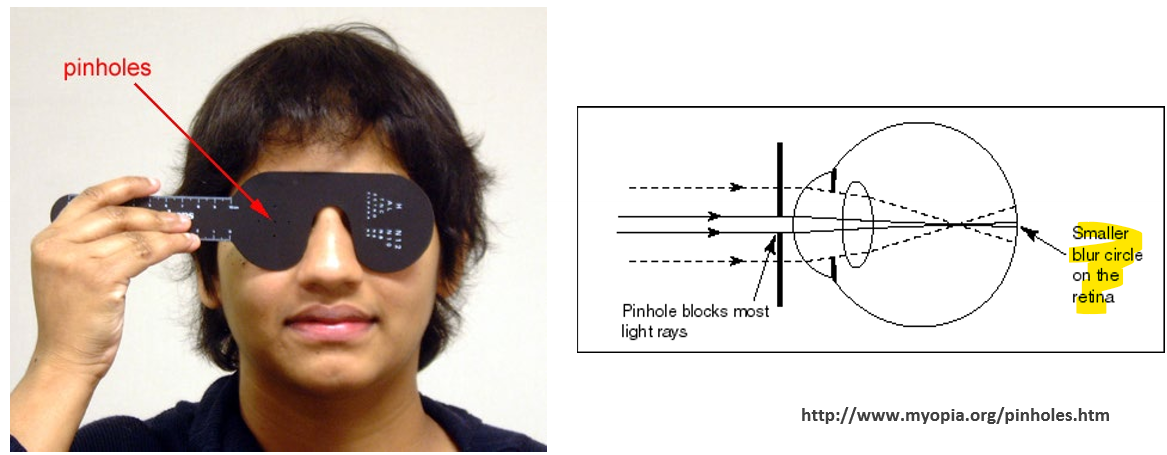
The Pinhole Test: This clinical test uses the principle of depth of focus to differentiate poor vision caused by uncorrected refractive errors from vision problems caused by pathology - if vision improves it identifies that there is a clear optical pathway and that the problem is refractive, if no improvement then there is a pathology.
Pathology = structural + physiological + neural problems.
Definition of Refractive Error: Any mismatch between the optical elements of the eye and the location of the sensors results in a refractive error.
Global Prevalence of Vision Impairment and Myopia
Reversible Blindness: Refractive error remains one of the leading causes of reversible blindness globally.
Projected Statistics: * By the year , it is projected that of the world's population will be short-sighted.
WHO Fact Sheet (11 October 2018): * Approximately people worldwide live with some form of vision impairment. * Distance Vision Impairment: * Mild impairment: people. * Moderate to severe impairment: people. * Blindness: people. * Near Vision Impairment: Approximately people live with near vision impairment. * Leading Causes: The primary causes are uncorrected refractive errors and cataracts. * Avoidability: Approximately of all global vision impairment is considered avoidable. * Demographics: The majority of people with vision impairment are over the age of years.
Anatomy and Optics of the Schematic Eye
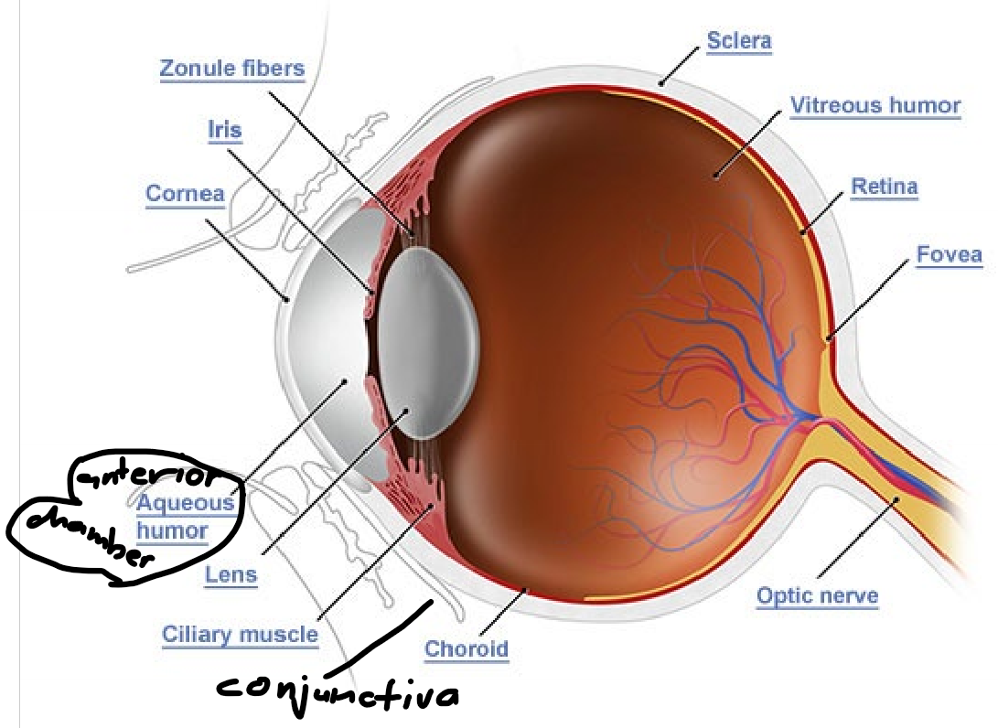
Anterior Optics: Comprised of the cornea and the crystalline lens.
Anatomical Structures: * Conjunctiva * Anterior chamber: Contains aqueous humor. * Iris and Pupil * Sclera and Choroid * Ciliary body: Includes the pars plicata and pars plana. * Vitreous humor * Retina and Macula * Optic nerve
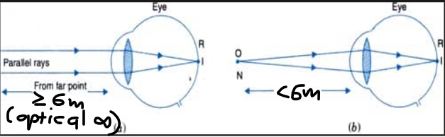
Incident Light and Divergence: The amount of divergence of light rays is inversely proportional to the distance of the target. * Far Target: When a target is far, there is effectively no divergence; the rays are parallel. * Optical Infinity: In optometry and ophthalmology, a distance of or more is considered to be "optical infinity." Rays a considered parallel after 6m.
Refractive Power: * The cornea and lens are curved to bend light and focus it on the retina. * The cornea provides approximately of the eye's total refractive power. * Posterior curvature also contributes to refractive power.
Optical Components and Axial Length: * Axial Length: Defined as the distance from the anterior corneal surface to the front of the retina. * Components of Axial Length: 1. Corneal thickness. 2. Anterior chamber depth. 3. Lens thickness. 4. Vitreous chamber depth.
Mechanisms of Accommodation
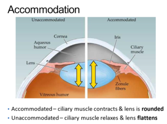
Ciliary Muscle Action: Ciliary muscles alter the anterior and posterior curvature of the crystalline lens, thereby changing its refractive power. * Muscle Contraction: When the ciliary muscle contracts, it slackens the lens ligaments. Ciliary muscle is a ring of muscle, therefore, contraction leads to movement inwards and smaller diameter ring resulting in slackened zonules. * Lens Shape Change: This allow the lens to take its preferred round shape, which results in greater refractive power.
Definition of Accommodation: This is the act of the eye's crystalline lens becoming more convex (if pliable enough) to increase the refractive power of the eye via the relaxation of ciliary ligaments.
Function: It is used to bring forward an image that would otherwise focus behind the retina (e.g., during near reading or to compensate for hyperopia).
Visual Function and Acuity (VA)
Visual Acuity: The first test of visual function, typically measuring the smallest letters a person can read.
Other Visual Integrity Tests: * Sensitivity to grey levels (contrast sensitivity). * Sensitivity to colours. * Ability to detect movement. * Sensitivity to light (threshold levels). * Distribution of viable photoreceptors (periphery test and OCT). * Speed of conduction of retinal nerve fibres (measure electrical signal).
Basics of Resolution: * If two point sources of light fall on two adjacent photoreceptors, they are perceived as one large light source. * The two lights are resolved (seen as separate) only if they fall on two photoreceptors separated by at least one non-illuminated receptor.

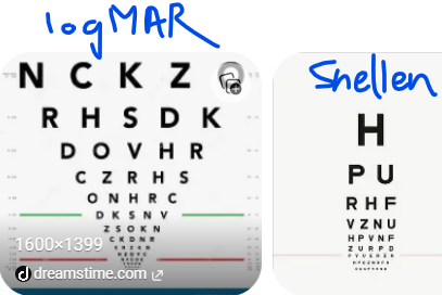
Visual Acuity Charts: Variety of charts including Snellen charts, LogMAR, and multi-language charts (Greek, Turkish, Italian, Macedonian, etc.).
Snellen Chart Design: * The width of critical detail (stroke width and gap width) is precisely of the total letter size.
Snellen Formula: * * : Standard viewing distance (usually ). * : The distance at which the letter subtends minutes of arc.
Entoptic Phenomena and the Purkinje Tree
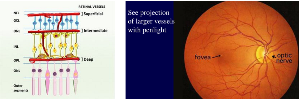
Retinal Blood Vessels: Photoreceptors lie behind the retinal blood vessels, which cast shadows on the retina.
Adaptation: These shadows are usually invisible because the visual system adapts to them.
Purkinje Tree: This refers to the image of the retinal blood vessels in one's own eye. Seeing them is an optic phenomenon occurring when movement or light positioning bypasses normal adaptation.
Development of the Eyeball (Emmetropisation)
Axial Length Sensitivity: A mere error in eye length can result in approximately of refractive error.
At Birth: There is a wide spread of eye sizes and refractive errors. The average error is approximately (hyperopia).
Through emmetropisation the eye grows toward 0.00D:
At Age 1 Year: For the majority, eye differences narrow significantly via the process of "emmetropisation."
Age 3: Approximately of individuals have emmetropised. The eye is roughly long. Minor lens changes continue, but the cornea is generally stabilised.
Age 6: The crystalline lens is stabilised and the eye reaches the adult size of approximately . Only minor changes occur thereafter unless disease (e.g., diabetes or cataract) is present.
Types of Refractive Error (Ametropia)
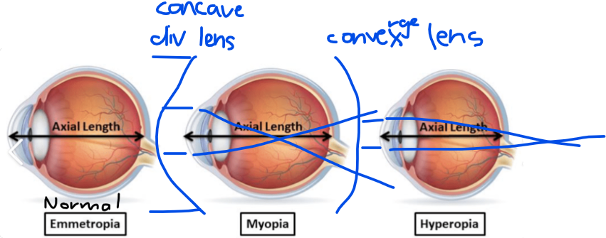
Emmetropia: The condition where parallel light is brought into focus clearly on the retina without the need for accommodation.
Ametropia: The condition where parallel light does not focus on the retina when accommodation is inactive. - Myopia & Hyperopia
Optical Lens Properties: * Light bends (refracts) when traveling between mediums of different refractive indices. * Slower light speed indicates a higher refractive index. * Refractive power increases with higher curvature.
Convex Lens (Plus Lens): * Converges incoming light to a focal point. * Thick in the middle; thin at the edges. * Magnifies the image.
Concave Lens (Minus Lens): * Diverges incoming light to an imaginary focal point. * Thin in the middle; thick at the edges. * Minifies the image.
Myopia (Short-Sightedness)
Mechanism: There is too much refractive power for the length of the eye. * Most common: Normal power but axial length is too large. * Alternatively: Normal axial length but too much refractive power.
Emergence: Generally emerges during childhood or early adulthood.
Symptoms: Near objects are clear; distance vision is blurred.
Correction: Uses a negative (concave/diverging) lens.
Visual Acuity: VA decreases as the error increases. Corrected VA may be slightly lower than expected due to the minifying effects of negative lenses.
Risk Factors: Highly associated with increased education, near-work activities, and limited time spent outdoors.
Hyperopia (Long-Sightedness)
Mechanism: Insufficient refractive power for the length of the eye. * Too little refractive power for a normal axial length. * Normal power but axial length is too small.
Stability: High hyperopia present at pre-school age often persists throughout life.
Symptoms: * Young people: Distance may be clear if they can accommodate, but near is blurred if accommodation is insufficient. * Older people: Distance is blurry and near vision is even worse due to loss of accommodation.
Asthenopia: Symptoms like headache, fatigue, or "pulling" of the eyes caused by sustained muscular effort of accommodation.
Correction: Uses a positive (convex/converging) lens.
Visual Acuity: Corrected VA might be slightly better than expected due to the magnifying effects of the positive lens. Modern young hyperopes may achieve VA similar to emmetropes due to natural accommodation.
Astigmatism
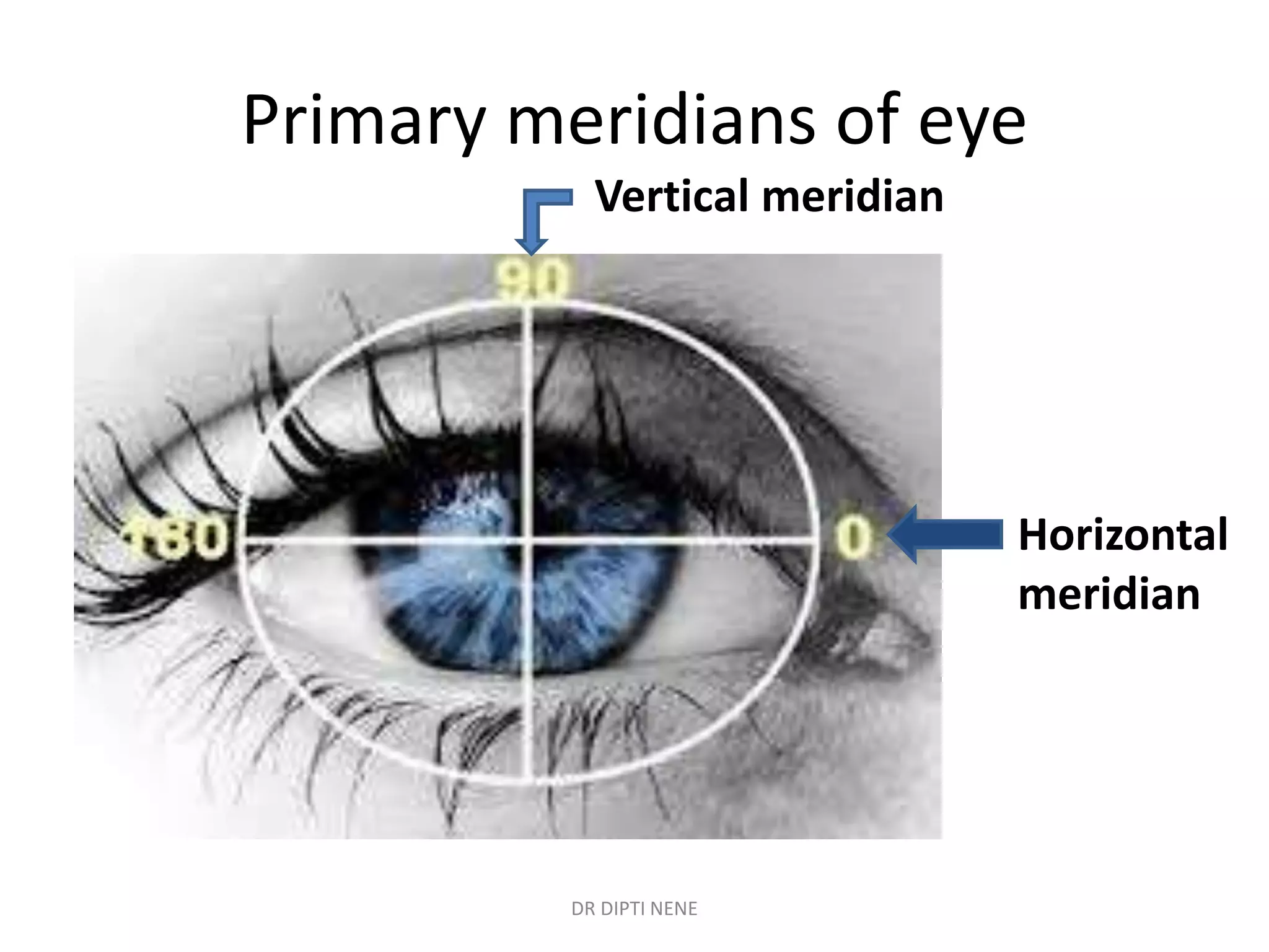
Mechanism: Refractive power in one meridian differs from that of the other meridian; light cannot be brought to a single point focus.
Causes: Usually found in the cornea, but the crystalline lens can also be a cause.
Symptoms: Blurred vision at both distance and near.
Types of Astigmatism: * Myopic astigmatism: Differing degrees of myopia in both meridians. * Hyperopic astigmatism: Differing degrees of hyperopia in both meridians. * Mixed astigmatism: One meridian is myopic, the other is hyperopic. * Simple astigmatism: One meridian has zero power, the other is myopic/hyperopic.
Correction: Uses a sphero-cylinder lens, where edge thickness changes around the circumference.
Effect on VA: Astigmatism elongates blur in one direction; its effect on VA varies based on the direction of astigmatism.
Presbyopia and Ageing of the Lens
Lens Flexibility Loss: Flexibility of the crystalline lens decreases from birth.
Accommodation Units: Flexibility measured in dioptres (). * Age 10: * Age 20: * Age 44: * Age 60: (no focusing power)
Calculating Accommodation: Divide by the nearest focus distance in while wearing distance correction.
Definition: Presbyopia (Greek for "old eyes") occurs when accommodative power is less than .
Correction: Positive lenses for near work are added to the distance correction (usually for a reading distance). * Age 40-45: May just hold material further away. * Age 45-50: Weak reading specs required. * Age 50-55: Moderate strength reading specs required. * Over 55: Full strength reading specs required.
Note: Presbyopia is not a category of refractive error; it is superimposed on top of existing refractive errors.
Presbyopia makes hyperopia worse. Presbyopia can correct near vision only for myopes.
Light and Dark Adaptation
Adaptation Definition: The ability of the visual system to adjust its sensitivity to function over a wide range of ambient light levels.
Dark Adaptation: The process of recovering sensitivity in the dark following bright light exposure. * Photopic Vision: Mediated by cones above unit for luminance. * Scotopic (Night) Vision: Mediated by rods below that level. * Mesopic Vision: A transition zone where both mechanisms work together.
Dark Adaptation Curve: Displays the "duplex nature" of vision. * The first curve reflects the cone mechanism. * The second curve reflects the rod mechanism. * Sensitivity of the rod pathway improves significantly after in the dark.
Factors Affecting Dark Adaptation: * Pre-adapting Light: Higher intensity and longer duration of pre-adapting light lead to a longer cone curve and a more delayed rod curve. * Retinal Location: Differences in photoreceptor topography (rod/cone density) across the retina affect the curve. * Test Spot Size: Larger test spots stimulate more rods/cones, providing greater sensitivity and allowing dimmer lights to be seen. * Wavelength (Colour): Rods are insensitive to long wavelengths. Therefore, red light () shows no "rod-
cone break" on the adaptation curve. That’s why red light/torch is used for night vision. Even in the dark red light can be seen by a person with poor dark adaption as it needs no rods.
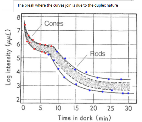
High on the y‑axis = LOW sensitivity ; Low on the y‑axis = HIGH sensitivity
Cones
Adapt quickly
Reach their best sensitivity in ~7 minutes
But they never get as sensitive as rods
So their curve drops fast, then plateaus
Rods
Adapt slowly
Keep improving for ~30 minutes
Become far more sensitive than cones
Their curve drops much lower on the y‑axis
Light Adaptation: Eye recovery sensitivity in light after dark exposure. * Plotted as increment threshold versus background luminance (tvi curve).
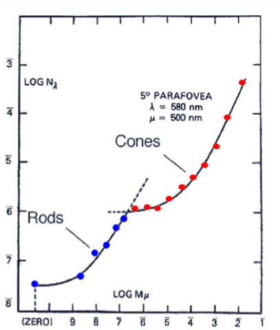
As the background gets brighter (x), how much extra light do rods or cones need to detect a target (y).
Mechanisms of Adaptation: 1. Pupil size regulation. 2. Switch between rods and cones. 3. Bleaching and regeneration of photopigment. 4. Feedback from horizontal cells.
Questions & Discussion
Question (Retina vs Sensor): What is a similarity between the retina and the digital sensor in a camera? * Answer: Both serve the primary function of converting light into electrical/electronic signals. In the retina, photoreceptors capture light for transmission to the brain; in a camera, photosensitive pixels detect light for processing by electronics.
Question (Pinhole): How does a pinhole improve vision for individuals with refractive errors? * Answer: It reduces the amount of unfocused light. It blocks peripheral light rays and allows only central, focused rays to enter, resulting in a clearer retinal image through a smaller blur circle.
Question (Astigmatism): What is characteristic of mixed astigmatism? * Answer: It is a combination of both myopia and hyperopia in the same eye; one meridian is nearsighted while the perpendicular meridian is farsighted.
Question (Wavelength): How does exposure to red light affect dark adaptation? * Answer: Red light enhances/accelerates dark adaptation. Because rods are insensitive to long wavelengths, red light minimizes the bleaching of rhodopsin while the rod sensitivity increases. Because rods barely absorb red light:
Rhodopsin is not bleached
Rod sensitivity is not reset
Rods remain in their fully dark‑adapted state