Bio25 Ch. 9 - Skeletal System: Articulation
Objectives
Types of Joints:
Structural:
Fibrous
Cartilaginous
Synovial
Functional:
Synarthrosis
Amphiarthrotic
Diarthrotic
Selected Joints:
Temporal
Elbow
Shoulder
Knee
Ankle
9.1 Classification of Joints
Joints (articulations): Points where bones, cartilage, or teeth come together.
Characteristics:
Weaker points in the skeleton but capable of resisting forces.
Classified by structure (composition) and function (movement).
Arthrology: Study of joints.
Structural Classifications
Fibrous Joints:
Connected by dense connective tissue.
Generally immobile or slightly mobile.
Cartilaginous Joints:
Connected by cartilage.
Generally immobile or slightly mobile.
Synovial Joints:
Connected by ligaments, featuring a fluid-filled cavity.
Functional Classifications
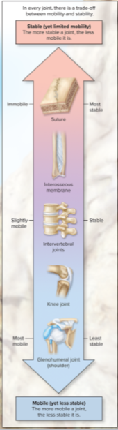
Synarthroses: Immobile joints (fibrous or cartilaginous).
Amphiarthroses: Slightly mobile joints (fibrous or cartilaginous).
Diarthroses: Freely mobile joints (all synovial JOINTS).
9.2 Fibrous Joints
Types:
Gomphoses: Joints between teeth and jawbone.
Sutures: Found in the skull, immovable.
Syndesmoses: Slightly movable joints, e.g., between certain long bones.
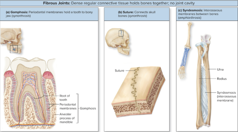
Connected by dense regular connective tissue
immobile or only slightly mobile
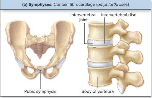
9.3 Cartilaginous Joints
Cartilaginous Joints: Cartilage between ends of articulating bones: no joint cavity
Types:
Synchondroses: Composed entirely of hyaline cartilage, immobile.
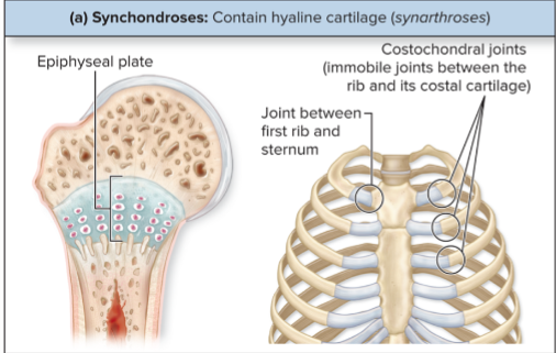
Symphyses: Composed of fibrocartilage, Compression. slightly movable (e.g., pubic symphysis).
9.4a Synovial Joints
Characteristics:
Freely mobile diarthroses featuring:
Articular capsule: Surrounds joints with two layers.
Synovial fluid: Lubricates and nourishes cartilage.
Lubricates articular cartilage
act as a shock absorber
nourished the chondrocytes
Articular cartilage: Reduces friction during movement. (Hyaline cartilage on bone surface)
Ligaments & nerves: Provide stability and feedback.
connect one bone to another
Extrinsic Ligament: physically separate from articular capsule.
Intrinsic Ligament: Located within the articular capsule, these ligaments help to connect bone ends and provide additional support during joint movement.
Tendons: dense regular connective tissue. Attach muscle to bone
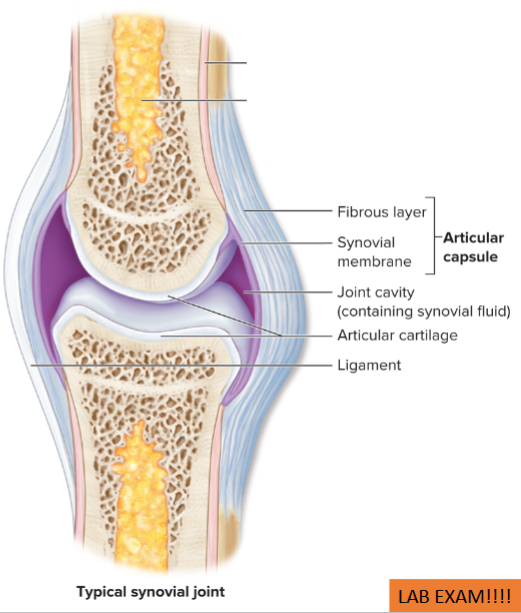
Articular Capsule
Outer Fibrous Layer: Composed of dense connective tissue to prevent bone separation.
Inner Synovial membrane Layer: Synovial membrane that produces synovial fluid.
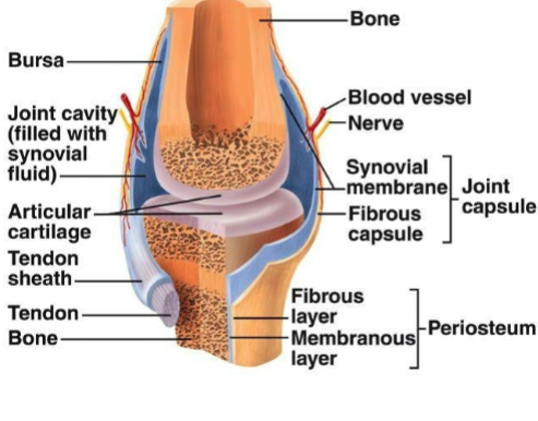
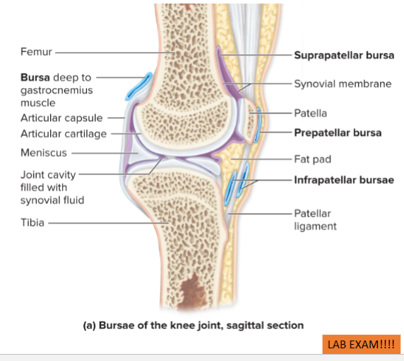
Additional Structures
Bursae: Sac-like structures reducing friction.
contains synovial fluid
connected to or separate from joint cavity
Fat pads: Protective packing material surrounding joints periphery.
Tendon sheaths: Encase tendons in areas of potential friction.
Sensory receptors and blood vessels
numerous in synovial joints
receptors detect pain stimuli.
They also provide information about joint position and movement, contributing to proprioception, which is essential for coordinated movement
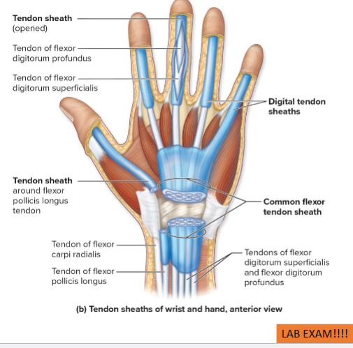
Tendon sheath: Elongated bursae wrapped around tendons where friction is excessive
Common in wrist and ankle
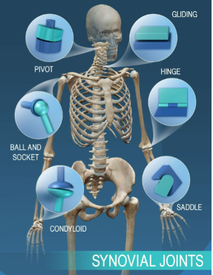
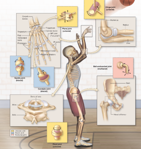
Classification of Synovial Joints
By Movement:
Uniaxial: Movement in one plane or axis
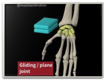
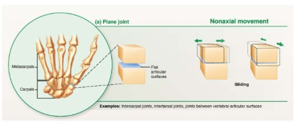
plane joint: flat articular surfaces
limited to (side to side gliding movements) in a single plane
ex: small ones of wrist and foot

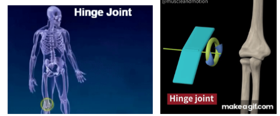
UNIAXIAL:
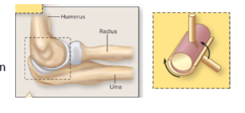
Hinge Joint: Convex surface within concave depression
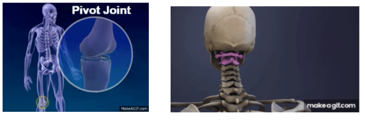
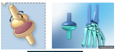
UNIAXIAL:
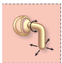
Pivot Joint: Rotational movement around a single axis.
Rotation on longitudinal axis, allowing for specific movements such as turning the head or pronating the forearm.

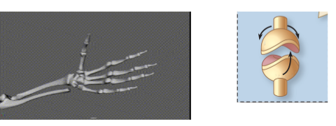
Biaxial: Movement in two planes or axes
Condylar Joint: Oval, convex surface articulating with concave surface, but NO ROTAATION
Biaxial:
Saddle Joint: Convex and Concave surface resemble saddle shape, Two directions: SOME ROTATION
EX: Thumb
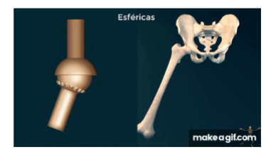
Multiaxial: Movement in multiple planes or axes
Ball and Socket Joint
Permit movement in 3 planes
Freely mobile type: EX- Shoulder and Hip
By Shape:
Plane
Hinge
Pivot
Condylar
Saddle
Ball-and-socket
9.5 Movements of Synovial Joints
Gliding Motion
Surfaces slide over each other (e.g., intercarpal joints). Carpals or Tarsals
back and forth
side to side
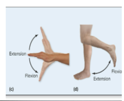
Angular Motion
Changes the angle between bones:
Flexion: Decreases angle.
Extension: Increases angle.
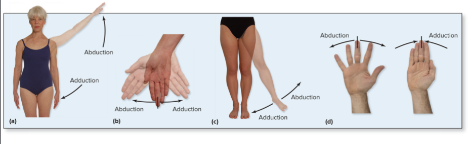
Abduction/Adduction: Movement away/closer to the midline.
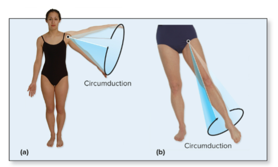
Circumduction: Circular movement.
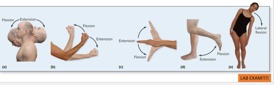
ANTERIOR - POSTERIOR PLANE:
Flexion: decreases the angle between bones
bring bones closer together
ex: bending fingers
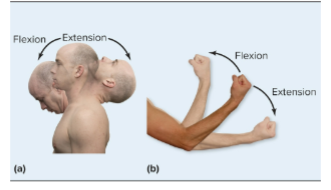
Extension: (Opposite of Flexion) Increases angle between articulating bones
ex: straightening fingers
Hyperextension: Extension beyond normal rang of motion
Not normal motion, Can result in injury
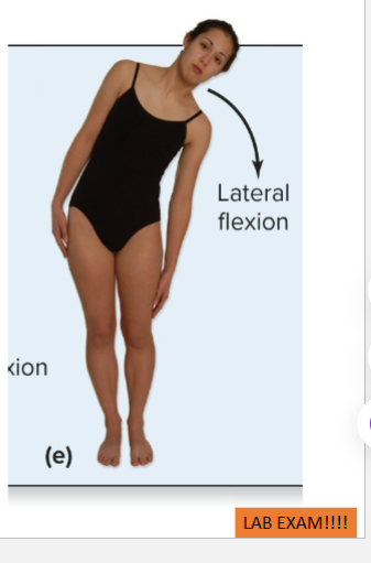
CORNAL PLANE
Lateral Flexion: trunk of body moving in coronal plane laterally.
occurs between vertebrae in cervical and lumbar region.
Abduction: Lateral movement of body part (away from midline)
Adduction: towards midline
Circumduction: Circular movement of a body part, combining flexion, extension, abduction, and adduction in a sequence that describes a cone shape.
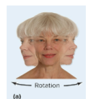
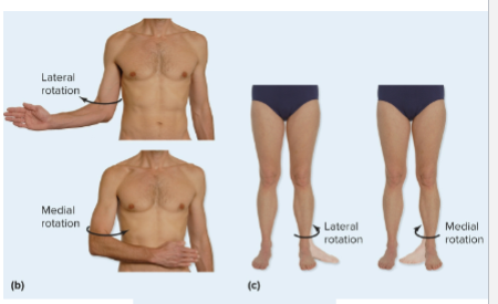
Rotational Motion
Bone rotates around its axis:
Medial: Turns anterior surface of bone medially
Lateral Rotation: turns anterior surface of the bone laterally, away from the midline of the body.
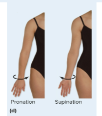
Pronation: medial rotation of forearm - palm of hand to posterior
Supination: lateral rotation of forearm - palm of hand to anterior
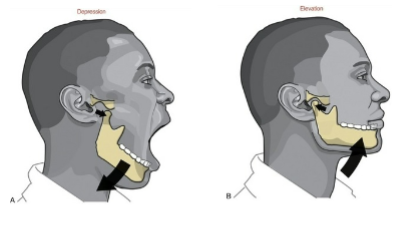
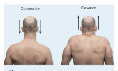
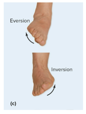
Special Movements
Elevation/Depression: Up/down movements (e.g., mouth opening/closing).
Eversion/Inversion: Foot movements.
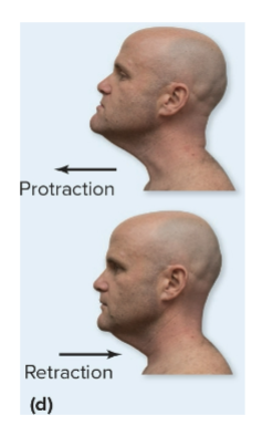
Protraction/Retraction: Forward/backward movements (e.g., jaw).
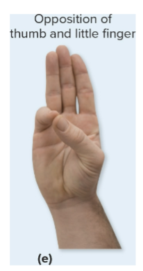
Opposition/Reposition: Thumb movements.
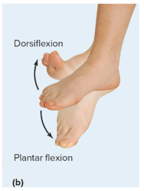
Dorsiflexion/Plantar Flexion: Upward/downward movements of the foot, crucial for walking and running.
9.6 Synovial Joints and Levers
Biomechanics: Applying mechanical principles to biological systems.
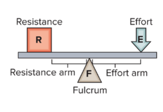
Levers: Composed of a
fulcrum (pivot point), between effort and resistance
effort (force),
resistance (load).
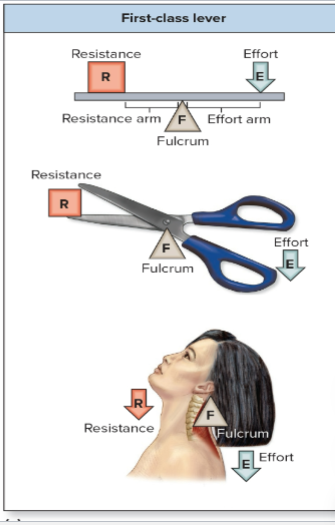
Types of Levers
First-Class Lever: Fulcrum is between effort and resistance (e.g., neck).
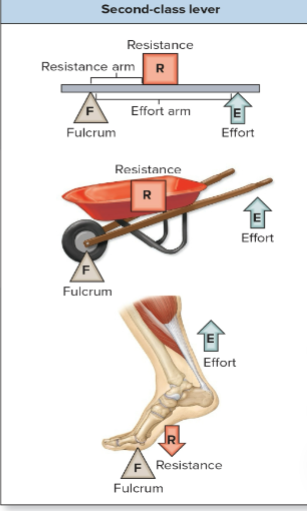
Second-Class Lever: Resistance is between fulcrum and effort (e.g., standing tiptoe).
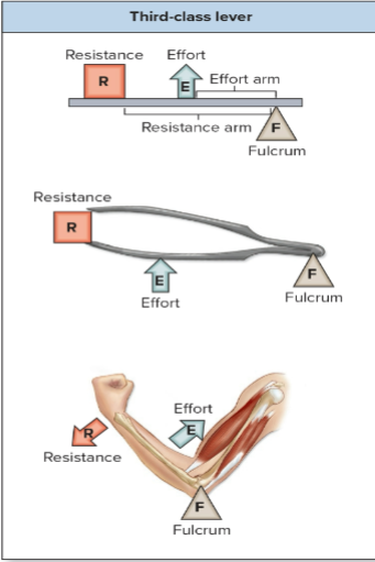
Third-Class Lever: Effort applied between fulcrum and resistance (e.g., elbow).
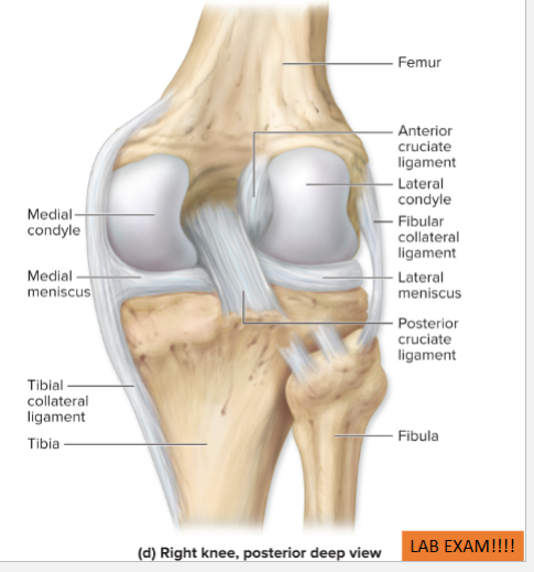
9.7 Knee Joint
Largest Diarthrosis: Primarily a hinge joint that allows slight rotation and gliding.
Composed of 2 separate articulations
Tibiofemoral joint: between condyles of femur and condyles of tibia
Patellofemoral joint: between patella and patellar surface of femur
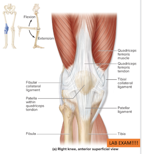
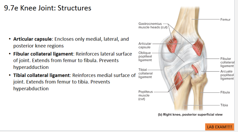
Structures of the Knee
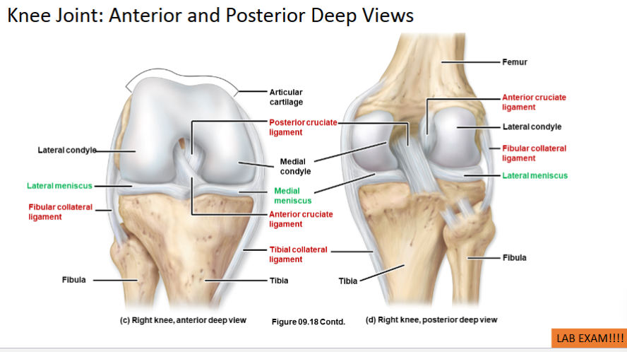
Support and Stabilization:
Quadriceps femoris muscle tendon: Passes over knee’s anterior surface, surrounds patella
Patellar ligament: extends rom patella to tibial tuberosity
Fibular Collateral ligaments (fibular & tibial) reinforce lateral surface of joint
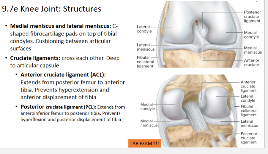
Menisci: C-shaped cartilages for cushioning.
Cruciate ligaments: ACL and PCL preventing excessive movements.
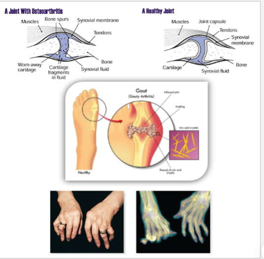
9.8 Joint Aging and Health
Arthritis: Group of conditions characterized by joint inflammation and degeneration.
Types include
Osteoarthritis: Degenerative joint conditions in older ppl
Gouty Arthritis: Inflammation due to high lvl of uric acid
Rheumatoid Arthritis: Autoimmune disorder causing joint inflammation and pain, often affecting both sides of the body symmetrically.
Joint Health:
Regular exercise enhances joint health but may exacerbate pre-existing conditions.
Clinical Notes
Injuries: Common injuries include collateral ligament injuries, sprains, Pott fractures, and meniscal tears.