MODULE 3
MCB 102
LECTURE
MODULE 3: virus structure, components, function
DEFINITION OF TERMS |
- subunit
- single folded polypeptide chain
- CAPSOMERE
- structural unit (protomer, asymmetric unit)
- one or more subunits
- unit from which capsids or nucleocapsids are built
- CAPSID
- capsid (“box” in latin)
- protein shell surrounding the genome
- nucleocapsid (core)
- nucleic acid and protein assembly within the particle
- used when substructure is discrete
- nucleic acid + capsid
- envelope (viral membrane)
- host-cell derived lipid bilayer
- virion
- infectious virus particle
virion size |
average size: 10 – 300 (or 400) nm in diameter
comparison
- Nanometer: 10-9 meters = 10 angstrom = 0.001 microns
- Alpha helix in protein: 1 nm in diamter
- DNA: 2 nm in diameter
- Ribosome: 20 nm in diameter
- Poliovirus: 30 nm in diameter
- Pandoravirus: 1000 nm in diameter
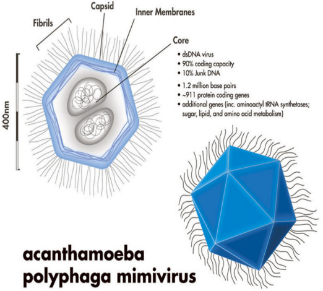
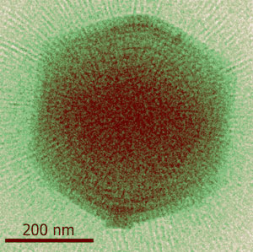
2003 - mimivirus |
- Giant MIMIVIRUS was described but was sequenced in 2004
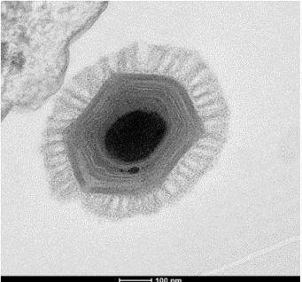
2011 – megavirus chilensis |
- Megavirus chilensis was discovered
- largest virus
- 440 nm
- most complex genome with 1.2 Mb
2013 – pandoravirus |
- infect amoeba
- larger than some bacteria
- size: 1.0 um / 1000 nm
- genome 2.8 Mb
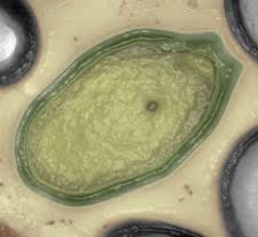
comparison – PITHOVIRUS SIBERICUM
- SIZE: 1.5 UM / 1500 NM
- GENOME: 0.6 Mb
SUMMARY OF VIRAL STRUCTURES
- VIRION
- CAPSID/PROTEIN COAT
- VIRION SYMMETRY
- VIRAL GENOME
- VIRAL PROTEINS
- ENVELOPE
- VIRAL CARBOHYDRATES
- OCCLUSION BODIES
structure of viruses |
PROTOMER
- Basic protein building block of the capsid
CAPSOMERE
- Morphological unit on the surface of the virus
- Either 5 protomers (PENTAMER) or
6 protomers (HEXAMER)
VIRION SYMMTERY
- Helix
- Icosahedral
- Rod
- Cone
Note: most common are helical and icosahedral
VIRION |
- Complete virus particle that represents the extracellular phase of the virus life cycle
- FUNCTIONS:
- Protects the genome
- Delivers the genome
- Delivers protein contained in the virion
- Interacts with the host
CAPSID/PROTEIN COAT |
- Constructed from many copies of one or few types of protein subunits (protomers)
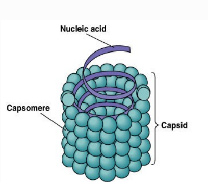
- CAPSOMERE: subunit (self assembles to form the capsid)
- CAPSID: structural unit
- FUNCTIONS:
- Protects the genome
- Recognizes and interacts with the host cell
- Facilitates the transfer of the viral nucleic acid
- Determines the antigenic characteristic of the virus
VIRION SYMMETRY |
ICOSAHEDRAL (CUBIC) |
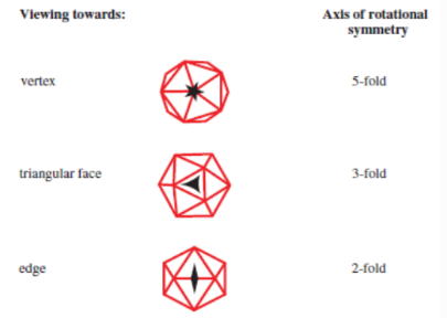
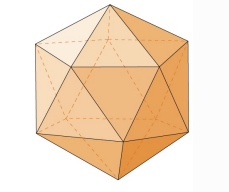
- 20 FACES (each an equilateral triangle)
- 12 VERTICES (where the vertices of 5 triangles meet)
- 30 EDGES (each of which the sides of two triangles meet)
capsids with icosahedral symmetry
- Exemplified by many common plants and animal viruses
- Constructed from identical protein molecules
- Pentamers are found at the VERTICES
- Hexamers are found at the FACES
ADVANTAGES of icosahedral symmetry
- Allows tight-packing of subunits
- Subunits can be smaller (focus on genetic info)
- Most efficient arrangement of subunits in a closed shell
(smallest number of subunits used to build a shell)
HELICAL |
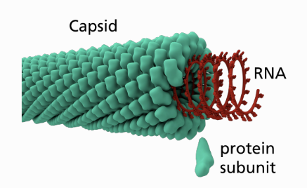
- Nucleic acid is coiled in the form of a helix
- Protein subunits are arranged helically like hollowed cylinders around a coil
- RIGID in plant virus
- LONG AND FLEXIBLE in animal viruses
- SIZE is determined by both NUCLEIC ACID and CAPSOMERES
- DIAMETER is dependent on SIZE and SHAPE PROTEIN INTERACTION
- LENGTH is determined by LENGTH of NUCLEIC ACID
viruses with helical symmtery
- Measles Virus
- Beet Yellows Virus Particle
 Tobacco Mosaic Virus
- Vesicular Stomatitis Virus
COMPLEX |
- ATYPICAL viruses
- Neither icosahedral nor helical
VIRUS | DESCRIPTION |
Poxviridae | Oval/Brick-shaped |
Bacteriophage T4 (E.coli) | Tailed phages |
HIV-1 | Conical capsids |
Baculovirus | Rod-shaped capsids |
Note: in HIV-1 and Baculovirus, virus genome is coated in highly basic protein (enveloped virions)
VIRAL GENOME |
- Stores the genetic information of the virus
- REQUIREMENT:
Genome MUST contain the information coded in a form that can be RECOGNIZED and DECODED by the cell parasitized
CATEGORIES OF VIRAL GENOMES
- Double-stranded DNA (dsDNA)
- Single-stranded DNA (ssDNA)
- Double-stranded RNA (dsRNA)
- Single-stranded RNA (ssRNA)
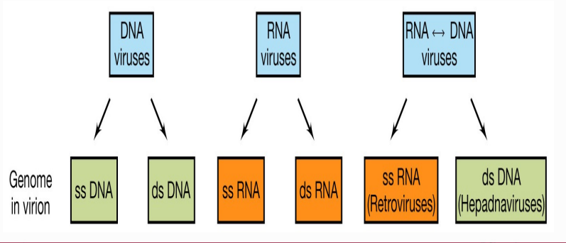
CLASSIFICATIONS:
- According to NUCLEIC ACID CONFIGURATION:
- LINEAR
- CIRCULAR
- SEGMENTED
- According to SINGLE-STRANDED (ss) viral genomes:
- POSTIVE SENSE
- NEGATIVE SENSE
- AMBISENSE
DNA GENOMES | EXAMPLES |
ss // linear | Parvovirus |
ds // linear | Poxvirus |
ss // circular | Phage Phi X174 |
ds // circular | Baculovirus |
RNA GENOMES | EXAMPLES |
ss // linear | Tobacco Mosaic Virus |
ds // linear | Reovirus |
ss // circular | Hepatitis delta virus |
IMPORTANT!!
- Most PLANT viruses have ssRNA genomes
- Most FUNGAL viruses have dsRNA genomes
- Most PROKARYOTIC viruses have dsDNA genomes
Reason: Diverse origin of viruses in different host types
GENOME SIZE |
- LARGE VIRUS GENOMES are composed of dsDNA
- Largest RNA genomes: CORONAVIRUSES (33 kb ssRNA)
- LARGEST VIRUS GENOME: Pandoravirus (2.8 Mb)
SEGMENTED GENOME |
- Genome DIVIDED INTO SEVERAL PARTS but enclosed into the same capsid
- Example:
INFLUENZA VIRUS (ORTHOMYXOVIRIDAE)
8 RNA segments
Each segment codes for one protein
SEGMENT 4 codes for HEMAGGLUTININ
MULTIPARTITE GENOMES |
- Also have SEGMENTED GENOMES but are not contained in the same capsid, rather each segment is packaged into A SEPARATE VIRUS PARTICLE
- Occurs in both RNA and DNA PLANT VIRUSES
ADVANTAGE OF MULTIPARTITE GENOMES
SOLVES THE PROBLEM OF BREAKAGES
DISADVANTAGE OF MULTIPARTITE GENOMES
THE HOST CELL MUST TAKE UP ALL VIRUS PARTICLES TO ESTABLISH A PRODUCTIVE INFECTION
IMPORTANT notes
SEGMENTED GENOMES:
- More common in RNA VIRUSES than in DNA VIRUSES
MULTIPARTITE GENOMES:
- More common among PLANT VIRUSES
VIRAL proteins |
STRUCTURAL PROTEINS |
- Proteins that are COMPONENTS OF THE VIRIONS
- FUNCTIONS:
- PROTECTION of the virus genome
- ATTACHMENT of the virion to the host cell (for many viruses)
- FUSION of the virion envelope to a cell membrane
- Provide STRUCTURAL SYMMETRY
- Determine ANTIGENIC CHARACTERISTICS of the virion
NON-STRUCTURAL PROTEINS |
- Proteins synthesized by the virus in an infected cell but are NOT VIRION COMPONENTS
- FUNCTIONS:
- ENZYMES (protease, reverse transcriptase)
- TRANSCRIPTION FACTORS
- PRIMERS for NUCLEIC ACID REPLICATION
- INTERFERENCE with host immune response
NOMENCLATURE OF VIRAL PROTEINS |
- STRUCTURAL PROTEINS: VP1, VP2, VP3…
note: VP = VIRAL PROTEIN
- NON-STRUCTURAL PROTEINS: NSP1, NSP2, NSP3
- STRUCTURAL CHARACTERISTIC:
G – GLYCOPROTEIN
P – PHOSPHOPROTEIN
- FUNCTION:
F – FUSION
P - POLYMERASE
RT – REVERSE TRANSCRIPTASE
ENVELOPE |
- LIPID-PROTEIN STRUCTURE that encloses the nucleocapsid
- FLEXIBLE MEMBRANOUS STRUCTURE (thick lipid bilayer)
- Derived from the HOST CELL MEMBRANE (Golgi, Nuclear Membrane, Cell Membrane)
- Released by BUDDING of the particle through the membrane
- Mostly found in ANIMAL VIRUSES
- FEW in plant and bacterial viruses
- Dissolved by ORGANIC SOLVENTS (e.g. ETHER)
ENVELOPE | EXAMPLES |
Helical Nucleocapsid | Influenza Virus |
Icosahedral Nucleocapsid | Herpesvirus |
ENVELOPE PROTEINS |
- GLYCOPROTEINS
- TRANSMEMBRANE PROTEINS anchored to the membrane by a HYDROPHOBIC DOMAIN
- Coded for by the VIRUS GENES
- TYPES:
- EXTERNAL GLYCOPROTEINS
Referred to as SPIKES or PEPLOMERS
Project about 10 nm from the surface at 7-8 nm intervals
FUNCTIONS:
- Major Ag of enveloped viruses
- Provide contact with EXTERNAL ENVIRONMENT
- NEURAMINIDASE and HEMAGGLUTININ for influenza virus
- TRANSPORT CHANNEL PROTEINS
FUNCTIONS:
- Enable virus to alter the PERMEABILITY OF THE MEMBRANE
- Form PROTEIN-LINED CHANNEL through the envelope
- Contain MULTIPLE HYDROPHOBIC TRANSMEMBRANE DOMAINS
- Allows MODIFICATION OF THE INTERNAL ENVIRONMENT of the virion
- MATRIX (M) PROTEINS
- INTERNAL virion proteins in the INNER SURFACE OF THE ENVELOPE (non-glycosylated)
- LINKS the INTERNAL NUCLEOCAPSID to the ENVELOPE for stability
- ABUNDANT because it encompasses 30% of the total weight of retroviruses
LOSS OF ENVELOPE LEADS TO LOSS OF VIRAL INFECTIVITY
REASONS:
- Lipid/lipoprotein may be required for attachment
- Loss of lipids may lead to loss of protection of nucleoproteins
- Solvents may extract lipids and denature proteins
VIRION SHAPE | EXAMPLES |
Sphere | Influenza virus |
Bullet | Rabies virus |
Rod | Baculovirus |
Thread | Ebola virus |
With Internal Lipid Membrane | Iridovirus |
VIRAL CARBOHYDRATES |
- Viral proteins have CARBOHYDRATE MOITIES found in the envelope
- Attach the virus to the cell by INTERACTING WITH A RECEPTOR
- Functions as an ANTIGEN
OCCLUSION BODIES |
- PROTEIN CRYSTALS produced by some viruses which provide ADDED PROTECTION outside the host
VIRION STRUCTURE | GENOMES |
Icosahedral Naked | All genomes |
Icosahedral Enveloped | Lacks ssDNA |
Helical Naked | Lacks dsRNA |
Helical Enveloped | ssRNA only |