Topic 4_Anatomy and Physiology of Joints
Anatomy & Physiology of Joints
Types of Joints: Classified by structure and mobility.
Fibrous Joints: Little to no movement (e.g., sutures in the skull).
Cartilaginous Joints: Limited movement (e.g., pubic symphysis).
Synovial Joints: Full range of movement, most common in limbs.
Objectives of the Study
Understand classification of joints:
Fibrous Joints: Describe structure and give examples.
Cartilaginous Joints: Outline general structure and types.
Synovial Joints: Discuss structure, stability factors, movement types, and major body joints.
Classification of Joints
Structural Classification:
Fibrous Joints: Two bones connected by dense connective tissue.
Cartilaginous Joints: Bones unified by cartilage.
Synovial Joints: Joint cavity present, allowing for significant movement.
Functional Classification:
Synarthroses: Immovable joints (e.g., sutures).
Amphiarthroses: Slightly movable joints (e.g., intervertebral discs).
Diarthroses: Freely movable (e.g., shoulder, elbow).
Fibrous Joints
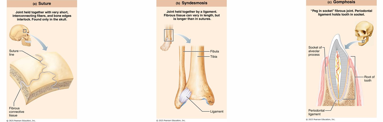
Types:
Synostoses (sutures): Immovable joints, only found in the skull.
Syndesmoses: Joints connected by ligaments; movement varies based on fiber length.
Gomphoses: Peg-in-socket (e.g., tooth in its socket).
Characteristics:
Connected by dense connective tissue.
No joint cavity present.
Movement is typically minimal or non-existent.
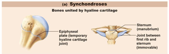
Cartilaginous Joints
Types:
Synchondroses: Bones connected by bar or plate of hyaline cartilage; mostly immovable (e.g., epiphyseal plates).
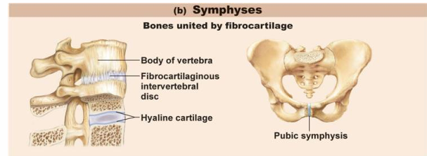
Symphyses: Bones connected by a plate of fibrocartilage; provide strength with flexibility (e.g., pubic symphysis).
Ends covered with hyaline, plate of fibrocartilage between
Characteristics:
No joint cavity.
Limited movement.
Synovial Joints Structure
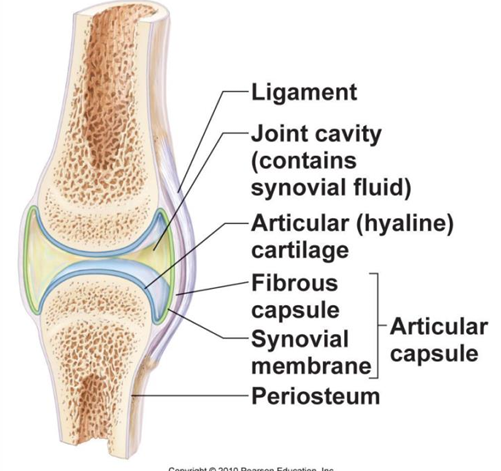
Features:
Articular Cartilage: Covers bone surfaces, cushions joints.
Joint Cavity: Small fluid-filled space reduces friction.
Articular Capsule: Two layers (outer fibrous layer and inner synovial membrane).
Synovial Fluid: Lubricates joints.
Reinforcing Ligaments: Add stability, can be capsular, extracapsular, or intracapsular.
Nerve and Blood Supply: Detects pain and monitors joint position.
Bursae and Tendon Sheaths
burase: bags of synovial fluid that act as a lubricating “ball bearing”
tendon sheath: elongated form that wraps around tendons
Ligaments
more lig. = more strength
ligaments can only stretch ~6% before break
Muscle tone
tendons of muscles crossing joints usually MOST IMPORTANTin stabilizing the joint, as they help maintain the position, keep taut by muscle tone
esp. shoulderknee, arches of foot
Stability:
-allow lots of movement, not as stable as fibrous cartilage/joint
Articular surfaces
Shape of articular surfaces of many joints are such that they dont contribute stability
deep ball and socket joints have good shape for mobility
Movement Types of Synovial Joints
Movement Categories:
Nonaxial: Gliding only.
e.g.
Uniaxial: Movement in one plane (e.g., hinge joints elbow).
Biaxial: Movement in two planes (e.g., condyloid joints knuckle).
Multiaxial: Movement in all three planes (e.g., ball-and-socket joints shoulder and hip).
General Movements:
Gliding Movements: Flat surfaces slide past each other.
Angular Movements: Increase or decrease the angle between two bones.
Rotation: Bone turns around its own axis.
Examples of Specific Movements
Flexion and Extension: Alter angle between bones in sagittal plane.
Abduction and Adduction:
Abduct: away from midline
Adduct: towards midline
Circumduction: Circular movement at joints.
Special Movements:
Pronation and Supination: Rotational movements of the forearm.
drink soup
Dorsiflexion and Plantarflexion: Foot movement towards or away from shin.
Opposition: Movement of thumb to touch fingers.
Movements of the Mandible: Protraction, retraction, elevation, and depression.
Feet
Dorsiflexion: Movement that brings the toes closer to the shin, allowing for activities such as walking on heels.
Plantar flexion: Movement that points the toes away from the shin, facilitating actions like standing on tiptoes or pushing off during running.
Inversion: turn the sole of the foot medially
Eversion turn the sole of the foot laterally
Jaw (mandible)
Elevation: up (close mouth)
Depression: down (open mouth)
Protraction: stick out jaw anteriorly
Retraction: pull jaw back posteriorly
Types of Synovial Joints
Based on Shape:
Plane (gliding movement).
two flat opposing surfaces
Hinge (flexion and extension).
cylinder into trough
flexion extension (elbow)
uniaxial
Pivot (rotation)
insertion into a ring or sleeve
between atlas and dens of axis; proximal radio ulnar joint
Condyloid (movement in all planes).
“knuckle joint'“
articulating surfaace like an oval
All planes of motion
Saddle (enhanced movement).
surface on one bone is saddle, other bone fits into
most freely moving
Ball-and-socket (most freely moving).
multiaxial joints
freely moving
e.g. shoulder/hips.
Specific Joint Examples
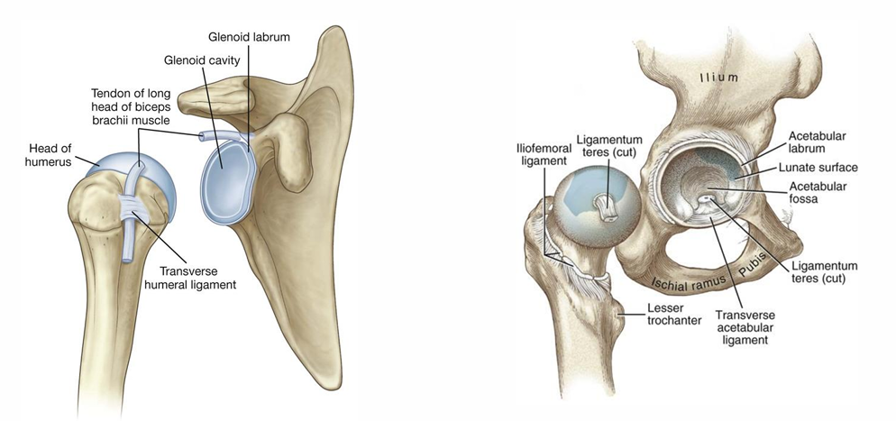
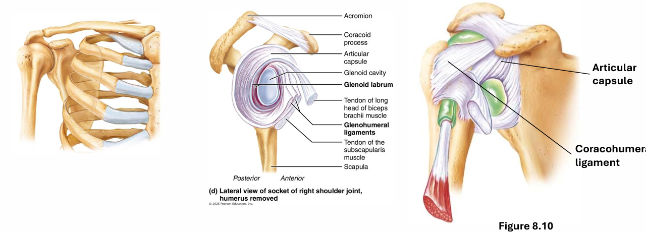
Shoulder Joint: Ball-and-socket; stabilized by rotator cuff tendons.
glenoid cavity broadened slightly by the glenoid labrum
largely the tendons of the rotator cuff muscles that stabilize this joint
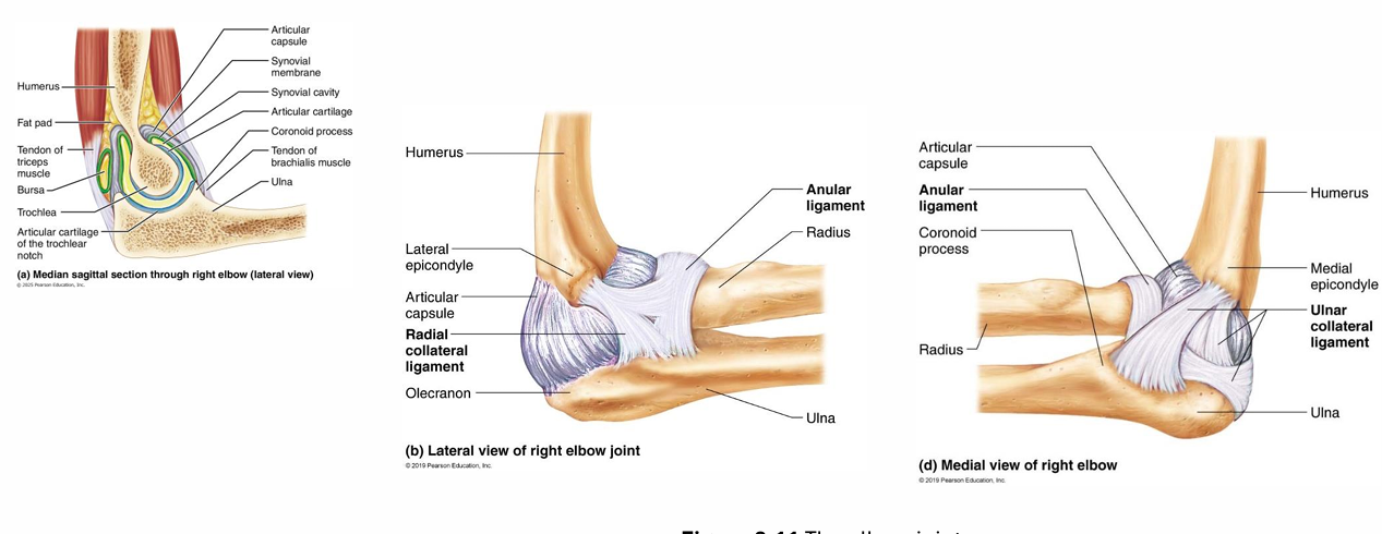
Elbow Joint: Hinge; primary movements are flexion and extension; stabilized by collateral ligaments.
humerus articulates with radius and ulna
annular ligament allows rotation of the radius during pronation and supination
ulnar collateral
radial collateral
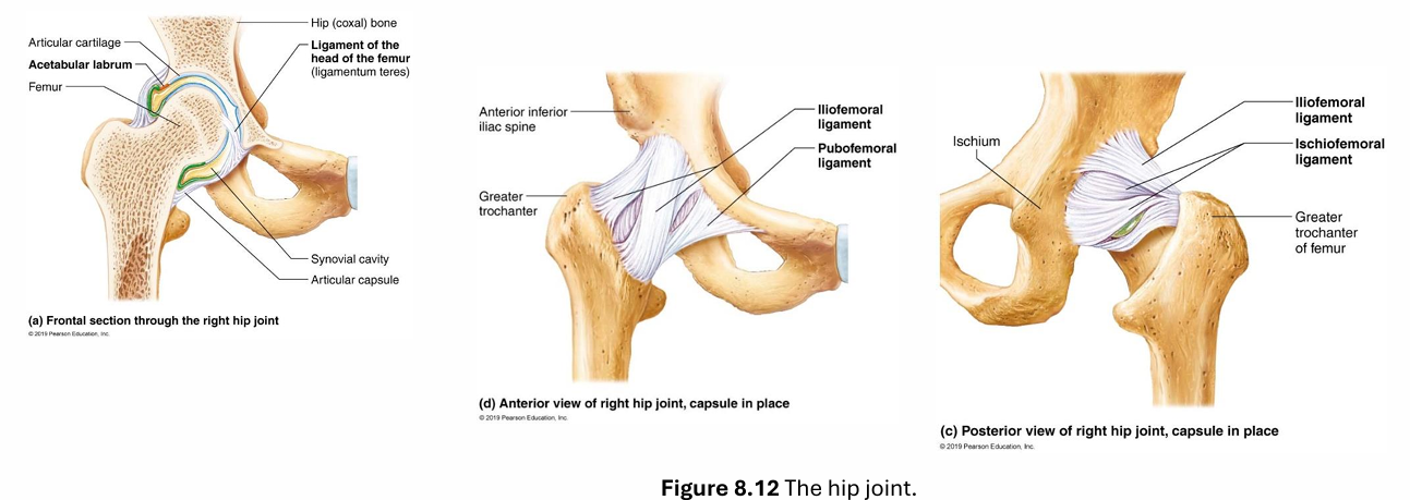
Hip Joint: Deep ball-and-socket; includes intracapsular and extracapsular ligaments.
deep ball and socket
intracapsular ligament that extends from the fovea capitis to ligamentum teres
extracapsular ligaments: iliofemoral, pubofemoral and ischiofemoral
the articular surface does provide some stability
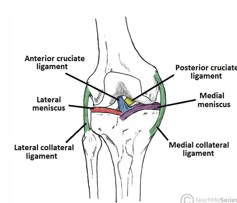
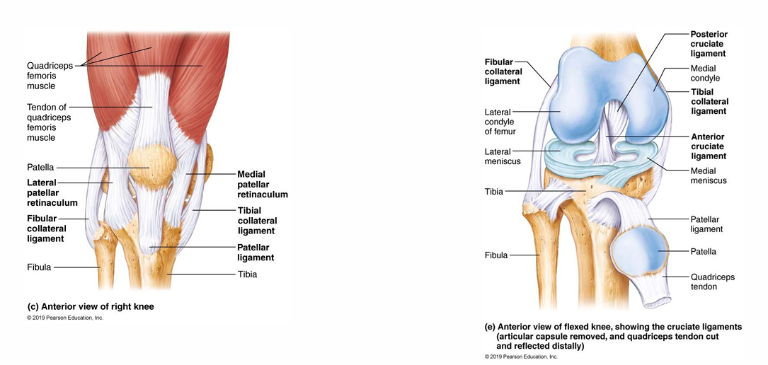
Knee Joint: Hinge; complex structure with multiple ligaments for stabilization.

largest and most complex
doesnt allow movementin multiple planes, primarily permitting flexion and extension.
when flex knee allows slight rotation
described as having 3 individual joints
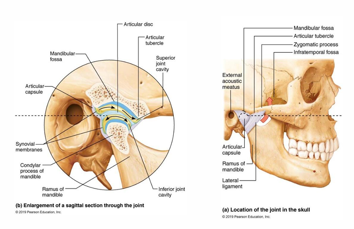
Temporomandibular Joint (TMJ): Modified hinge; allows for opening/closing and side-to-side jaw movement.
has articular disc
serves to divide joint capsule into superior and inferior parts
initial jaw opening is hingeded, while further opening involves a gliding motion facilitated by the articular disc, allowing for a full range of motion during activities such as chewing and speaking.
Summary of Midterm Evaluation
Content for Midterm 1 includes information discussed in lectures and slides; not responsible for extraneous details from the textbooks.