8.2 Spleen and Pancreas Anatomy and Functions
Spleen
Description
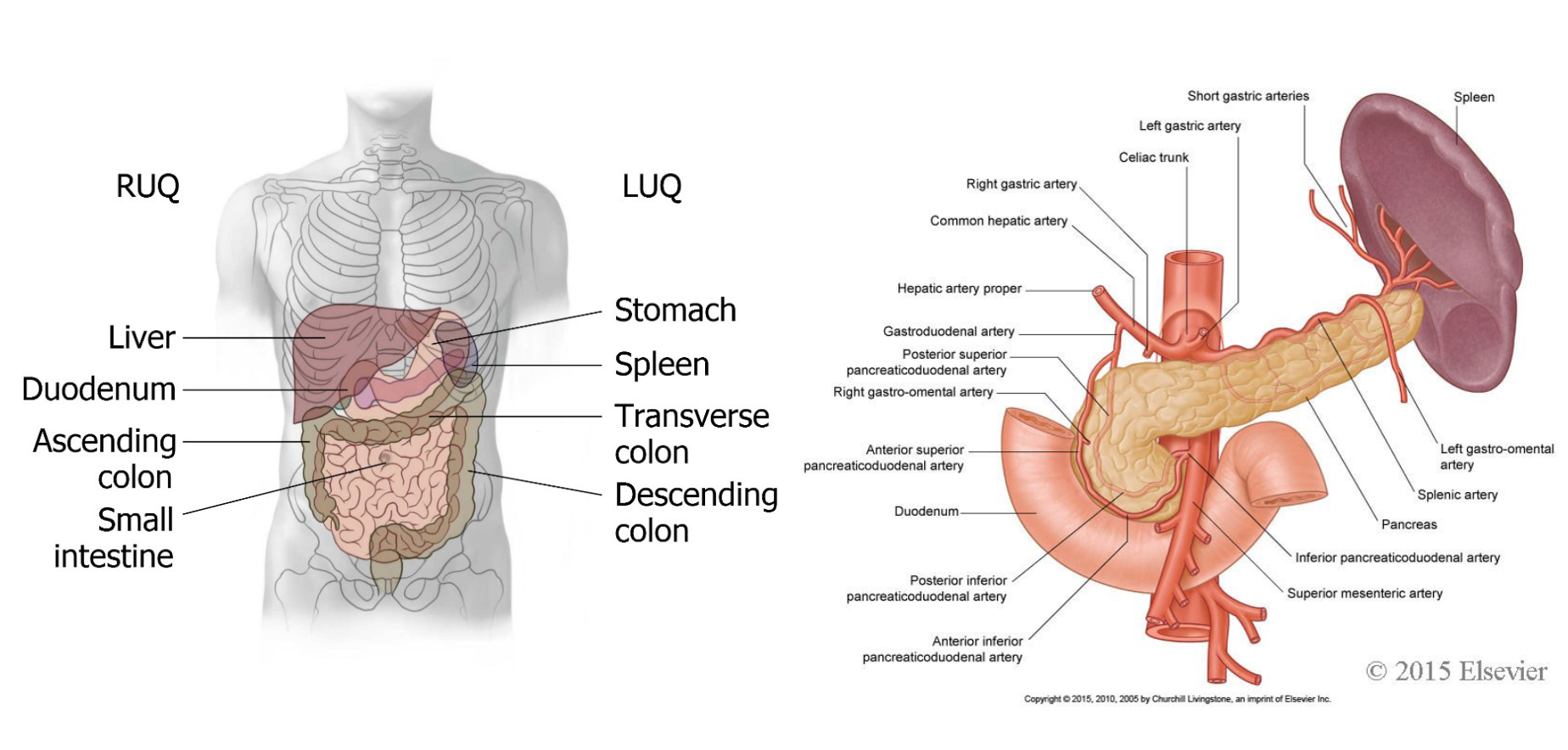
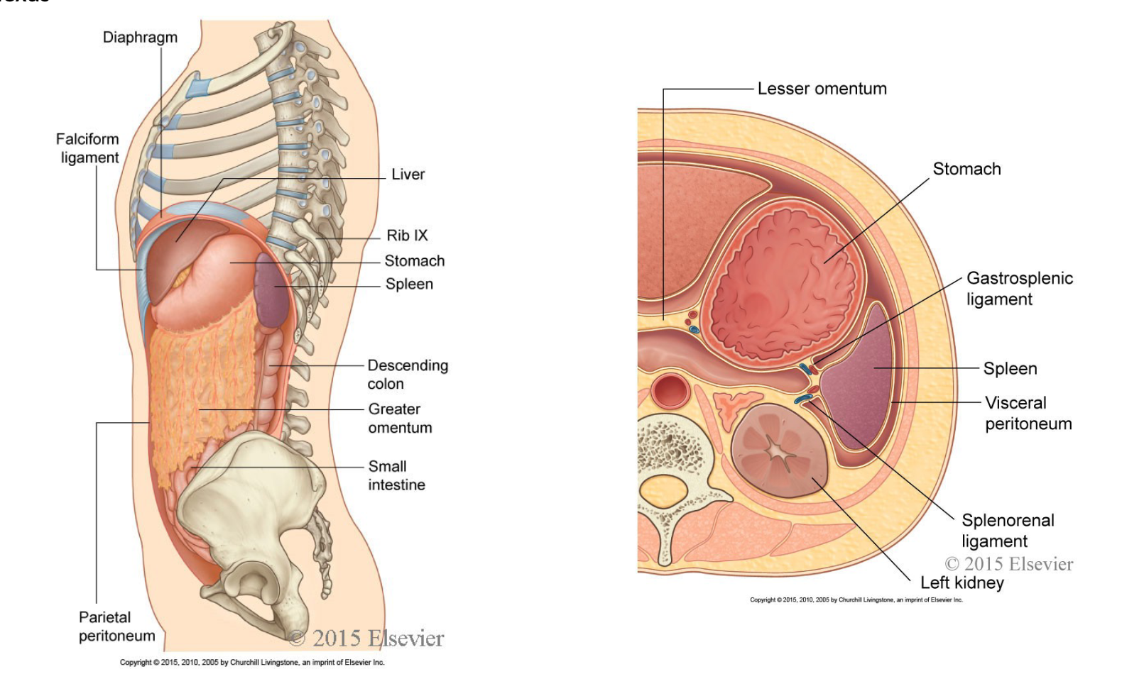
Intraperitoneal organ covered with visceral peritoneum, except at the hilum; non-vital organ located in the left upper quadrant (LUQ), beneath the diaphragm.
Accessory spleen presence in approximately 10% of people.
Protectively encased in a connective tissue capsule.
intraperitoneal organ, so all of its surfaces are covered with visceral peritoneum. Only the hilum of the spleen, the site through which the splenic artery and vein pass, is peritoneum-free attached to splenorenal and gastrosplenic ligaments
Structure
Has an anterior and posterior segment.
Size: Approximately 10-12 cm, weight 150-200 g.
Located between the fundus of the stomach and diaphragm, between the 9th and 11th ribs. left side of the abdomen, inferior to the diaphragm (left upper quadrant, left hypochondriac region)
Neurovascular: artery – splenic artery, Vein – splenic vein; lymph – celiac lymph node, innovation – celiac plexus
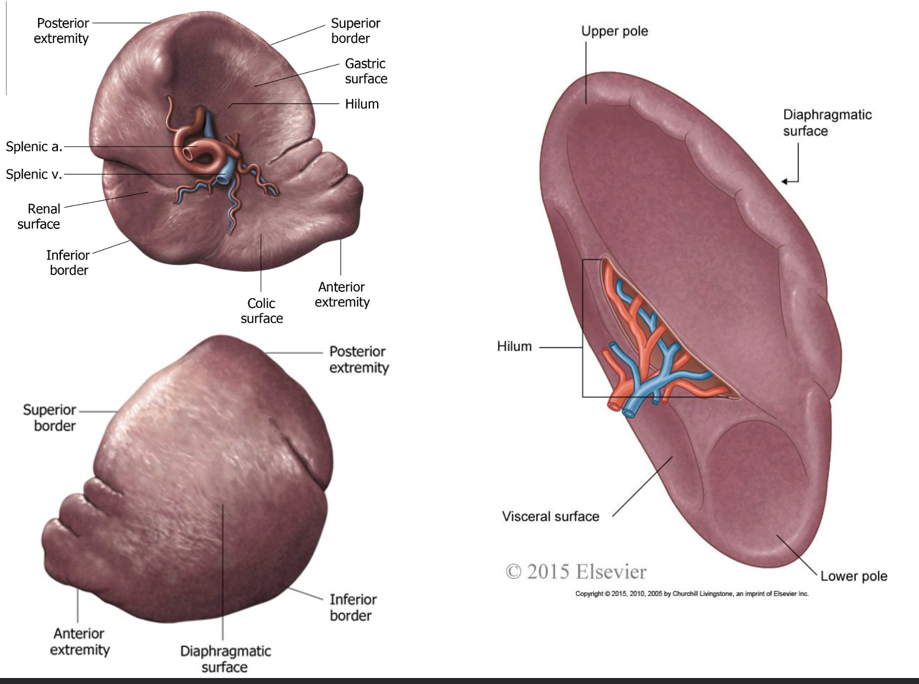
Borders and Surfaces
Borders: Superior, inferior, and intermediate, with superior border having an anterior notch.
Surfaces: Visceral (concave with impressions from surrounding organs) and diaphragmatic (convex).
Lobules
Subdivided into lobules, consisting of red pulp and white pulp.
Red pulp: filters blood, removes aged RBCs, and serves as a storage area for blood cells.
White pulp: involved in the immune response, particularly in the production of lymphocytes (B and T cells).
Functions
Blood filtration: removal of microbes and damaged RBCs.
Activation of immune response via WBC production and antibody synthesis.
RBC storage, especially during injury or blood loss.
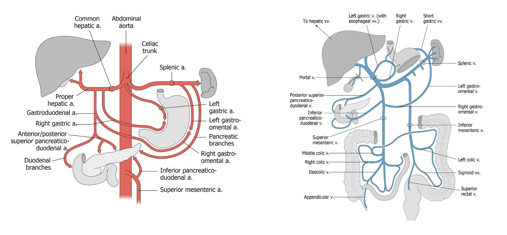
Blood Supply
Arterial Supply: Via the celiac trunk, specifically the splenic artery.
Venous Drainage: Through the splenic vein into the portal vein.
Lymphatic Drainage: Through celiac lymph nodes.
Innervation
Receives nerves from the celiac plexus.
Sympathetic Supply: splanchnic nerves T1 to L2; Parasympathetic: Vagus nerve (CN X).
Pancreas
Description
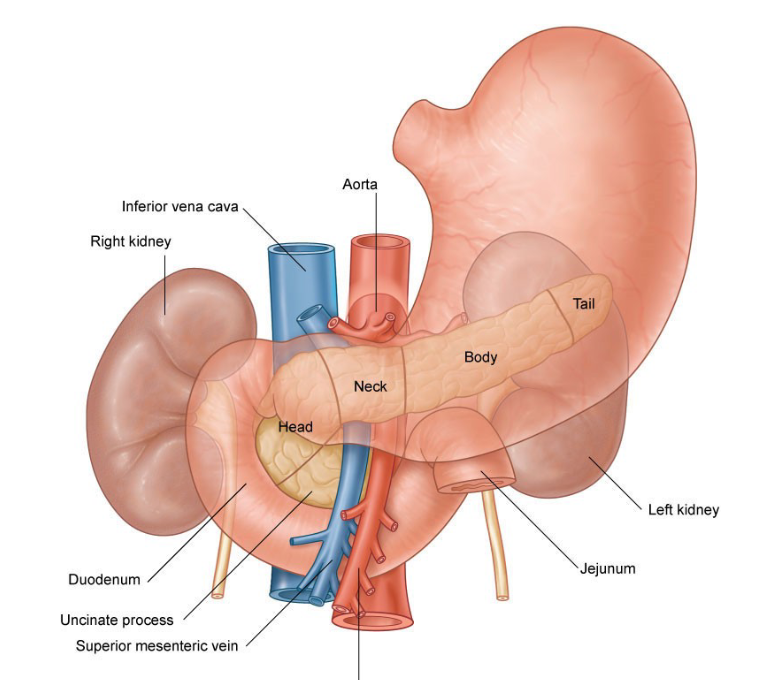
Located behind the stomach; functions both as an exocrine and endocrine gland, transpyloric plane
Consists of four parts: head, neck, body, and tail.
Exocrine Functions
Secretes pancreatic juices (containing enzymes like amylase and lipase) through the main pancreatic duct into the small intestine, aiding digestion.
Endocrine Functions
Secrete hormones (insulin and glucagon) from islets of Langerhans (cell type in pancreas) directly into the bloodstream, regulating blood glucose levels.
Insulin: Lowers blood glucose levels when high.
Glucagon: Raises blood glucose levels when low.
Structure
Main pancreatic duct merges with the bile duct to form the ampulla of Vater, regulated by sphincters to control the flow.
Blood Supply
Arterial Supply: Supplied by Splenic a via celiac trunk and pancreaticoduodenal via superior mesenteric artery
Venous Drainage: Portal vein via splenic vein and superior mesenteric vein
Lymphatic Drainage
Follows similar lymphatic pathways as the spleen; drains into pancreatic lymph nodes, celiac lymph nodes, and intestinal trunks.
• Pancreatic lymph nodes
• Celiac lymph nodes
• Intestinal trunks
Innervation
Receives autonomic innervation from the celiac and mesenteric plexuses.
Sympathetic: Splanchnic nerves; Parasympathetic: Vagus nerve (CN X).
Hormonal secretion primarily regulated by digestive hormones from the duodenum rather than direct neural input.