Cardiovascular System
REMEMBER!!!
pulmonary = to lungs, systemic = to the body, coronary= to the heart
diagrams DO NOT show your left and right → shows patients so everything is flipped
Coverings of the Heart
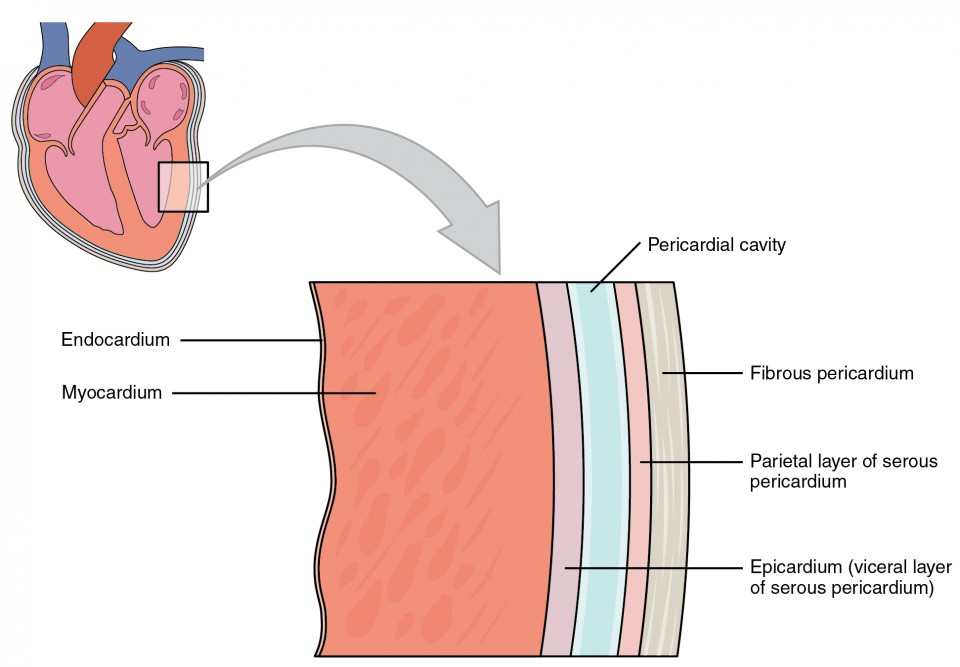
Acronym: FSPVME
Pericardium: sac that encloses heart composed of
fibrous pericardium
serous pericardium
visceral pericardium (epicardium)
parietal pericardium
fibrous pericardium (tough and dense connective tissue)
Pericardial cavity contains serous fluid to reduce friction
Wall of the Heart:
epicardium- connective tissue with epithelium
capillaries and nerve fibers, fat along coronary arteries and cardiac veins
myocardium- cardiac muscles that pumps blood out of heart chambers
endocardium- epithelium and underlying connective tissue
Purkinje Fibers
Specialized cardiac muscle fibers that conduct electrical impulses in the heart
Play a significant role in the coordinated contraction of heart chambers
Located in the inner ventricular walls of the heart to facilitate synchronized heartbeats.
Heart Chambers & Valves
Atria
upper chambers → atria (thin walls and receive blood returning)
auricles: ear like projections that extend from atrioventricular orifice and guarded by A-V valve
Ventricles
Valves of the Heart
tricuspid valve (right atrioventricular orifice) - prevents blood from moving from right ventricle into right atrium during ventricular contraction
pulmonary valve (entrance to pulmonary valve) - prevents blood from moving from pulmonary trunk into right ventricle during ventricular relaxation
mitral valve (left atrioventricular orifice) - prevents blood from moving from left ventricle into left atrium during ventricular contraction
aortic valve (entrance to aorta) - prevents blood from moving from aorta into left ventricle during ventricular relaxation)
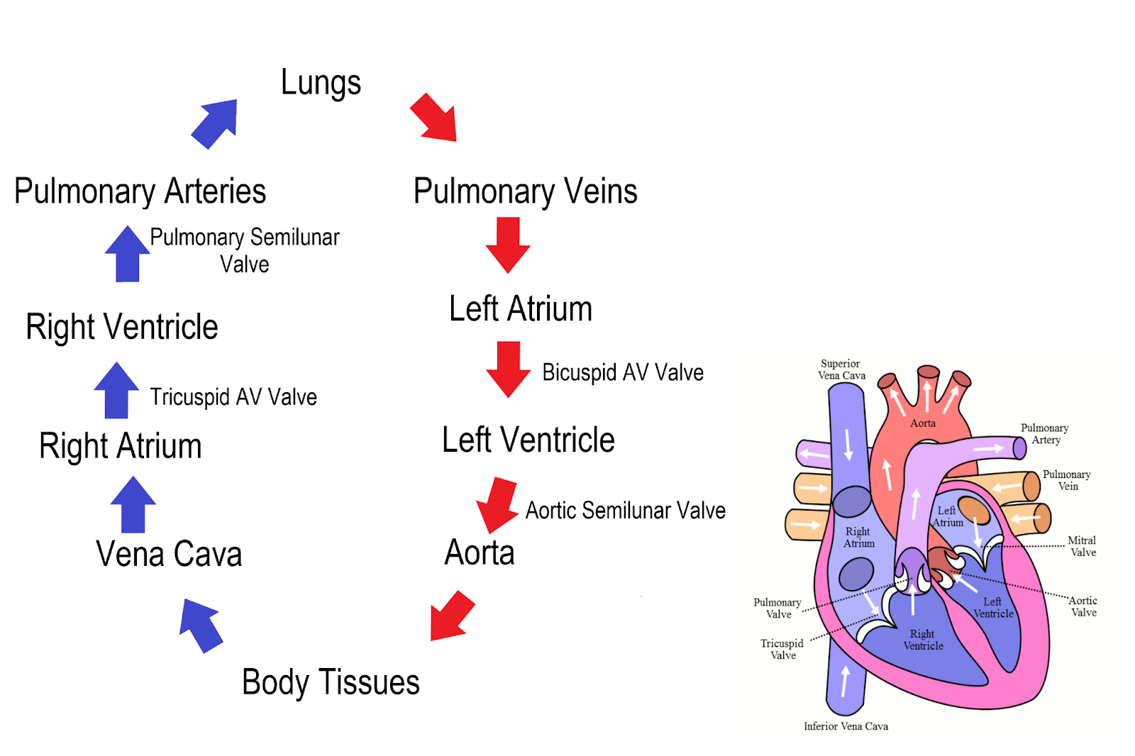
Blood Flow Circulation
deoxygenated blood enters right atrium through superior and inferior vena cava
blood passes tricuspid valve into right ventricle
right ventricle contracts and pushes through pulmonary valve
pulmonary arteries
pulmonary arteries then sends to lungs where blood receives oxygen
then goes through pulmonary veins back to left atrium
mitral (bicuspid ) valve opens for left atrium contraction and flows into left ventricle
left ventricle contracts and blood is pushed into aortic valve
oxygenated blood enters aorta and sent out to body
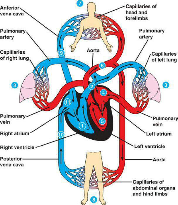
Blood Vessels
veins → to heart, arteries → to body
arteries and arterioles
strong and elastic
3 layers
Tunic Interna (innermost simple squamous with smooth surface)
Tunic Media (tube of smooth muscle fibers)
Tunica Extrema (thin connective tissue of collagen and elastin)'
capillaries
connects to arterioles (extension of inner lining) and venules
semipermeable layer allows exchange of oxygen, carbon dioxide, metabolites, and nutries
venules and veins
parralllels arteries back to heart
3 layers like arteries but
thinner middle layer
less elastic
lumen has greater diameters and contains flaplike valves to stop blood backing up
Heart Actions (Cardiac Cycle + Sounds)
What is a cardiac cycle versus blood circulation?
cardiac cycle : events that occur during one complete heartbeat
blood circulation: continuous movement of blood
systole: contraction
diastole: relaxation
atrial systole while ventricular diastole and ventricular systole while atrial diastole
heart sound: lubb - dupp, causes by vibrations of heart tissues and opening and closing of the valves
lubb- ventricular systole
dupp- ventricular diastole
murmur: abnormal sound and can be detected using a stethoscope
Cardiac Conduction System
cardiac conduction system: A network of specialized muscle cells is found in the heart's walls. These muscle cells send signals to the rest of the heart muscle causing a contraction
a "node" refers to a specialized area of tissue within the heart that generates and conducts electrical signals
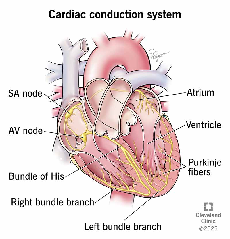
S-A Node (sinoatrial node)- small specialized cardiac muscle below epicardium in right atrium, near opening of superior vena cava
decrease in potassium permeability
over 80 times in a minute (rhythmic)
known as the “pacemaker”
A-V Node (atrioventricular node)- inferior portion of septum and beneath endocardium
(gatekeeper between atria and ventricles)
only normal conduction between atrial and ventricular syncytia
impluse delayed as moved away from A-V node
A-V Bundle (aka. Bundle of His)- large fibers and divides right and left bundle branches
Purkinje Fibers- carry impluses to distant regions
Electrocardiograms, Pulse Rate, and Blood Pressure
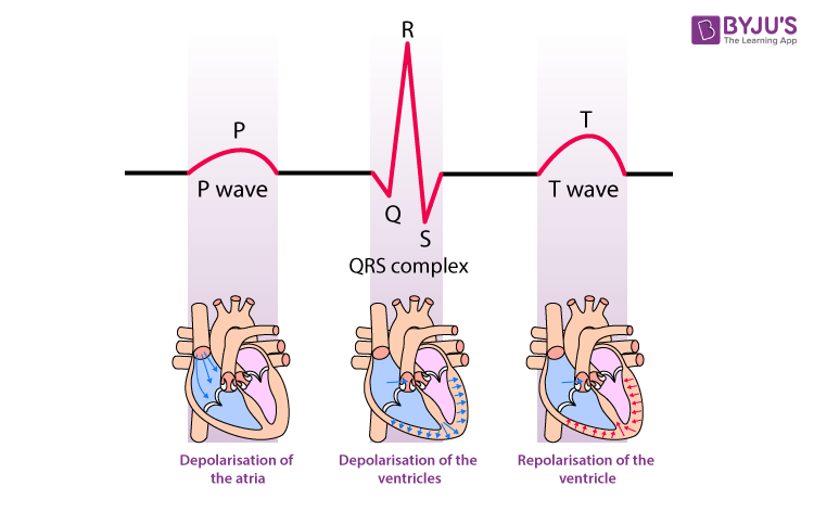
Electrocardiogram
an electrocardiogram is a recording of the electrical changes that occur in the myocardium during a cardiac cycle using electrodes placed on skin
How it works?
the S-A node triggers a cardiac impulse
the atrial fibers depolarize → produces electrical change
pen moves producing a P wave → represents depolarization of atrial fibers that will lead to contraction of the atria
when reaches ventricular fibers, depolarization of ventricular fibers→ QRS complex
depolarization of ventricular walls (contraction) → T wave
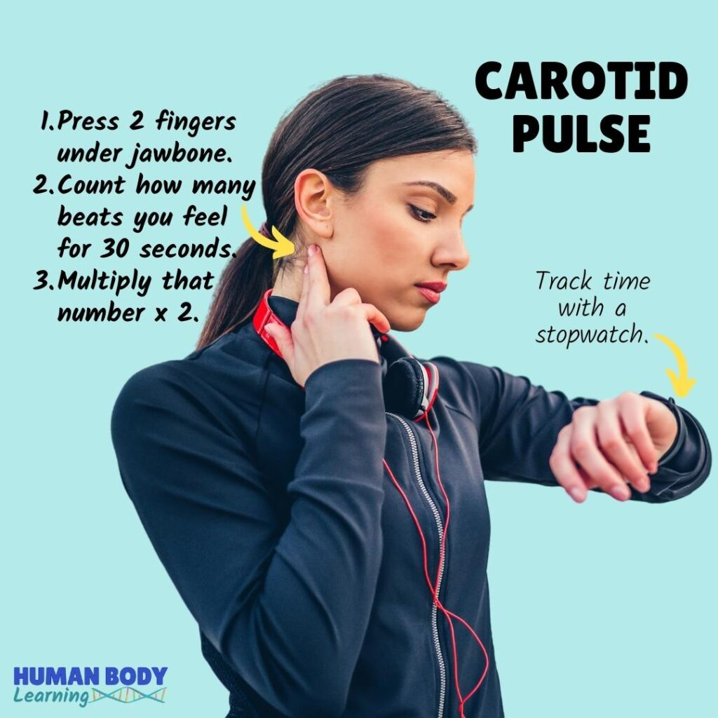
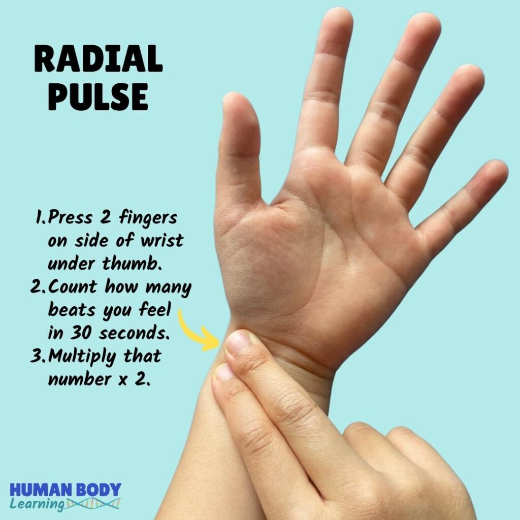
Lab on Pulse and Blood Pressure
blood pressure is the force exerted against the walls of the arteries
blood pressure refers too systemic arterial pressure
maximum pressure achieved during ventricular contraction is called systolic pressure
lowest pressure that remains in arterial system during ventricular relaxation is called diastolic pressure
a pulse is caused by the closing of the aortic valve
the brachial artery in the arm is the standard systemic artery in which we measure blood pressure
the first sound of the cardiac cycle occurs when the atrioventricular valves are closing
the second sound of the cardiac cycle occurs when the semilunar valves are closing
Reading blood pressure:
top number - systolic pressure (contraction)
lower number - diastolic pressure (relaxation)
Heart Issues & Concerns
Endocarditis- inflammation of the endocardium
Hypertension- high blood pressure
potential
causes: kidney disease, sodium, obesity, stress, arteriosclerosis, essential, primary or idiopathic hypertension
enlargement and weakening of heart due to increased leftt venricle pumping
Atherosclerosis- build up of fats or cholesterol around artery walls
can be developed from coronary thrombosis, coronary embolism,s troke
Extra Quizizz Review!!
blood pressure is lowest in vena cava
the valve between the left ventricle and aorta is the aortic
capillaries is smallest vessel
EKG= PQRST
coronary is the circulation that brings blood from aorta to myocardium and back to right atrium through coronary sinus
ventricular fibrillation refers irregular contraction of the ventricle, heartbeat fast and ineffective heart rhythm that may result in death
tachycardia refers to heart rate that is fast
heat may cause vasodilation
angina is spasm of chest pain due to decrease in blood flow to myocardium
medical term for heart attack is myocardial infraction
renin regulated blood