HA Test 2 prep
Bone shapes, Components and Markings
The skeletal system
Consists of bones and associates cartilage connected to form articulations (joints)
The axial skeleton
80 bones that form the
Sull
Vertebral column
Ribs
Sternum
Forms the longitudinal axis of the body and protects the brain, spinal cord and organs in the thorax
The appendicular skeleton
Bones of the upper and lower limbs
Bone
Definition: bone is a calcified living connective tissue that forms the majority of the skeleton
Compact = dense bone that forms the outer shell of all bones and surrounds spongy
Bone
Spongy = highly pours (this is the spongy appearance)
Classifications of bones
Long bones | A shaft with two distinct ends Shaft = Diaphysis Ends = Epiphysis Epiphyseal growth plate = Made of cartilage | Femur Humerus
|
Irregular bone | Have weird shapes | Vertebra Face bones |
Short bones | Squar in shape | Navicular |
Flat bones | Two thin pieces of compact bone with a thin layer of spongy bone in-between | Sternum Skull
|
Bones have 3 landmarks
Articular surfaces
Areas of the bone that will form a joint
Usually smooth and covered with hyaline cartilage
Protrusions
Boney landmarks that stick out
Function as bony landmarks for tendons and ligaments to attach to
Depressions (holes)
Boney landmarks that provide space for another structure like blood vessels or nerves
Bone Development
Ossification = A process of bone formation
Bone of the skeleton develop in two methods
Endochondral ossification
Hyaline cartilage turning into bone
Long bone, short bones and most irregular bones
Intramembranous ossification
Bone develops withing a membrane
Flat bones
Bones grow in length
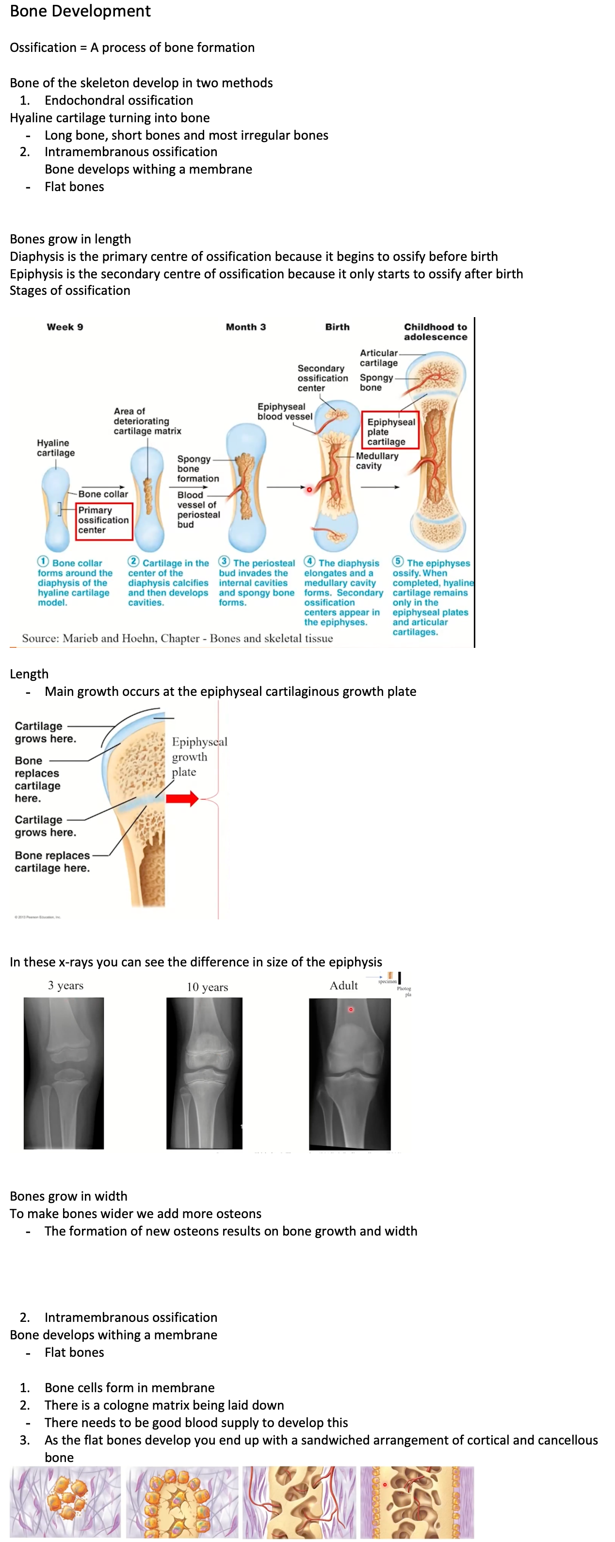
Diaphysis is the primary centre of ossification because it begins to ossify before birth
Epiphysis is the secondary centre of ossification because it only starts to ossify after birth
Stages of ossification
Length
Main growth occurs at the epiphyseal cartilaginous growth plate
In these x-rays you can see the difference in size of the epiphysis
Bones grow in width
To make bones wider we add more osteons
The formation of new osteons results on bone growth and width
Intramembranous ossification
Bone develops withing a membrane
Flat bones
Bone cells form in membrane
There is a cologne matrix being laid down
There needs to be good blood supply to develop this
As the flat bones develop you end up with a sandwiched arrangement of cortical and cancellous bone
Bone Remodelling
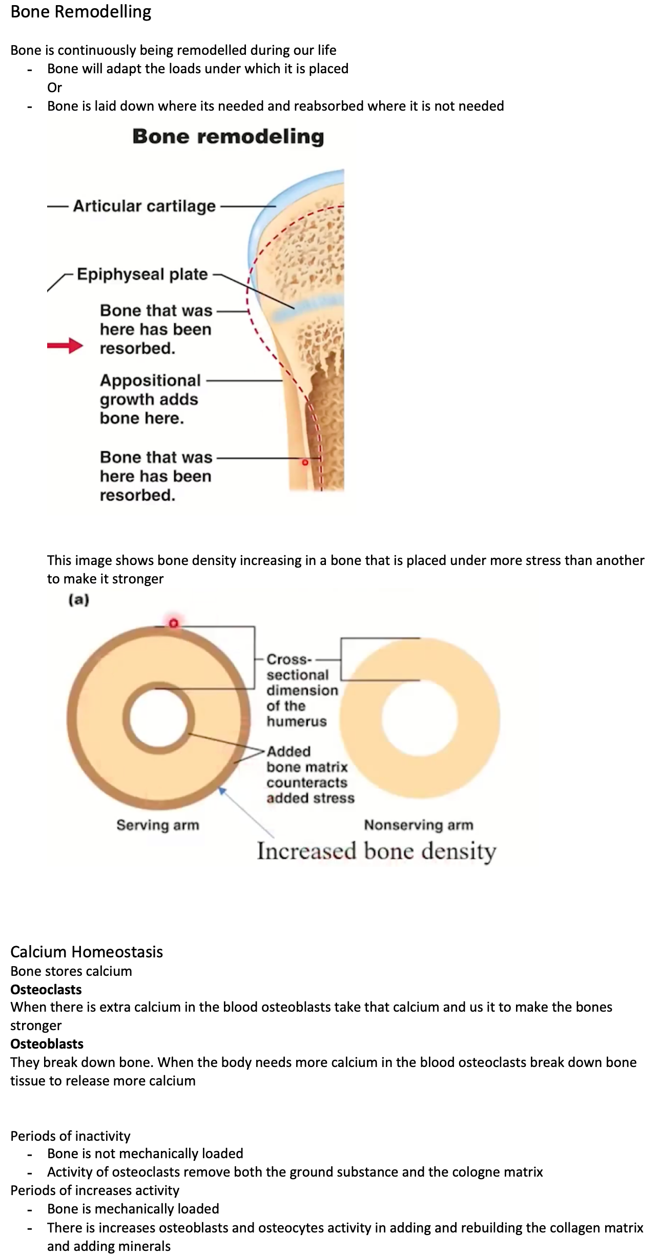
Bone is continuously being remodelled during our life
Bone will adapt the loads under which it is placed
Or
Bone is laid down where its needed and reabsorbed where it is not needed
This image shows bone density increasing in a bone that is placed under more stress than another to make it stronger
Calcium Homeostasis
Bone stores calcium
Osteoclasts
When there is extra calcium in the blood osteoblasts take that calcium and us it to make the bones stronger
Osteoblasts
They break down bone. When the body needs more calcium in the blood osteoclasts break down bone tissue to release more calcium
Periods of inactivity
Bone is not mechanically loaded
Activity of osteoclasts remove both the ground substance and the cologne matrix
Periods of increases activity
Bone is mechanically loaded
There is increases osteoblasts and osteocytes activity in adding and rebuilding the collagen matrix and adding minerals
Bone Fracture and Repair
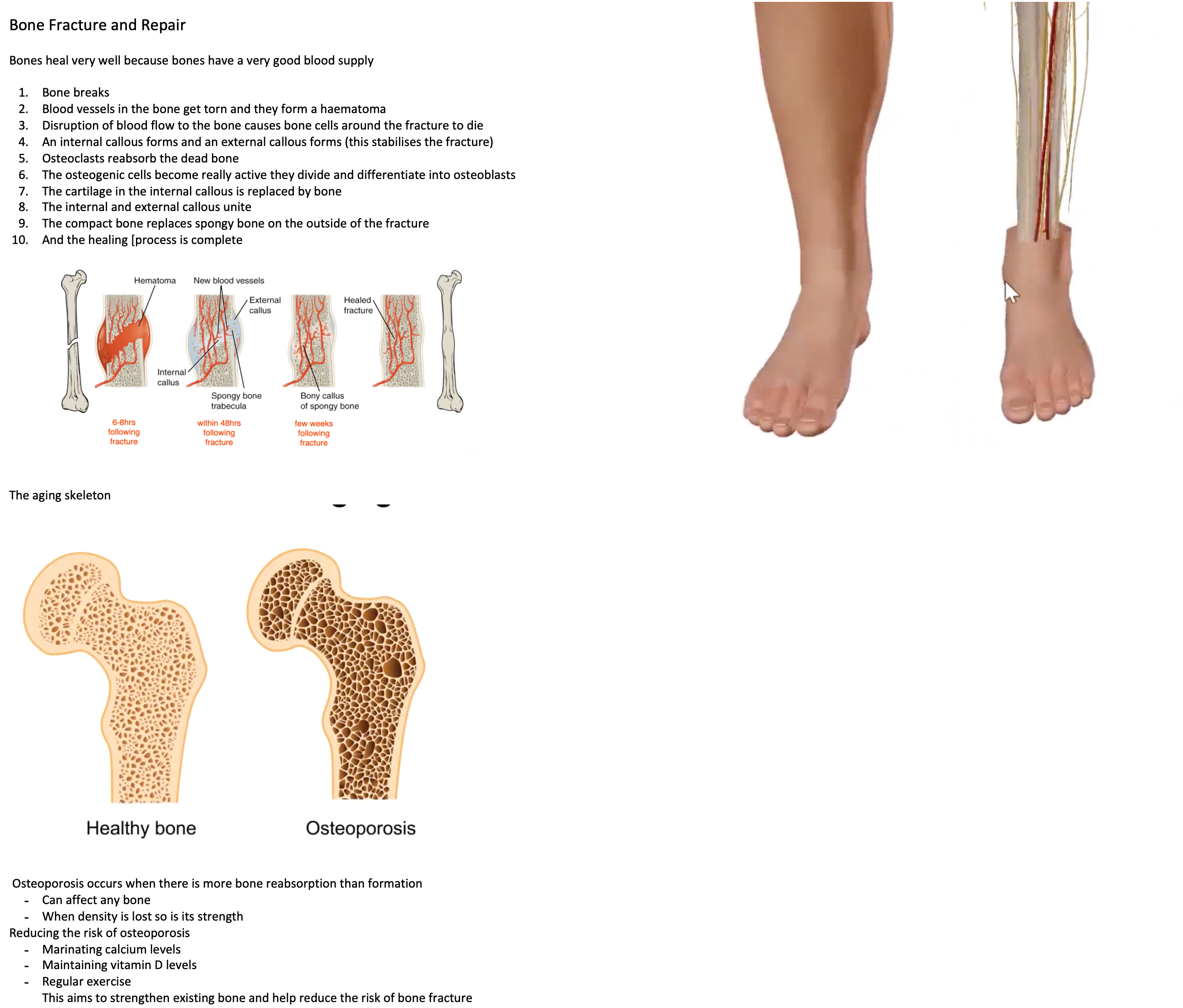
Bones heal very well because bones have a very good blood supply
Bone breaks
Blood vessels in the bone get torn and they form a haematoma
Disruption of blood flow to the bone causes bone cells around the fracture to die
An internal callous forms and an external callous forms (this stabilises the fracture)
Osteoclasts reabsorb the dead bone
The osteogenic cells become really active they divide and differentiate into osteoblasts
The cartilage in the internal callous is replaced by bone
The internal and external callous unite
The compact bone replaces spongy bone on the outside of the fracture
And the healing [process is complete
The aging skeleton
Osteoporosis occurs when there is more bone reabsorption than formation
Can affect any bone
When density is lost so is its strength
Reducing the risk of osteoporosis
Marinating calcium levels
Maintaining vitamin D levels
Regular exercise
This aims to strengthen existing bone and help reduce the risk of bone fracture
Surface Anatomy - Bones and Bony Landmarks
The mastoid process
A bony landmark behind the ear
Medial aspect of the clavicle (Collar bone)
How to find it
The notch is at the front and bottom of my neck
(anatomical terminology) Palpate the notch at the medial, anterior and inferior aspect of the neck. The medial aspect of the clavicle sits on either side of this notch.
Medial epicondyle
How to find it
On the inside of the elbow joint
(anatomical terminology) Palpate the distal end of the humerus on the medial side
Anterior superior iliac spine
How to find it
The upper part of the pelvis on the side
(anatomical terminology) Palpate the lateral side of the pelvis to its most anterior point
In-between these landmarks is the appendix
Patella
How to find it
To the outside of the knee cap
(anatomical terminology) Palpate the anterior distal aspect to the thigh. Palpate laterally to the inferior part of the patella. This is the head of the fibula
Or
Proximal aspect of the fibula found laterally
Medial malleolus
How to find it
In the inside of the ankle
(anatomical terminology) Palpate the distal aspect of the tibia on the most medial side

Articulate = When bones join with each other
Articulations = Joints
Movement always occurs at a joint
Joints can be classified by the type of connective tissue that unites them. This will have a direct correlation to how mobile they will be.
Immobile
Partly mobile
Highly Mobile
We have 3 different histological joint classifications
Fibrous joints
Held together by fibrous connective tissue
Little to no movement
There are 3 types of fibrous tissue
Sutures
Gomphoses
Syndesmoses
Cartilaginous joints
United by a pad or disc of cartilage
A little bit of movement
2 types
Primary cartilaginous joints
United by hyaline cartilage
Secondary cartilaginous joints
Thin layer of hyaline cartilage over the articulating bone surfaces and then a pad or dics of fibro cartilage in between
Synovial joints
Features
Boney components are indirectly connected to one another by a joint casual that encapsules the joint
Lining the inner surface of this casual is the synovial membrane that produces synovial fluid
The ends of the bones are also covered in hyaline cartilage
Functional joint classifications
Hinge - one plane
Pivot - one plane
Condyloid
Saddle
Plane or gliding
Ball and socket
The shape of the joint will also determine how many axis the joint can move around
Joints that only move in one plane = uniaxial and include a hinge joint
Joints that move in 2 planes = biaxial and include a condyloid joint
Joints with the most movement = triaxial or Multiaxial
Simple joints
Function for stability
Complex joints
Function to achieve mobility

Locating joints using surface anatomy
What is it? | Where is it | Photo |
Sternoclavicular | The medial end of the clavicle |
|
Acromioclavicular | Lateral end of the clavicle |
|
Elbow superior Radioulnar joint
Inferior radioulnar joint | Feel for the medial and lateral epicondyle Move distally from the lateral epicondyle and there will be a space before another bony prominent (the head of the radius) The head of the radius articulates with the ulnar |
|
Metacarpophalangeal | Metacarpal bones articulate with our phalanges Feel the knuckle while you flex and extend |
|
Hip | The head of the femur articulating with the hip bone (ball and socket joint)
Locate the Anterior superior iliac spine Move inferiorly and locate ethe hip joint
Locate the greater trochanter Move medially and locate the hip joint |
|
Knee | Located between the tibia and the femur
Feel for a gap between the bumpy landmarks of the distal femur and the proximal tibia
|
|
Ankle | Locate the lateral malleolus and move medially and feel a gap where the ankle joint is found
|
|
1st metatarsal | Find the mesotarsal bone on the medial side of the foot Distally to that there will be a bony landmark and even more distally to that there will be a gap that is the joint |
|
Manubriosternal 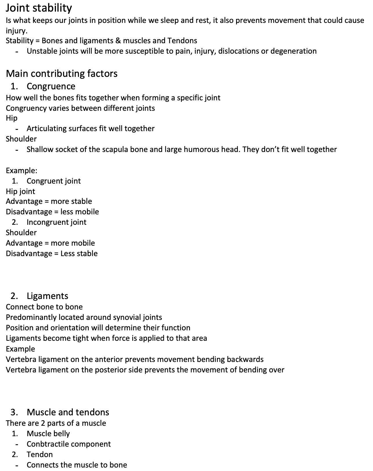 | Find the manubrial. Inferiorly to that and find a small prominence identifying the Manubriosternal joint |
|
Intervertebral joints | locate the spina's process (posterior bony landmark)
|
|
Joint stability
Is what keeps our joints in position while we sleep and rest, it also prevents movement that could cause injury.
Stability = Bones and ligaments & muscles and Tendons
Unstable joints will be more susceptible to pain, injury, dislocations or degeneration
Main contributing factors
Congruence
How well the bones fits together when forming a specific joint
Congruency varies between different joints
Hip
Articulating surfaces fit well together
Shoulder
Shallow socket of the scapula bone and large humorous head. They don’t fit well together
Example:
Congruent joint
Hip joint
Advantage = more stable
Disadvantage = less mobile
Incongruent joint
Shoulder
Advantage = more mobile
Disadvantage = Less stable
Ligaments
Connect bone to bone
Predominantly located around synovial joints
Position and orientation will determine their function
Ligaments become tight when force is applied to that area
Example
Vertebra ligament on the anterior prevents movement bending backwards
Vertebra ligament on the posterior side prevents the movement of bending over
Muscle and tendons
There are 2 parts of a muscle
Muscle belly
Conbtractile component
Tendon
Connects the muscle to bone
Skeletal muscle / Voluntary muscle functions
Produce movement
Maintain posture
Stabilise joints
Generate heat
Protect organs
Connect bone to bones with tendons and they cross joints to produce movement
With an exception of facial muscles which attach bones to skin (wrinkles)
2 principles that determine how muscles work
A muscle must cross a joint in order to act at that joint
The position of a muscle at a joint determines the plane and direction of movement it can produce
To see what plane a movement will be in you just need to see where the muscle is crossing the joint of interest and then we can work out the action
The muscle compartment rule
Muscles that have a common function at a joint are usually located together in a muscle compartment, innervated by the same nerve and share the same arterial supply and venous drainage
How to apply this?
Lower limb example:
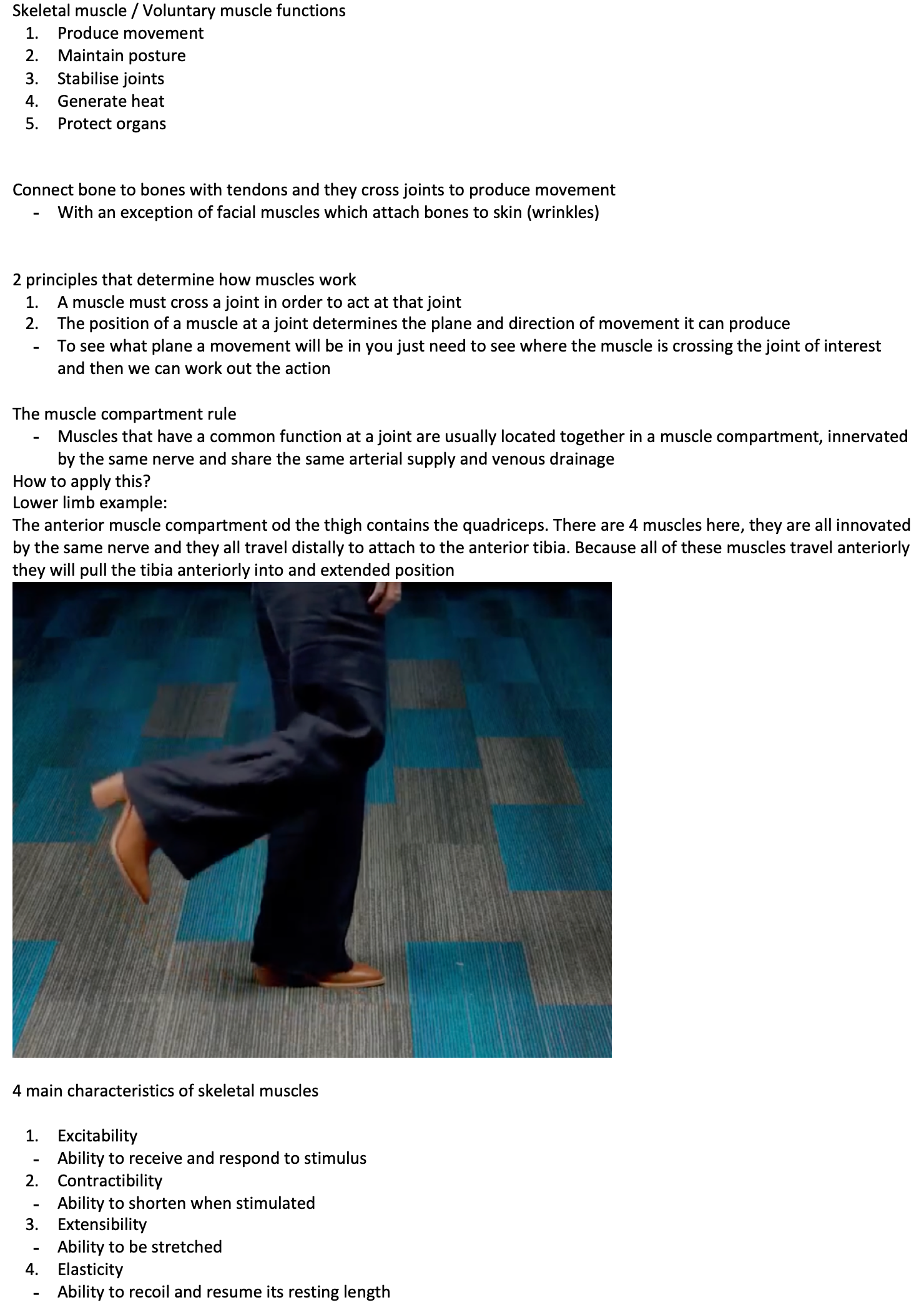
The anterior muscle compartment od the thigh contains the quadriceps. There are 4 muscles here, they are all innovated by the same nerve and they all travel distally to attach to the anterior tibia. Because all of these muscles travel anteriorly they will pull the tibia anteriorly into and extended position
4 main characteristics of skeletal muscles
Excitability
Ability to receive and respond to stimulus
Contractibility
Ability to shorten when stimulated
Extensibility
Ability to be stretched
Elasticity
Ability to recoil and resume its resting length
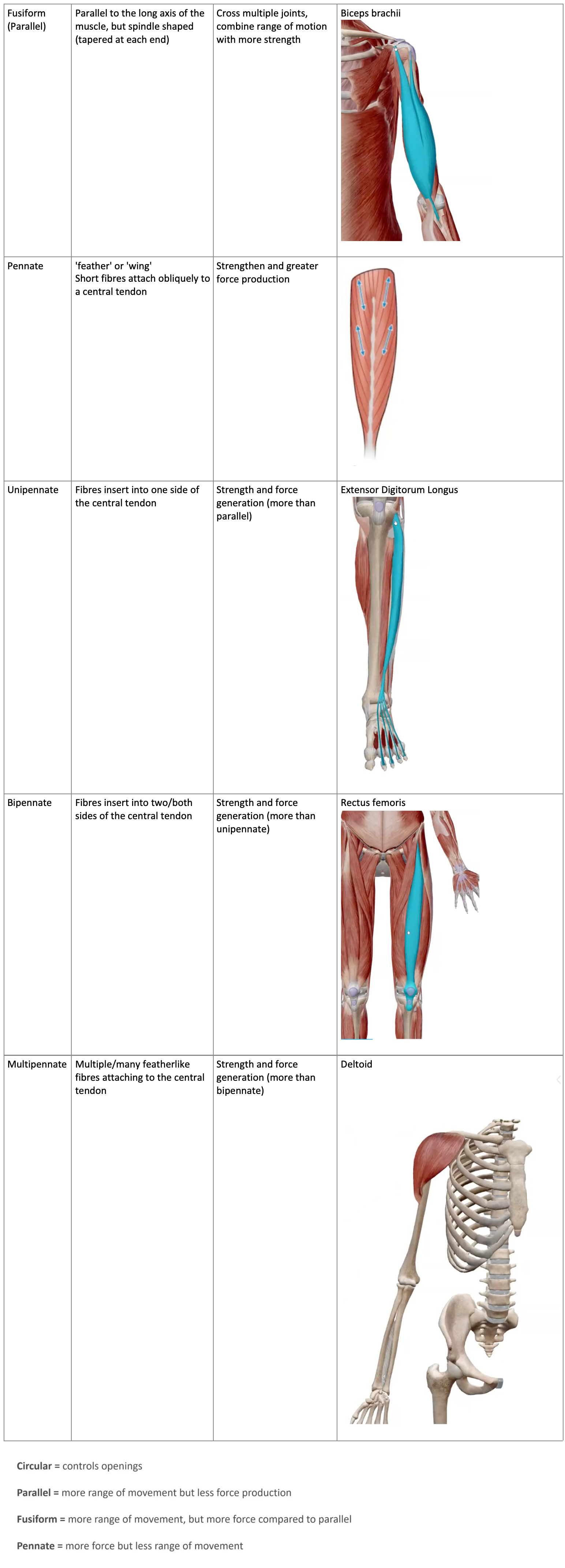
Fusiform (Parallel) | Parallel to the long axis of the muscle, but spindle shaped (tapered at each end) | Cross multiple joints, combine range of motion with more strength | Biceps brachii
|
Pennate | 'feather' or 'wing' Short fibres attach obliquely to a central tendon | Strengthen and greater force production |
|
Unipennate | Fibres insert into one side of the central tendon | Strength and force generation (more than parallel) | Extensor Digitorum Longus
|
Bipennate | Fibres insert into two/both sides of the central tendon | Strength and force generation (more than unipennate) | Rectus femoris
|
Multipennate | Multiple/many featherlike fibres attaching to the central tendon | Strength and force generation (more than bipennate) | Deltoid
|
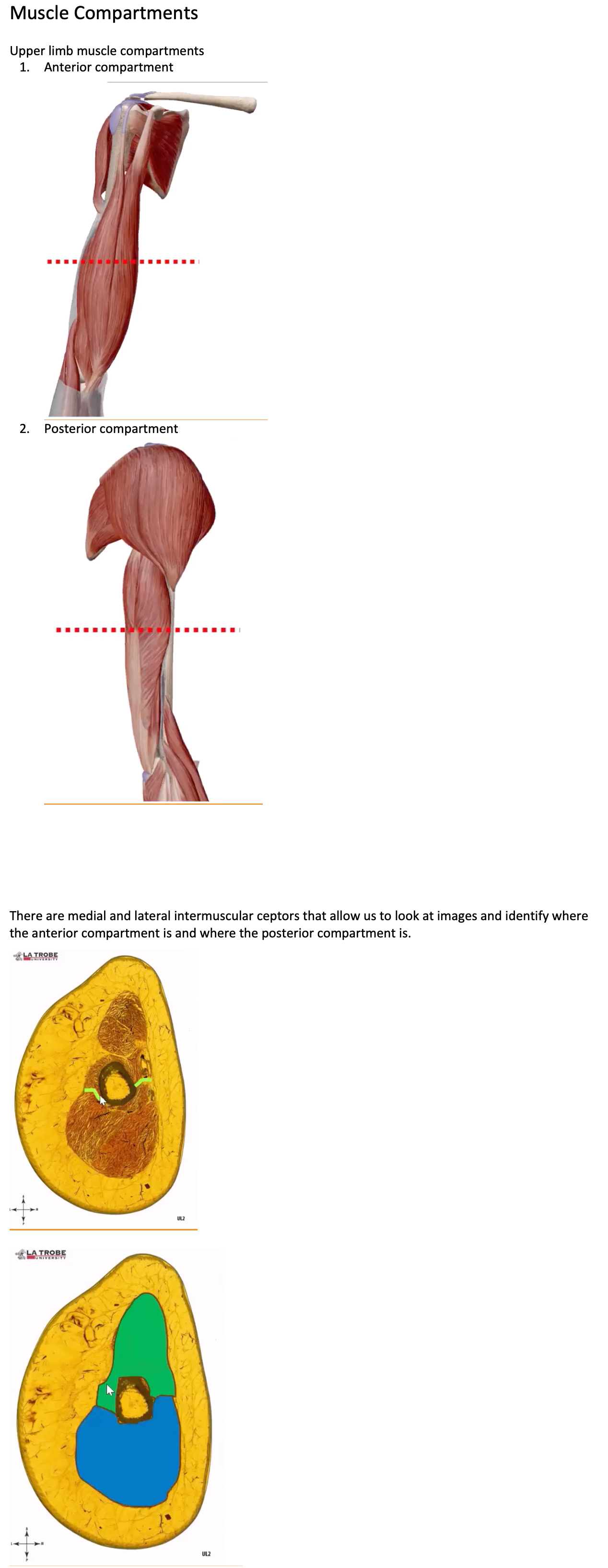
Muscle Compartments
Upper limb muscle compartments
Anterior compartment
Posterior compartment
There are medial and lateral intermuscular ceptors that allow us to look at images and identify where the anterior compartment is and where the posterior compartment is.
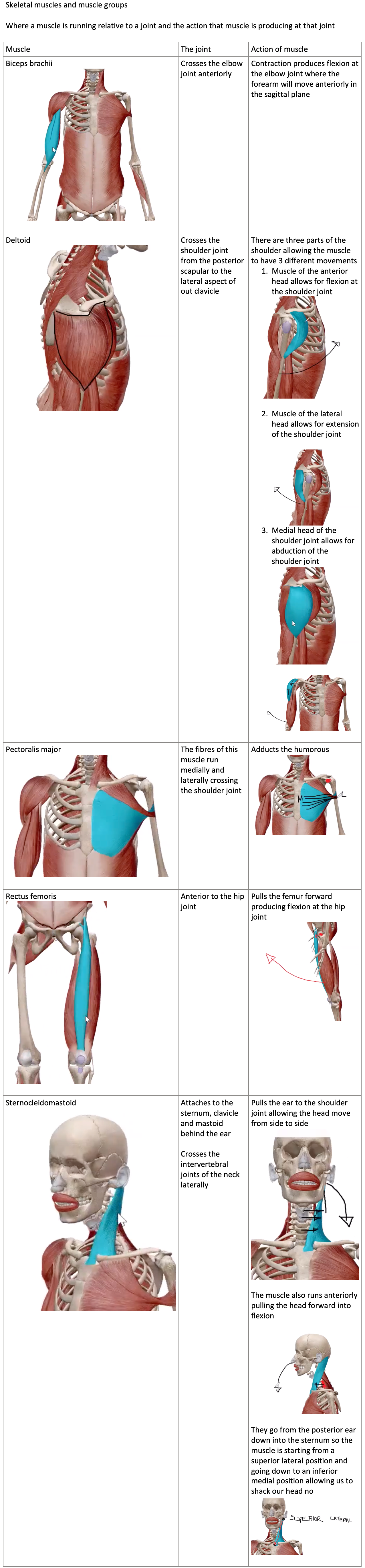
Skeletal muscles and muscle groups
Where a muscle is running relative to a joint and the action that muscle is producing at that joint
Muscle | The joint | Action of muscle |
Biceps brachii
| Crosses the elbow joint anteriorly
| Contraction produces flexion at the elbow joint where the forearm will move anteriorly in the sagittal plane |
Deltoid
| Crosses the shoulder joint from the posterior scapular to the lateral aspect of out clavicle | There are three parts of the shoulder allowing the muscle to have 3 different movements
|
Pectoralis major
| The fibres of this muscle run medially and laterally crossing the shoulder joint | Adducts the humorous
|
Rectus femoris
| Anterior to the hip joint | Pulls the femur forward producing flexion at the hip joint
|
Sternocleidomastoid
| Attaches to the sternum, clavicle and mastoid behind the ear
Crosses the intervertebral joints of the neck laterally | Pulls the ear to the shoulder joint allowing the head move from side to side
The muscle also runs anteriorly pulling the head forward into flexion
They go from the posterior ear down into the sternum so the muscle is starting from a superior lateral position and going down to an inferior medial position allowing us to shack our head no
|
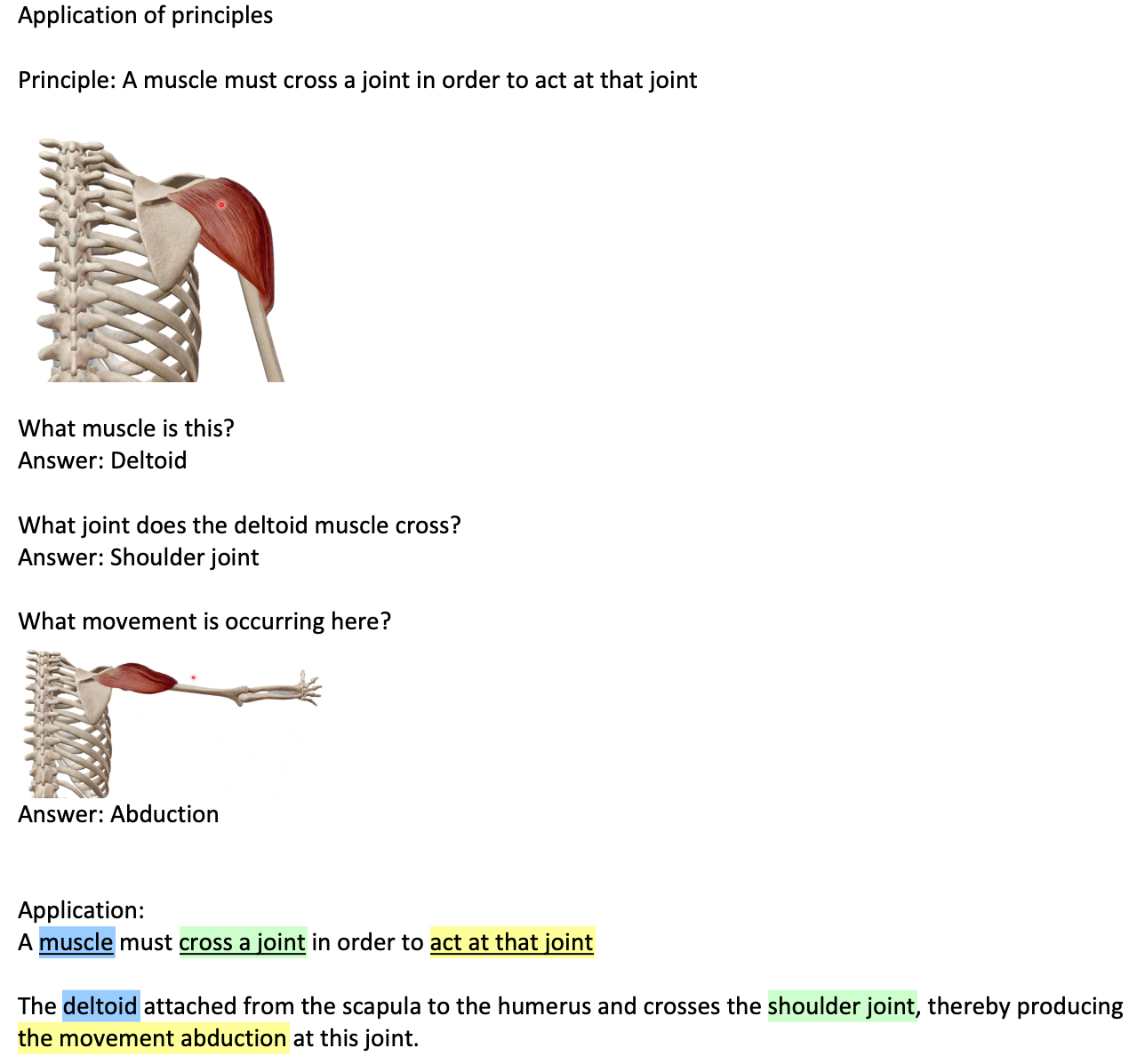
Application of principles
Principle: A muscle must cross a joint in order to act at that joint
What muscle is this?
Answer: Deltoid
What joint does the deltoid muscle cross?
Answer: Shoulder joint
What movement is occurring here?
Answer: Abduction
Application:
A muscle must cross a joint in order to act at that joint
The deltoid attached from the scapula to the humerus and crosses the shoulder joint, thereby producing the movement abduction at this joint.
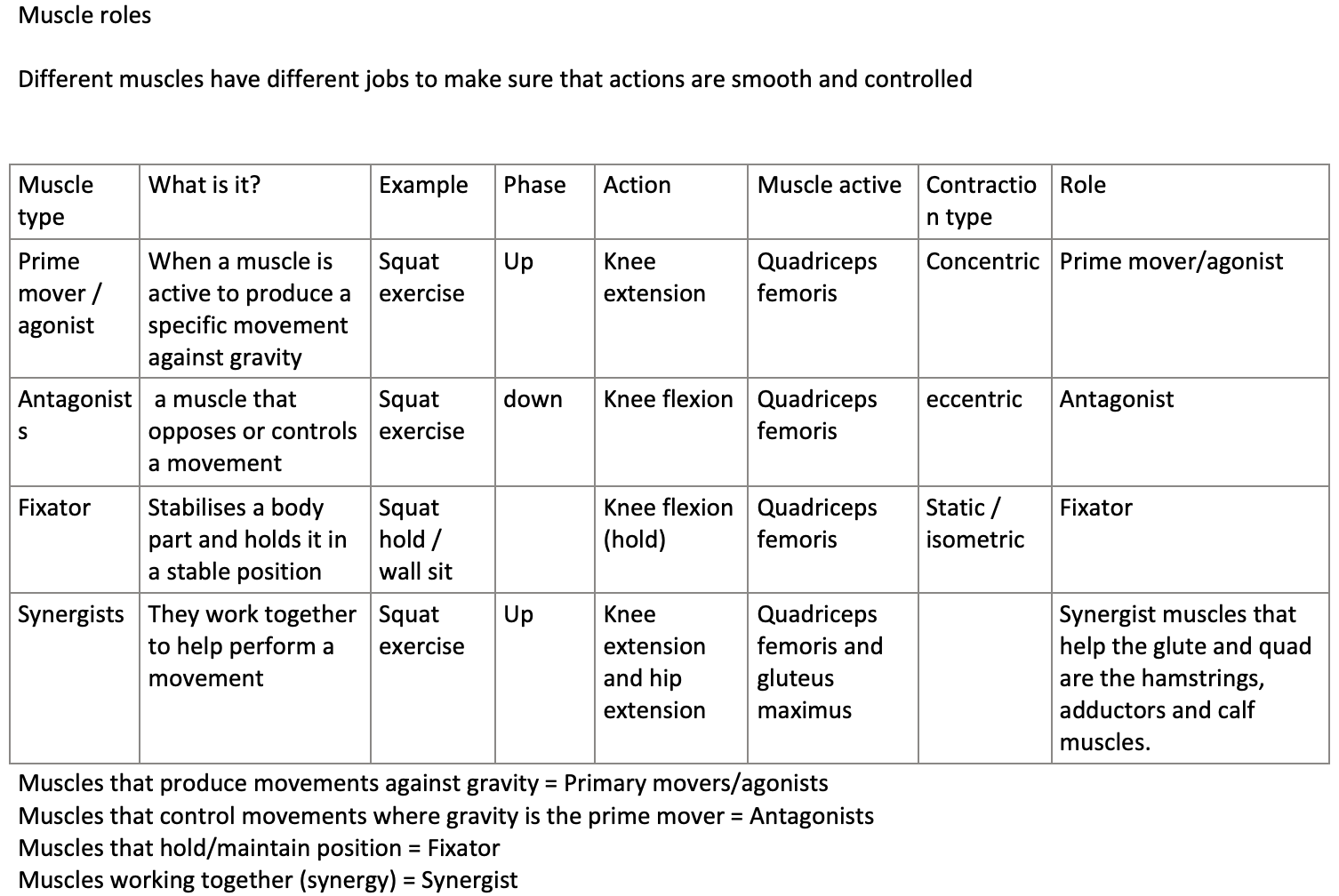
Muscle roles
Different muscles have different jobs to make sure that actions are smooth and controlled
Muscle type | What is it? | Example | Phase
| Action | Muscle active | Contraction type | Role |
Prime mover / agonist | When a muscle is active to produce a specific movement against gravity | Squat exercise | Up | Knee extension | Quadriceps femoris | Concentric | Prime mover/agonist |
Antagonists | a muscle that opposes or controls a movement | Squat exercise | down | Knee flexion | Quadriceps femoris | eccentric | Antagonist |
Fixator | Stabilises a body part and holds it in a stable position | Squat hold / wall sit |
| Knee flexion (hold) | Quadriceps femoris | Static / isometric | Fixator |
Synergists | They work together to help perform a movement | Squat exercise | Up | Knee extension and hip extension | Quadriceps femoris and gluteus maximus |
| Synergist muscles that help the glute and quad are the hamstrings, adductors and calf muscles. |
Muscles that produce movements against gravity = Primary movers/agonists
Muscles that control movements where gravity is the prime mover = Antagonists
Muscles that hold/maintain position = Fixator
Muscles working together (synergy) = Synergist
Types of muscle contractions
Muscle tension
Force generated by muscle contracting
Load does not need to move necessarily
Isotonic contraction
Where tension in the muscle remains constant
What changes
The length of muscle either shorter or longer
Eccentric = longer
Concentric = shorter
Isometric
When muscles produce tension without changing the length of the joint or the length oof the muscle
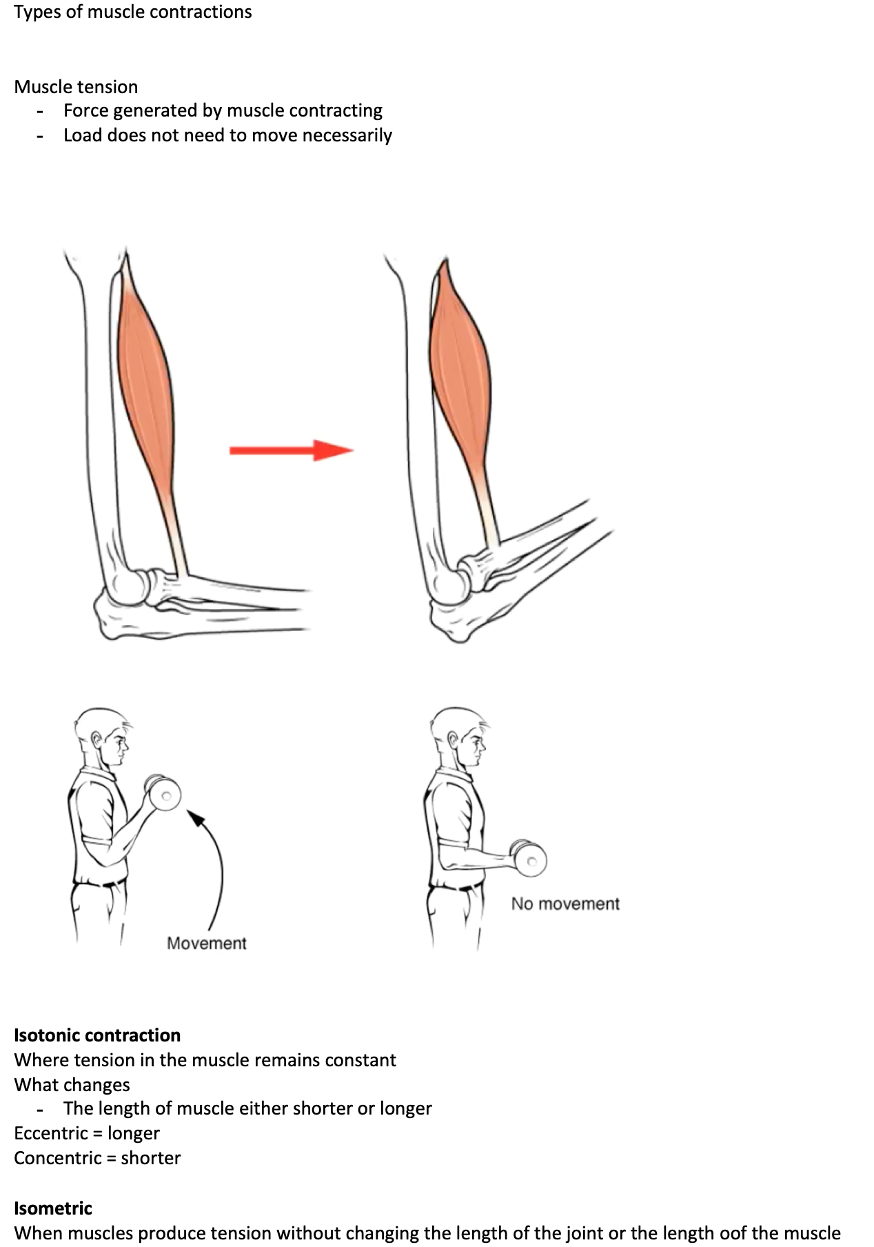
Opened and closed chain movements
When we move our bodies our limbs interact with the environment in different ways. Sometimes our hand is free to move through space and other times it is fixed against a surface.
Open chained movements
When the distal part of the body is free to move while the proximal part is fixed
Closed chain movements
When the distal end of a body segment is fixed and the proximal end of the body if free to move
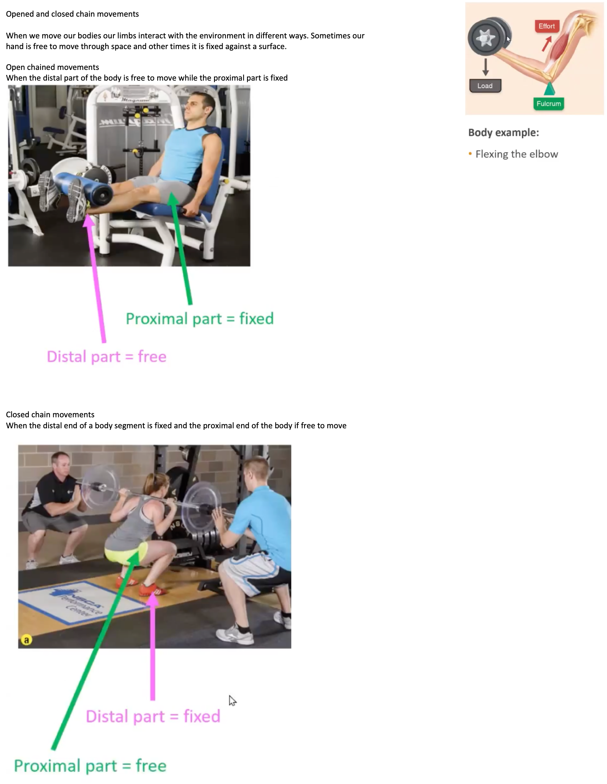
Levers
A lever is a rigid bar that pivots on a fulcrum when force is applied. In the body, joints serve as fulcrums and bones act as levers. Muscles contract to create effort at their insertion points, moving the load—which includes the bone, tissues, and any object being lifted.
First class lever
Arrangement
Effort applied at one end
Load at the other end
Fulcrum can sit anywhere in-between
Second class lever
Arrangement
Effort applies at one end
Fulcrum is at the other end
Load sits anywhere in between
Third class lever
Arrangement
Load at one end
Fulcrum is at the other end
Effort is applied anywhere in between
