Lesson 14: Coronary Circulation
Lesson 14: Coronary Circulation Overview
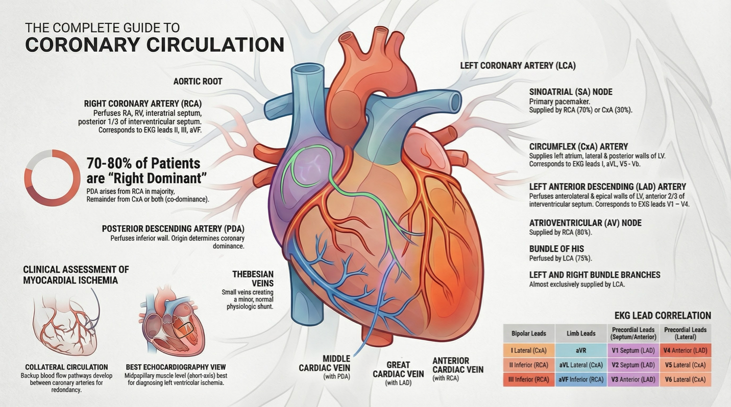
Coronary Artery Origins: Left and right coronary arteries (LCA and RCA) arise from the aortic root.
Coronary Arteries
LCA: Divides into left anterior descending (LAD) and circumflex arteries.
LAD: Supplies anterolateral and apical walls of left ventricle, anterior two-thirds of interventricular septum (EKG leads V1-V4).
Circumflex: Supplies left atrium, lateral and posterior walls of left ventricle (EKG leads I, aVL, V5-V6).
RCA: Supplies right atrium, right ventricle, interatrial septum, and posterior third of interventricular septum (EKG leads II, III, aVF).
Posterior Descending Artery (PDA): Supplies inferior wall, defines coronary dominance (usually arises from RCA in 70-80% of patients).
Coronary Venous Circulation
Main Veins:
Great cardiac vein (associated with LAD)
Middle cardiac vein (associated with PDA)
Anterior cardiac vein (associated with RCA)
Coronary Sinus: Most blood returns here, located on posterior right atrium.
Thebesian Veins: Drain into all cardiac chambers, contributing to small anatomical shunt.
Ischemia Assessment
Epicardial Vessels: RCA, LAD, and CxA; subject to vascular stenosis due to atherosclerosis.
Collateral Circulation: Provides redundancy of blood flow; risk if occlusion occurs proximal to collateral branches.
Key Areas of Supply and EKG Leads:
Septum: LAD (Bipolar leads)
Inferior Region: RCA (aVF leads).
Echocardiography
Myocardial Ischemia Diagnosis: Best view is midpapillary muscle level in short-axis; second best is apical segment in short-axis.