SON 100 Chapter 1
Sonographers
Sonographers work with ultrasound imaging equipment to produce cross-sectional images of anatomy and diagnostic data.
They must have:
Excellent communication skills
Strong computer skills
Responsibilities of the Sonographer
• Obtaining and recording patient data pertinent to the ultrasound study
Proper use of the ultrasound system, including care and cleanliness of the machine
Providing quality patient care
Having knowledge of standard precautions
Acquire, analyze, modify and select images to store and present to the interpreting physician for diagnosis
Use ultrasound terminology to document the technical summary of ultrasound findings
Assist with ultrasound-guided invasive procedures
Standard Precautions
Require treating all patients as if they are infected, ensuring safety precautions
Developed by OSHA (Occupational Safety and Health Administration
Designed to protect healthcare workers from transmission of diseases
Bloodborne Pathogens – Strategies to Reduce Exposure
OSHA Guidelines
Establishing engineering controls to limit risk of exposure, such as autoclaves and sterilization methods, self-sheathing needles
Employee work practice protocols such as proper handwashing, gloving with each patient and using additional PPE as necessary
Housekeeping controls such as keeping equipment contaminant-free, proper disposal of sharps and laundry and maintaining high-level disinfection
Offering vaccination against Hep B (recommended but not required) and Flu (annually)
Documentation for Ultrasound Exams
BEFORE the exam
Review the ultrasound request aka the doctor’s order
Patient identifiers: Full name and DOB, could also use the MRN (medical record number) – if checking an arm band
Examination priority: Routine, Urgent, STAT – must be completed within 30 - 60 mins
Patient precautions:
Isolation status
Name, address and phone number of referring physician
Essential clinical facts explaining the reason for the exam as well as clinical symptoms
Relevant medical history
Physician signature with date
Area for barcode
Examination type check boxes
Additional exam information: Sonographer’s name/initials, exam room number/location and data
Gather patient history
EMR (electronic medical record)
Assessment notes
Laboratory results
Previous imaging reports
Brief medical history from the patient
Clinical symptoms
Previous surgeries
Type, location and duration of symptoms
DURING the exam
Accomplished scanning techniques are a result of practice and knowledge of the following:
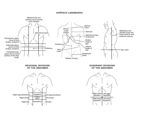
Location of anatomy. Body structures are accurately identified on ultrasound images by their location and landmarks.
Sonographic appearance of normal body structures and terms to describe their appearance. Knowing what normal looks like, will allow you to easily recognize abnormalities.
Imaging criteria:
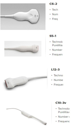
Begin with a transducer best suited to the structures of interest.
High MHz = superficial
Low MHz = deep
Use a coupling agent between the transducer and the skin surface to remove air.
Apply pressure with the transducer, but varies with:
Exam being performed
Patient body habitus and bowel patterns
Patient tolerance of probe pressure
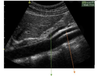
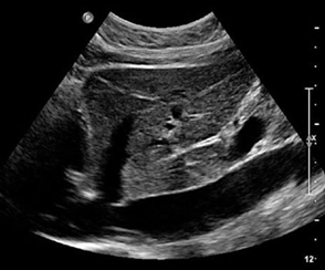
Perform comprehensive scanning surveys with detailed and inclusive observations. Investigate the area of interest and immediate adjacent structures in both longitudinal and axial (transverse) scan planes. No images are taken during this part of the exam.
Optimize image with keyboard and touchscreen controls.
Adjust field size and depth of penetration to best view the area of interest.
Focus near and far gain settings to enhance visualization of organs.
Adjust contrast to help differentiate structures.
Follow a scanning protocol which ensures standardization and that no images are missed.
AFTER the exam
Complete the examination worksheet: medical history, clinical symptoms, pertinent laboratory or imaging test results and describe ultrasound findings
Provide a written technical observation summary of the ultrasound findings using sonographic terminology. This should include echo pattern (composition), origin or location, number and size of finding(s). This is a permanent part of the patient’s chart and observations should be documented in a way as to not be legally compromising.
Legal Guidelines: If a sonographer fails to make an accurate description in the written observation summary, but demonstrates the findings on the images, he or she has performed within legal guidelines of scope of practice for diagnostic medical sonographers. Therefore, you cannot be held legally liable for forgetting to include pathology in your report if you have images of it – it is the radiologist’s job to review all images.
Body Planes
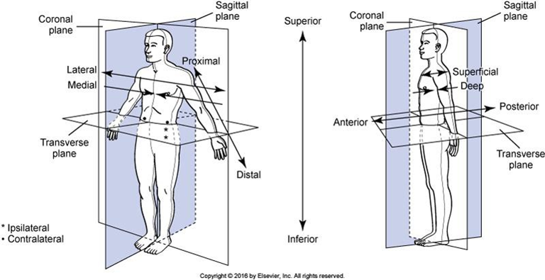
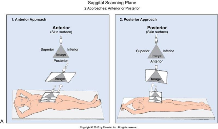
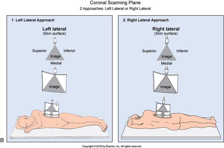
Scanning planes in sonography are the same as anatomic body planes, but their interpretations depend on the location of the transducer and the sound wave approach (where the sound waves enter the body).
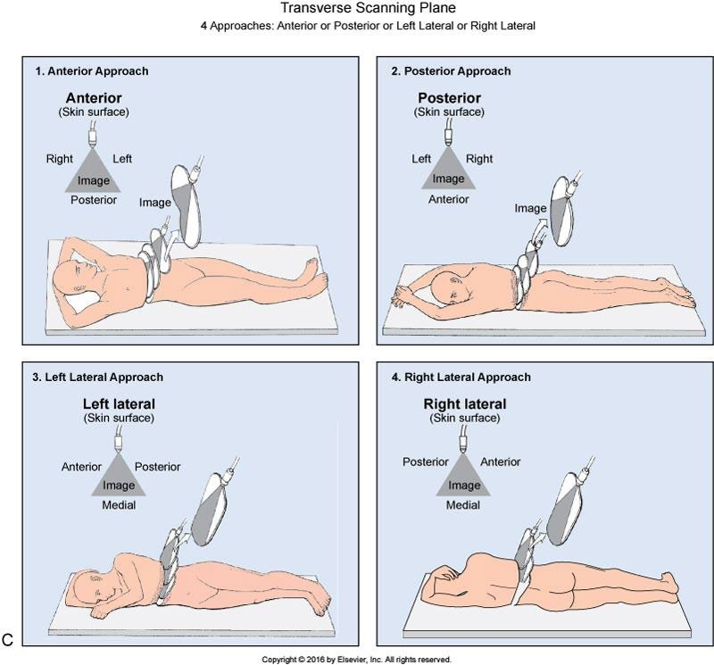
Scan planes: Sagittal (Longitudinal), Transverse (Axial) and Coronal
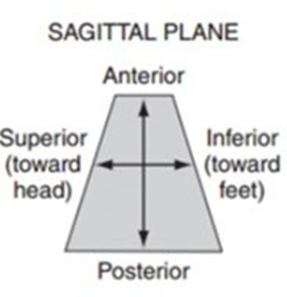
Sagittal is the plane that is parallel to the long axis of the body, meaning it is running along side or lengthwise to the body, dividing in to right and left sections.
Midsagittal – divides into equal right and left halves
Parasagittal – divides into unequal right and left sections
Coronal is the plane that is parallel to the long axis of the body and perpendicular to the sagittal scan plane, meaning it is dividing the body into anterior (front) and posterior (back) sections.
Midcoronal – divides into unequal anterior and posterior sections
Transverse is the plane that is perpendicular to the long axis of the body and separates the body into unequal superior (toward the head) and inferior (toward the feet) sections.
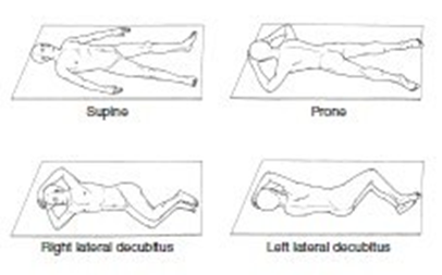
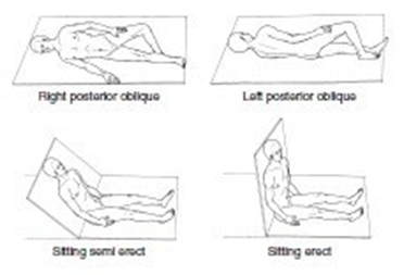
Patient Positions
Descriptive Terminology
Sonographers describe; we do not diagnose! However, ultrasound provides the interpreting physician with comprehensive sonographic details of a study upon which a diagnosis is based. So, differentiating abnormal echo patterns from normal echo patterns, documenting any differences in echo pattern appearance and describing any difference in echo pattern appearance using sonographic terminology is essential.
Every structure seen on ultrasound has soft tissue descriptors including:
Size, shape, contour
Position within the body
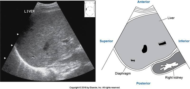
Parenchymal texture
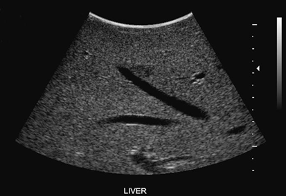
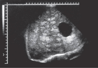
Homogenous = uniform in texture
Heterogeneous = irregular in texture
Diffuse disease (infiltrative) means it is diseased throughout the organ
Localized disease means there is a mass or masses in specific spots, not throughout the organ
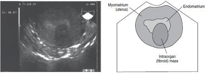
Echogenicity = the quantity of echoes
Hyperechoic – abundant/many echoes, appears bright on the image
Hypoechoic – fewer echoes, appears dark gray on the image
Anechoic – no echoes, appears black on the image
Solid (mass, lesion or structure): echogenic shades of gray, made up of tissue, not fluid
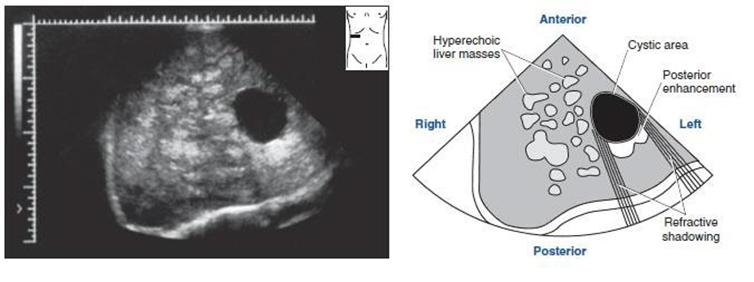
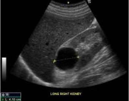
Cystic: 3 components
1. Anechoic
2. Smooth, defined walls
3. Posterior through transmission
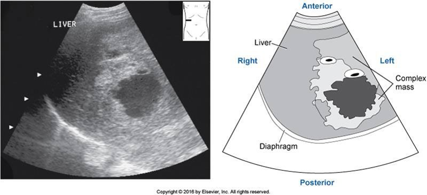
Complex: containing both fluid and tissue; appearance of internal composition may vary with time when related to vascular masses as blood appears anechoic at first but thickens and changes in composition to more of a solid, gray echogenicity once the clot forms.
Homogeneous vs Heterogeneous
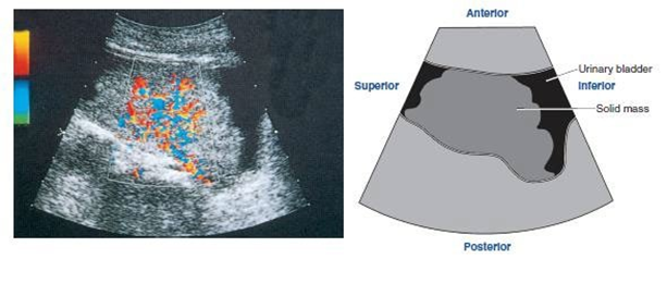
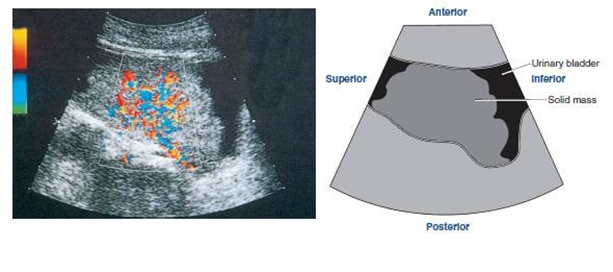
Irregular Borders vs Smooth Borders
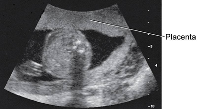
Posterior Shadowing: occurs when the sound beam strikes a strongly attenuating structure, such as bone, or a highly reflecting structure, such as bowel
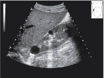
Posterior Enhancement or Through Transmission: occurs when the sound beam passes through a weakly attenuating structure, such as fluid
Directional Terms
Superior (cranial) – toward the head
Inferior (caudal) – toward the feet
Lateral – away from the midline of the body or structure of interest
Medial – toward the midline of the body or structure of interest
Anterior (ventral) – toward the front side of the body
Posterior (dorsal) – toward the back side of the body
Proximal – situated nearer to the center of the body or point of attachment
Distal – situated away from the center of the body or point of attachment
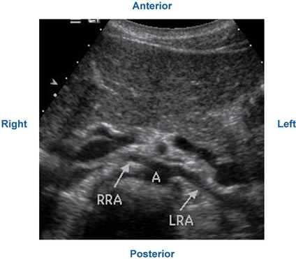
Transverse Imaging Plane
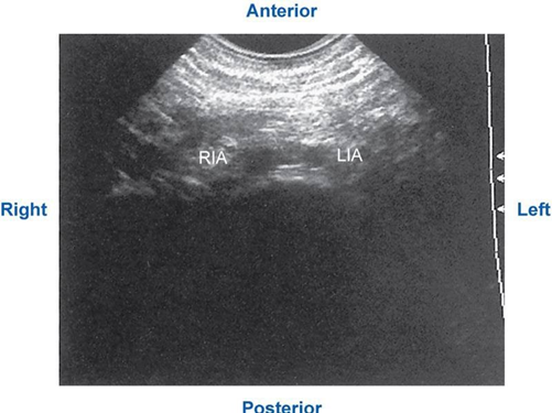
Sagittal Imaging Plane