Lecture 2: Clinical Tractography for Neurosurgical Planning
How diffusion MRI and tractography are used to help neurosurgeons avoid damaging important white matter pathways during surgery. The technique is especially important when lesions are close to areas responsible for movement, language, or vision.
Why Neurosurgical Planning Matters
The main aim of neurosurgery is to remove as much abnormal tissue as possible while preserving normal brain function. Surgeons therefore need detailed information about:
where the lesion is located
where important functional brain regions are located
where critical white matter pathways travel
Damage to eloquent brain regions can produce severe post-surgical deficits such as:
weakness or paralysis
speech and language problems
visual field loss
sensory deficits
Standard imaging methods help identify lesions and cortical function, but they provide limited information about white matter connectivity. Tractography fills this gap by visualising white matter pathways.
Imaging Methods Used in Neurosurgical Planning
Several imaging techniques are combined during surgical planning.
Structural MRI
T1-weighted MRI
Used to identify anatomy and lesion structure. Particularly useful for tumours and cortical abnormalities.
FLAIR MRI
Provides complementary contrast and is useful for detecting abnormal white matter changes, cortical dysplasia, oedema, or calcification.
These scans define lesion morphology but do not show functional pathways.
Functional Imaging Techniques
Functional MRI (fMRI)
Measures blood oxygenation changes associated with neural activity. Used to identify functional cortex such as language or motor areas.
Magnetoencephalography (MEG)
Measures magnetic fields produced by brain activity more directly than fMRI.
Transcranial Magnetic Stimulation (TMS)
Stimulates cortex non-invasively to identify regions involved in movement or language.
Cortical and Subcortical Stimulation
Electrodes are used during surgery or epilepsy monitoring to identify eloquent tissue.
Awake Craniotomy
Allows direct monitoring of patient function during surgery.
These methods mainly focus on grey matter function rather than white matter connectivity. Tractography therefore becomes essential for understanding how regions are connected.
A major limitation of these methods is that they mainly focus on grey matter. They provide little direct information about white matter pathways. Surgeons may know approximately where major tracts such as the corticospinal tract should be located anatomically, but this is not sufficiently precise for surgery. Tractography was developed to solve this problem by visualising white matter pathways directly.
Diffusion MRI: The Basis of Tractography
Tractography is based on diffusion MRI, which measures how water molecules move through tissue.
Brownian Motion
Water molecules are constantly moving randomly due to thermal energy. This random motion is called Brownian motion. Over time, molecules spread further from their original position.
The longer water molecules are allowed to move, the further they spread from their starting point. The amount of spreading depends on both time and the diffusion coefficient, which reflects how freely water can move in a particular environment.
The diffusion coefficient depends on:
temperature
molecular species
surrounding environment
In free water at body temperature, diffusion is relatively unrestricted.
How MRI Detects Diffusion
MRI scanners apply magnetic gradients during imaging.
If water molecules remain stationary:
the effects of gradients cancel out
If water molecules move:
signal loss occurs
Greater diffusion causes greater signal attenuation.
Diffusion-weighted imaging compares:
a non-diffusion-weighted image (b=0)
diffusion-weighted images (higher b-values)
Higher b-values increase sensitivity to diffusion.
Diffusion-weighted MRI compares how much signal is lost when water molecules move. Greater water movement causes greater signal loss, and this information is calculated for each voxel to create diffusion maps such as ADC maps.
Apparent Diffusion Coefficient (ADC)
In biological tissue, diffusion is restricted by:
cell membranes
axons
myelin
intracellular structures
Water therefore appears to diffuse less freely than in pure water. This measured value is called the Apparent Diffusion Coefficient (ADC).
Low ADC values indicate restricted diffusion.
High ADC values indicate freer diffusion.
Anisotropy
Diffusion in the brain is direction-dependent.
Isotropic Diffusion
Water diffuses equally in all directions.
Typical of:
cerebrospinal fluid
grey matter
Anisotropic Diffusion
Water diffuses preferentially in one direction.
Typical of:
organised white matter tracts
Axons constrain water movement across fibres while allowing easier movement along fibre direction.
This directional property is fundamental for tractography because it allows estimation of fibre orientation.
Diffusion Tensor Imaging (DTI)
DTI models diffusion in three dimensions using a diffusion tensor.
The tensor:
is a 3×3 matrix
describes both magnitude and direction of diffusion
At least:
6 diffusion-weighted images
plus 1 unweighted image
are required to estimate the tensor, although 20–30 directions are typically acquired for better accuracy.
Eigenvectors and Eigenvalues
The diffusion tensor can be decomposed into:
3 eigenvectors = diffusion directions
3 eigenvalues = diffusion magnitudes
The principal eigenvector indicates the dominant fibre orientation.
This allows white matter direction to be estimated within each voxel.
Important DTI Metrics
Fractional Anisotropy (FA)
FA measures how directional diffusion is.
High FA:
highly organised white matter
strong directional diffusion
Low FA:
isotropic diffusion
damaged or disorganised tissue
Mean Diffusivity (MD)
MD measures the overall magnitude of diffusion regardless of direction.
High MD:
freer diffusion
tissue loss or oedema
Low MD:
restricted diffusion
How Pathology Affects DTI Metrics
Pathology commonly causes:
Reduced FA
Because tissue organisation breaks down and barriers restricting diffusion are lost.
Increased MD
Because water can diffuse more freely in damaged tissue.
Examples include:
tumours
degeneration
oedema
white matter injury
Colour FA Maps
Colour-coded FA maps show fibre orientation.
Typical colour convention:
red = left-right
green = anterior-posterior
blue = superior-inferior
These maps provide intuitive visualisation of major white matter tracts.
Limitations of DTI
DTI assumes only one dominant fibre orientation per voxel.
This creates problems in regions with:
crossing fibres
branching fibres
kissing fibres
The tensor therefore becomes inaccurate in complex white matter regions.
Constrained Spherical Deconvolution (CSD)
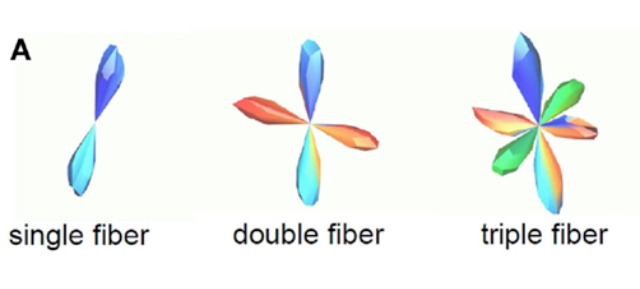
CSD is an advanced diffusion MRI method used to estimate multiple fibre directions within a single voxel, making it much more useful for tractography than standard DTI in regions where fibres cross, branch, or curve.
Unlike DTI, which assumes there is only one dominant fibre direction in each voxel, CSD assumes that several fibre bundles may pass through the same voxel in different directions. This is important because most white matter voxels actually contain complex fibre arrangements rather than one single tract.
CSD works by analysing the full diffusion signal measured across many directions and comparing it to an idealised diffusion pattern from a single fibre bundle, known as the response function. It then mathematically separates the mixed diffusion signal into multiple probable fibre orientations within the voxel.
The output of CSD is called a fibre orientation distribution (FOD), which is a spherical model showing the likely directions of fibres. Each peak in the FOD represents a different fibre population travelling through the voxel.
This allows tractography algorithms to follow pathways through crossing-fibre regions much more accurately. For example, instead of incorrectly averaging two crossing tracts into one false diagonal direction like DTI often does, CSD can separate the two directions individually.
CSD is particularly useful for reconstructing complex pathways such as:
optic radiations
Meyer’s Loop
arcuate fasciculus
corticospinal tract.
It is preferred over DTI for tractography because it provides:
more anatomically realistic pathways
better handling of crossing fibres
fewer false tract terminations
improved continuity of streamlines through complex white matter regions.
In neurosurgery, this improves surgical planning because surgeons can estimate tract locations more accurately and reduce the risk of damaging important pathways during resections.
However, CSD is more computationally demanding and requires:
higher quality diffusion data
more diffusion directions
better signal-to-noise ratio
more advanced processing pipelines.
It is also more sensitive to noise, meaning false fibre orientations or spurious streamlines can sometimes be produced if data quality is poor.
CSD scans often take longer to acquire than standard DTI scans, which can be difficult in:
children
unwell patients
intraoperative MRI settings.
Interpretation is also more complex because fibre orientation distributions are harder to analyse than simple tensors and usually require greater expertise.
Importantly, CSD still does not directly visualise axons. Like all tractography methods, it estimates likely fibre pathways indirectly from patterns of water diffusion.

Tractography
Tractography reconstructs white matter pathways by connecting voxels with similar diffusion directions.
The aim is to move beyond looking at diffusion in single voxels and instead estimate:
how brain regions are connected
where important white matter bundles travel
whether pathways are close to lesions or surgical targets.
This is especially important in neurosurgery because damage to white matter tracts can produce major deficits even if cortical regions are preserved.
For example:
damaging the corticospinal tract can cause paralysis
damaging optic radiations can cause visual field loss
damaging the arcuate fasciculus can impair language.
Basic Tractography Algorithm
Seed Point
Tractography starts from a seed point or seed region.
The algorithm:
takes a small step in the principal diffusion direction
recalculates direction in the next voxel
continues repeatedly through neighbouring voxels.
This process gradually creates streamlines representing estimated white matter pathways.
Tracking is usually performed in both directions from the seed point so that the full pathway can be reconstructed.
Why Termination Criteria are Needed
Without stopping rules, streamlines would continue into biologically meaningless regions.
Tractography is an estimation process, so the algorithm needs rules to prevent anatomically impossible pathways.
Low Fractional Anisotropy (FA)
Tracking stops when FA becomes too low.
Low FA usually means:
diffusion is no longer strongly directional
tissue is no longer organised white matter.
This commonly occurs when entering:
grey matter
CSF
damaged tissue.
Why this matters
If tracking continued into isotropic tissue, streamline directions would become random and biologically meaningless.
Exit Brain Mask
Tracking stops when the streamline exits the brain.
Why this matters
White matter pathways cannot continue outside brain tissue.
This prevents false tracts extending into non-brain regions.
Sharp Turns
Tracking stops if the next step involves a sharp turn.
Sudden turns are biologically implausible because real white matter fibres usually follow smooth trajectories.
Why this matters
Sharp turns often occur because:
the algorithm jumps between tracts
noise disrupts direction estimates
the streamline enters CSF or pathological tissue.
Stopping tracking reduces false-positive pathways.
Streamlines
Tractography outputs 3D lines called streamlines. The colourful lines. They are mathematical reconstructions based on water diffusion patterns.
In motor tractography:
streamlines often travel from motor cortex to brainstem
most follow anatomically sensible paths.
Some spurious streamlines still occur because:
diffusion models are imperfect
fibre crossings complicate tracking
MRI data contains noise.
Why this matters
Tractography should always be interpreted cautiously.
Individual streamlines are not proof of real anatomical fibres.
Instead, the overall pattern of pathways is most important clinically.
Regions of Interest (ROIs)
ROIs are binary masks used to guide tractography.
ROI placement is one of the most important factors affecting tractography results.
Why this matters
Different ROI strategies can produce very different tract reconstructions in the same patient.
Poor ROI placement can:
miss important fibres
include false pathways
mislead surgical planning.
Types of ROIs
Seed ROI
Starting region for streamlines.
Why it matters
Determines which pathways are reconstructed.
Different seed locations emphasise different parts of the tract.
Waypoint / Inclusion ROI
Streamlines must pass through this region to be included.
Why it matters
Removes anatomically implausible streamlines.
Improves specificity of tract reconstruction.
The transcript compares this to marathon checkpoints:
if a runner misses checkpoints, they likely took the wrong route.
Exclusion ROI
Streamlines entering this region are discarded.
Why it matters
Helps eliminate false-positive pathways.
Example:
when reconstructing motor pathways in one hemisphere
exclusion ROIs can remove streamlines incorrectly crossing the midline.
Iterative Refinement
Iterative refinement means repeatedly adjusting and improving the tractography reconstruction step-by-step until the pathways look anatomically reasonable and clinically useful.
Researchers repeatedly:
inspect pathways
adjust ROIs
add exclusions
test different thresholds.
Why this matters
There is rarely one perfect tractography solution.
Exploring multiple reconstructions helps avoid false conclusions.
This is particularly important in surgical planning where missing fibres could harm patients.
ROI Definition Methods
Manual ROIs
ROIs can be manually drawn using imaging software.
Advantages
flexible
tailored to individual anatomy
useful in abnormal brains.
Limitations
time consuming
operator dependent.
Why this matters
Manual methods are often essential in pathology because automated tools may fail.
Automated Parcellation
Software such as FreeSurfer automatically segments cortical and subcortical regions.
Advantages
fast
reproducible
useful in healthy anatomy.
Limitations
relies on relatively normal brain structure
can fail in:
tumours
previous resections
cortical malformations
severe pathology.
Why this matters
In neurosurgical patients, anatomy is often distorted.
Automated segmentation errors can directly affect tractography accuracy.
Functional Activation ROIs
Functional imaging activations from scans such as fMRI can be used as tractography seed regions so that white matter pathways are reconstructed from brain areas that are actually active during a task.
Why this matters
Allows researchers to investigate white matter pathways connected to functional cortex.
Limitation
If fMRI activation is inaccurate:
tractography errors become compounded.
Some researchers therefore prefer:
analysing functional imaging separately
then comparing it with tractography to check whether findings agree.
Seed Strategy Differences
Cortical Seed
A cortical seed means the tractography starting point is placed near the motor cortex on the brain surface.
Because the seed starts close to cortex, the resulting streamlines spread out more broadly across cortical regions, showing detailed connections near the brain surface.
Why this matters surgically
When surgery is close to cortex, surgeons need to know:
how far the motor fibres spread
which cortical areas are connected
where the safest entry route is.
So broader projections give more detailed local information near the cortical surface.
Deeper in the brain, tracts are usually more tightly bundled together, which is why peduncle seeds often produce narrower streamline bundles.
Peduncle Seed
A peduncle seed means the tractography starting region is placed lower down in the corticospinal tract, usually around the cerebral peduncle in the brainstem.
Instead of starting near the broad fan-like fibres at the cortex, the algorithm starts in a deeper region where the motor fibres are already tightly bundled together.
Because the fibres are more compact there, the resulting streamlines are narrower and more focused.
Why this matters
This makes it easier to visualise the main deep motor pathway without lots of broad cortical branching.
Peduncle seeds are especially useful for deep lesions, such as thalamic tumours, because surgeons mainly need to know:
where the deep motor tract is travelling
whether it is displaced by the lesion
how close surgery will come to the tract.
So:
cortical seeds give more detailed information near the cortical surface
peduncle seeds give clearer reconstruction of the deeper compact motor pathway.
Streamlines Follow the Path of Least Resistance
Streamlines tend to follow smooth directional continuity.
Why this matters
Algorithms naturally prefer smooth curves over abrupt directional changes.
This can bias tracking toward some pathways while missing others.
In oedema or distorted anatomy:
streamlines may “wrap around” lesions
pathways may become artificially displaced.
Validation of Tractography
One major limitation is that tractography has no perfect gold standard.
Why this matters
Researchers cannot directly prove every reconstructed tract is anatomically correct.
Validation studies are therefore extremely important.
Lawes et al. (2008)
Compared tractography of the inferior fronto-occipital fasciculus with anatomical dissection.
Findings
Similar:
tract curves
bends
shapes
posterior anatomy.
Importance
Provided evidence that tractography can reflect real white matter anatomy.
Increased confidence that reconstructed pathways are biologically meaningful.
Limitation
Similarity does not prove perfect accuracy.
Tractography remains an indirect estimation method.
Important Conceptual Point
Tractography does NOT visualise individual axons.
Instead:
it estimates the average direction of fibre bundles across many axons.
Why this matters
Tractography is a probabilistic reconstruction technique, not a microscopic imaging method.
Clinical Tractography
Main Clinical Tracts
Motor Pathways
preserve movement.
Optic Radiations
preserve vision.
Arcuate Fasciculus
preserve language.
Why these matter
These functions have major impacts on quality of life.
Neurosurgical planning focuses heavily on avoiding injury to these pathways.
Kinoshita et al. (2005)
Early glioma tractography study.
Findings
One patient developed severe deficits after surgery.
Tractography underestimated tract extent.
Important technical issue
Used a very high FA threshold (0.3).
the algorithm only allows tracking through voxels with fairly strong directional diffusion.
So:
FA > 0.3 → tracking continues
FA < 0.3 → tracking stops.
Example:
a crossing-fibre voxel with FA = 0.25 might still contain important fibres
but tractography would stop there if the threshold is 0.3.
An FA threshold of 0.3 is relatively strict because tractography will only continue through voxels with strongly directional diffusion, meaning real fibres in crossing-fibre, curved, or damaged regions may be excluded and tracts may appear smaller than they really are.
Why This Matters Clinically
In the glioma study:
the high FA threshold likely caused tractography to underestimate the motor tract extent
surgeons may have thought fibres were further away from the tumour than they actually were
one patient developed severe deficits after surgery.
So choosing the FA threshold is a balance between:
avoiding false-positive tracts
but not accidentally removing real fibres.
Clinical implication
Incorrect tractography parameters can directly affect patient outcomes.
Demonstrated the importance of:
careful parameter selection
testing multiple reconstructions
understanding limitations of the method.
FA THRESHOLD
An FA threshold is a cutoff value used during tractography to decide whether tracking should continue through a voxel or stop.
FA (fractional anisotropy) measures how directional diffusion is:
high FA = water mainly moves in one direction
low FA = water moves more equally in all directions.
FA Range
FA values always range from:
0 → completely isotropic diffusion
1 → perfectly directional diffusion.
In reality, brain tissue usually falls somewhere between these extremes.
There is no single “correct” FA value because it depends on the tissue and brain region, but roughly:
CSF → around 0.0–0.1
(very isotropic)Grey matter → around 0.1–0.2
(less organised)Crossing-fibre regions → often around 0.2–0.4
(real white matter but diffusion directions are mixed)Healthy major white matter tracts → often around 0.4–0.8
(strongly directional)Very coherent white matter (e.g. corpus callosum) → can be even higher.
During tractography, the algorithm checks the FA value in each voxel.
If the FA:
stays above the threshold → tracking continues
falls below the threshold → tracking stops.
Why This Is Needed
Low FA means the diffusion direction is less reliable.
If tracking continued through very low FA regions:
streamlines would become random
false pathways would appear.
So thresholds help prevent biologically unrealistic tracts.
Mikuni et al. (2007)
Mikuni et al. (2007) investigated whether tractography accurately reflected real motor pathways in patients with gliomas undergoing surgery.
The study included 40 glioma patients and compared:
pre-operative motor tractography
with intraoperative subcortical stimulation during surgery.
During surgery, surgeons electrically stimulated tissue near the tumour to test whether it was functionally connected to motor pathways.
If stimulation produced a motor evoked potential (MEP), such as movement or muscle activity, this indicated the tissue was functionally important motor white matter.
The study found that in 18 out of 40 patients, motor evoked potentials occurred within 1 cm of the tractography-defined motor pathways.
Why this is important
This showed reasonably strong agreement between:
tractography predictions
and real functional motor tissue identified during surgery.
In other words, the reconstructed corticospinal tracts were often close to the actual motor pathways.
This was important because one major criticism of tractography is that it is only an indirect estimate based on water diffusion.
By comparing tractography with direct electrical stimulation, the study provided evidence that tractography can reflect clinically meaningful anatomy.
Clinical implication
Suggested tractography is clinically useful rather than purely experimental.
Also showed tractography and direct stimulation complement each other rather than replacing one another.
Tractography
provides a pre-operative anatomical estimate of pathway location
helps surgeons plan surgical approach
shows how tracts may be displaced by tumours.
Subcortical stimulation
provides real-time functional confirmation during surgery
tests whether tissue is actually involved in motor function at that exact moment.
Stimulation:
Intraoperative stimulation is the broad category of electrical stimulation performed during brain surgery to map brain function. It includes two phases: cortical (mapping the surface of the brain) and subcortical (mapping deeper white matter tracts).
Interoperative Subcortical stimulation is a specific type of intraoperative stimulation used to measure the distance to critical motor or language pathways deep inside the brain while a surgeon removes a tumor.
Kamada et al. (2007)
Kamada et al. (2007) investigated whether tractography of the arcuate fasciculus (AF) accurately reflected real language pathways during neurosurgery.
The arcuate fasciculus is a major white matter tract involved in language, particularly communication between frontal and temporal language regions.
The researchers reconstructed the arcuate fasciculus using tractography before surgery and imported it into the neuronavigation system used by the surgeons. This meant the surgeons could see the estimated language tract location during the operation itself.
During surgery, electro-cortical stimulation was performed. This involves electrically stimulating regions of cortex while monitoring the patient for language disruption.
If stimulation caused speech arrest or language problems, the stimulated area was assumed to be functionally important for language.
The study found that stimulation sites producing language disruption were within 6 mm of the reconstructed arcuate fasciculus pathways.
Why this matters
Demonstrated close agreement between tractography and functional language mapping.
Clinical implication
Supports use of tractography for protecting language pathways during surgery.
Why stimulation is still needed
Tractography estimates anatomy indirectly from water diffusion.
Electrical stimulation tests real functional tissue directly in the patient during surgery.
Therefore:
tractography provides anatomical guidance
stimulation provides functional confirmation.
The two techniques are most effective when combined together.
Epilepsy Surgery Case
Clinical Background
Patient had:
left frontal polymicrogyria
seizures
right hemiparesis before surgery.
Required left frontal lobectomy.
Surgical Outcome
Patient became seizure free.
Right arm strength improved significantly.
Why this is important
Demonstrates that removing epileptic tissue can improve function rather than worsen it.
Seizure activity itself had likely been disrupting motor cortex recovery.
Clinical implication
Careful tractography-guided surgery can preserve function while improving neurological outcome.
Why Intraoperative MRI is Needed → Brain Shift
Before surgery, patients usually have a high-quality pre-operative MRI with CST (corticospinal tract) tractography to map important motor pathways relative to the lesion.
These tractography maps are loaded into neuronavigation systems so surgeons can avoid damaging critical white matter during surgery.
Brain Shift
During surgery:
the skull is opened
pressure changes
CSF drains
tissue is removed
gravity affects the brain.
Because brain tissue is soft and deformable, the brain physically moves during surgery. This is called brain shift.
Why This Matters
Pre-operative tractography only reflects anatomy before surgery begins.
As the brain shifts:
tract locations may no longer align with the real anatomy
neuronavigation becomes less accurate.
A motor tract that originally appeared safely away from the lesion may move closer to the surgical cavity during resection.
This increases the risk of damaging important pathways.
Intraoperative MRI
Intraoperative MRI acquires updated scans during surgery so surgeons can see the brain’s current anatomy after brain shift has occurred.
Updated intraoperative CST tractography can then be generated using the new scan, improving:
tract localisation
surgical guidance
safety.
Challenges of Intraoperative Tractography
Intraoperative diffusion MRI is difficult because:
scans must be very fast
image quality is lower
surgical cavities cause distortions
patient positioning is unusual
real-time processing is required.
Despite these challenges, intraoperative MRI helps reduce navigation errors caused by brain shift and improves preservation of important white matter tracts during surgery.
Thalamic Pilocytic Astrocytoma Case
Clinical Problem
Deep tumour close to motor pathways.
Why this is challenging
White matter anatomy becomes distorted.
Internal capsule location may be unclear.
Surgeons need tractography to estimate:
pathway displacement
safest surgical entry route.
Tractography Findings
Streamlines displaced posteriorly.
Pathways wrapped around the tumour.
Oedema caused streamlines to corkscrew around the lesion.
Why this matters
Pathology can distort tractography dramatically.
Requires iterative testing of different ROI strategies and thresholds.
Outcome of Astrocytoma Case
Tumour successfully debulked.
Patient showed:
improved strength
better hand use
good functional recovery.
Why this matters
Demonstrates how tractography can help:
guide safer surgical approaches
preserve important pathways
improve post-operative outcomes.
Streamline Density Maps
Streamline density refers to how many streamlines pass through a particular brain region on a tractography map.
Areas with more streamlines are shown as brighter or denser regions on the image.
Brighter colours
more streamlines
greater confidence in tract location.
Limitation
Density naturally decreases with distance from the seed region.
Why this matters
High density does not necessarily mean the tract is biologically stronger.
Interpretation requires caution.
Optic Radiations
The optic radiations are major white matter pathways involved in vision.
Visual information travels:
from the eyes
through the optic nerves
into the optic chiasm
through the optic tracts
into the optic radiations
finally reaching primary visual cortex (V1).
Visual fields are crossed:
left visual field projects mainly to the right hemisphere
right visual field projects mainly to the left hemisphere.
Why this matters clinically
Damage to optic radiations can produce visual field deficits.
Even if surgery successfully removes a lesion, injury to visual pathways may significantly affect quality of life.
Meyer’s Loop
One particularly important part of the optic radiation is Meyer’s Loop.
Meyer’s Loop:
curves sharply anteriorly through the temporal lobe
then loops back toward occipital cortex.
Why this is important
Temporal lobe resections are common in epilepsy surgery.
If surgeons resect too far posteriorly or laterally:
they may cut Meyer’s Loop
causing visual field deficits.
This makes accurate tractography extremely important during temporal lobe surgery.
Why Optic Radiation Tractography is Difficult
Segmentation of optic radiations is particularly challenging.
Main reasons
Sharp Curvature
Meyer’s Loop bends sharply.
Tractography algorithms prefer smoother trajectories.
Crossing Fibres
Complex fibre architecture exists near the lateral geniculate nucleus (LGN).
Path of Least Resistance Problem
Streamlines tend to continue in straighter directions.
Instead of following the sharp curve of Meyer’s Loop, streamlines may incorrectly continue forwards.
Why this matters
Tractography may underestimate the anterior extent of Meyer’s Loop.
Surgeons could falsely assume tissue is safe to remove.
Epilepsy Case: Right Parietal Occipital Craniotomy
Clinical Background
Patient required:
right parietal-occipital craniotomy
removal of a tuber associated with epilepsy.
The lesion was close to the optic radiation.
Why this is important
Surgery near visual pathways risks post-surgical visual deficits.
Surgeons therefore needed tractography to estimate tract location before surgery.
Tractography Findings
Optic radiation tractography was reconstructed around the lesion.
The posterior part of the tract was particularly important in this case.
Post-operative imaging suggested:
most of the optic radiation was preserved
only a small region appeared affected.
Surgical Outcome
Patient had:
no post-operative seizures
reasonably preserved unaided vision.
Unfortunately:
no formal visual field map was available post-operatively.
A formal visual field map is a detailed clinical test that measures exactly which parts of a person’s visual field they can or cannot see.
After surgery near the optic radiations, doctors ideally perform this test to check whether any visual pathways were damaged.
The patient may feel their vision is “mostly normal,” but subtle visual field deficits can still exist.
Some deficits are difficult for patients to notice themselves.
Why this case is important
Demonstrates how tractography can guide resections near critical visual pathways.
Shows that preserving most of the tract may help preserve visual function.
Limitation
Without formal visual field testing:
exact visual outcome cannot be fully confirmed.
In This Case
The surgeons believed most of the optic radiation had been preserved because:
tractography suggested only small overlap with the surgical area
the patient reported reasonably good vision.
However, without a formal visual field test:
they could not objectively confirm whether small visual deficits were present.
Lacerda et al. (2020)
Aim of the Study
Investigated visual pathway damage following paediatric epilepsy surgery.
Researchers:
reconstructed optic radiations using tractography
segmented resection cavities
“Segmented” means the researchers used imaging software to identify and outline that cavity on the MRI scan.
measured overlap between the two.
Methodology
The optic radiation was rotated into the axial plane to allow measurement of:
cross-sectional overlap area
between the tract and resection cavity.
The study then compared:
overlap percentage
with post-surgical visual field deficits.
Findings
Patients with greater overlap between:
optic radiation
and resection cavity
were more likely to develop visual field deficits.
Examples from the table:
Patient 15 had 100% overlap and persistent right homonymous hemianopia
Patients with 0% overlap often retained normal vision.
Why this study is important
Provides evidence that tractography findings relate to real clinical outcomes.
Supports the idea that:
damage predicted by tractography
corresponds to actual visual deficits after surgery.
Clinical implication
Surgeons can potentially estimate visual risk before surgery.
Helps guide:
surgical planning
patient counselling
informed consent discussions.
Broader significance
This is important because one criticism of tractography is that it lacks a gold standard.
Outcome studies like this help validate its clinical usefulness.
Arteriovenous Malformations (AVMs)
What is an AVM?
An AVM (arteriovenous malformation) is an abnormal tangle of blood vessels where blood flows directly from arteries into veins without passing through normal capillaries.
This creates a high-pressure shortcut called a shunt, causing the vessels to become fragile and increasing the risk of bleeding, seizures, and neurological problems.
The central tangled mass of abnormal vessels is called the nidus.
Why AVMs are Dangerous
AVMs are fragile and prone to bleeding.
Surgical removal is extremely delicate because surgeons must:
disconnect feeding arteries first
disconnect draining veins afterwards.
Why order matters
If venous drainage is removed first:
pressure builds within the nidus
the AVM may rupture catastrophically.
AVM Tractography Case
In this case:
the AVM was close to the left optic radiation.
Tractography included:
left optic radiation
right optic radiation for comparison.
Why the Contralateral Side Was Included
Researchers noticed signal dropout near the AVM.
Signal dropout means part of the MRI image loses signal and appears darker or missing.
Near an AVM, this can happen because:
blood is flowing very fast
the abnormal vessels distort the magnetic field
MRI signals become unstable or disappear.
This is especially common in diffusion MRI because diffusion scans are very sensitive to magnetic distortions.
Why this matters
Streamlines appeared artificially displaced away from the AVM.
This could falsely suggest the tract was safer than it really was.
Including the opposite hemisphere allowed:
comparison with normal anatomy
checking whether tract displacement was biologically plausible.
Important implication
Tractography can be distorted by pathology itself.
Absence of streamlines does not necessarily mean absence of fibres.
Clinical Importance of the AVM Case
Tractography helped surgeons assess:
proximity of visual pathways
risk to patient vision.
Important point
The tractography itself did not directly guide the surgical dissection.
Instead, it improved:
surgical planning
risk assessment
patient counselling.
Surgeons could explain:
where the AVM was
where the visual pathways were
what level of visual risk existed.
Focal Epilepsy Case Near Three Major Tracts
Clinical Background
Patient had focal epilepsy with a lesion near:
arcuate fasciculus
motor tract
optic radiation.
Why this is difficult
Multiple eloquent pathways were crowded around the lesion.
Damaging any of them could impair:
language
movement
vision.
Surgical Planning
Surgeons reconstructed all three tracts simultaneously using tractography so they could visualise:
where each pathway travelled
how close they were to the lesion
which surgical routes might avoid them.
“All three tracts simultaneously” means the surgeons reconstructed and viewed all three white matter pathways together at the same time on the tractography images.
It lets surgeons see how the pathways are positioned relative to each other and to the lesion.
Why this matters
Demonstrates that neurosurgical planning often requires preserving multiple systems simultaneously.
Surgeons do not only protect one tract in isolation.
By visualising all three pathways together, the surgeon identified a narrow route between the tracts that allowed access to the lesion while minimising damage to surrounding white matter.
Outcome
Patient became seizure free one week after surgery.
Mild weakness remained in the:
left hand
left foot.
Post-operative imaging showed:
the motor tract was very close to the resection cavity.
Why this is important
Demonstrates that even careful surgery near eloquent pathways still carries risk.
Small shifts in anatomy or tractography inaccuracies may still produce subtle deficits.
Clinical implication
Tractography reduces risk but cannot eliminate it completely.
Major Limitations and Challenges of Tractography
Technical Challenges
crossing fibres
low resolution
susceptibility artefacts
image distortion
Biological Confounds
oedema
degeneration
compression
tumour infiltration
Interpretation Challenges
no true gold standard
false positives
false negatives
Clinical Challenges
requires expert knowledge
time consuming
difficult integration into surgical workflows
Important Conceptual Point
Tractography does NOT show individual axons.
Instead, it estimates likely pathways based on average diffusion patterns across many fibres. This is a major misconception.
Overall Significance
Tractography has become an increasingly important tool in neurosurgery because it allows surgeons to visualise critical white matter pathways before operating. It improves surgical planning and helps reduce functional deficits by identifying pathways that must be preserved.
What does ADC measure?
The overall magnitude of water diffusion within tissue.
What is fractional anisotropy (FA)?
A measure of how directional diffusion is within a voxel.
What does high FA indicate?
Highly organised white matter with strongly directional diffusion.
What does low FA indicate?
More isotropic diffusion, often seen in grey matter, CSF, oedema, or damaged tissue.
What is DTI?
A diffusion MRI model that estimates the main direction and magnitude of diffusion in 3D.
How many directions are minimally needed for DTI?
At least 6 diffusion-weighted directions plus 1 unweighted image.
What are eigenvectors in DTI?
The main diffusion directions within a voxel.
What are eigenvalues in DTI?
The magnitude of diffusion along each eigenvector.
What does the principal eigenvector represent?
The dominant fibre orientation in a voxel.
What is tractography?
A technique that reconstructs estimated white matter pathways using diffusion directions.
What are streamlines?
Computer-generated 3D lines representing estimated white matter pathways.
What is CST tractography?
Tractography of the corticospinal tract used to map motor pathways.
What is a seed region in tractography?
The starting region from which streamlines are generated.
What is a waypoint ROI?
A region that streamlines must pass through to be included.
What is an exclusion ROI?
A region where any streamline entering it is discarded.
What are termination criteria in tractography?
Rules that determine when streamline tracking should stop.
Why does tracking stop at low FA?
Because diffusion is no longer strongly directional and fibre orientation becomes unreliable.
Why is DTI limited in crossing-fibre regions?
Because DTI assumes only one dominant fibre direction per voxel.
What is CSD?
An advanced diffusion model that estimates multiple fibre directions within a voxel.
Why is CSD better than DTI for tractography?
Because it handles crossing fibres more accurately and produces more realistic pathways.
What is a fibre orientation distribution (FOD)?
A spherical model showing likely fibre directions within a voxel.
What is Meyer’s Loop?
A sharply curving part of the optic radiation that travels through the temporal lobe.
Why is Meyer’s Loop clinically important?
Damage during temporal lobe surgery can cause visual field deficits.
What is brain shift?
Movement and deformation of brain tissue during surgery.
Why is brain shift a problem for tractography?
Pre-operative tractography may no longer align with the real anatomy during surgery.
Why is intraoperative MRI used?
To update anatomy and tractography after brain shift occurs.
What is an AVM?
An abnormal tangle of blood vessels where arteries connect directly to veins without capillaries.
What is the nidus in an AVM?
The central tangled mass of abnormal vessels.
What is signal dropout?
Loss of MRI signal causing regions to appear dark or missing.
Why is signal dropout problematic in tractography?
It can make tracts appear falsely absent or displaced.
What is segmentation?
Separating and labelling brain regions or structures on MRI images.
What is a resection cavity?
The empty space left after surgical removal of brain tissue.
What does “segmented resection cavity” mean?
The cavity has been outlined and labelled on MRI for analysis.
What is a cortical seed?
A tractography seed placed near cortex, producing broader cortical projections.
What is a peduncle seed?
A seed placed lower in the corticospinal tract, producing narrower deep tract reconstructions.
Why are cortical seeds useful?
They provide detailed information about pathways near cortical surgical targets.
Why are peduncle seeds useful?
They better visualise deep compact motor pathways near deep lesions.
What is iterative refinement in tractography?
Repeatedly adjusting ROIs and parameters to improve tract reconstruction.
What did Mikuni et al. (2007) show?
Motor stimulation sites were often within 1 cm of tractography-defined motor tracts, showing good agreement with functional anatomy.
What did Kamada et al. (2007) show?
Language stimulation sites were within 6 mm of arcuate fasciculus tractography, supporting tractography for language preservation surgery.
What did Lacerda et al. (2020) show?
Greater overlap between optic radiations and resection cavities predicted visual field deficits after surgery.
What is the major limitation of tractography?
It estimates pathways indirectly from water diffusion and does not directly visualise axons.
Tractography uses diffusion MRI to estimate white matter pathways by following the direction water diffuses through organised axonal fibres.
Water diffuses anisotropically in white matter because axons and myelin restrict movement across fibres but allow easier movement along them.
DTI models diffusion in 3D using tensors and produces measures such as:
FA = directionality of diffusion
MD = overall magnitude of diffusion.
High FA indicates organised white matter; low FA indicates isotropic or damaged tissue.
The principal eigenvector represents the dominant diffusion direction and is used to estimate fibre orientation.
Tractography reconstructs pathways as streamlines by following diffusion directions voxel-to-voxel.
Tracking stops when diffusion direction becomes unreliable, such as in low-FA regions.
ROIs guide tractography:
seed ROIs start tracking
waypoint ROIs force streamlines through regions
exclusion ROIs remove false pathways.
Cortical seeds produce broader cortical projections useful near the brain surface; peduncle seeds produce narrower deep motor tract reconstructions useful for deep lesions.
DTI is limited because it assumes one fibre direction per voxel and performs poorly in crossing-fibre regions.
CSD improves tractography by estimating multiple fibre directions within a voxel using fibre orientation distributions (FODs).
CSD provides more anatomically realistic tractography and better reconstruction of complex pathways such as Meyer’s Loop and arcuate fasciculus.
Tractography is used clinically to preserve:
motor pathways
optic radiations
language pathways during neurosurgery.
Optic radiation tractography is important because damage to Meyer’s Loop during temporal lobe surgery can cause visual field deficits.
Brain shift occurs during surgery because tissue moves after the skull is opened and tissue is removed, reducing the accuracy of pre-operative tractography.
Intraoperative MRI can update anatomy and tractography during surgery but has challenges including lower image quality, distortion, and limited processing time.
Tractography is useful but indirect:
it estimates pathways from water diffusion
it does not directly visualise axons.
Validation studies showed good agreement between tractography and intraoperative stimulation:
Mikuni et al. (2007) → motor pathways
Kamada et al. (2007) → language pathways.
Lacerda et al. (2020) showed greater overlap between optic radiations and resection cavities predicted post-surgical visual field deficits.
Major tractography limitations include:
crossing fibres
noise
signal dropout
brain shift
lack of a true gold standard
dependence on ROI placement and tracking parameters.