cardiac cycle
arteries:
thick walls made up of: more collagen, elastic fibres, and smooth muscle to withstand high pressure
narrow lumen to maintain high blood pressure - more muscle tissue to maintain pressure in the aorta
smooth endothelium reduces resistance to blood flow
no valves because blood is always being forced forwards by the heart an elastic recoil of the arteries
outer layer of connective tissue with fibres of collagen, a fibrous protein that makes the outer wall tough
veins:
thin walls made up of: less collagen, elastic fibres, smooth muscle to withstand low pressure
wide lumen ( thin layer of elastic tissue) to maintain low blood pressure
smooth endothelium reduces resistance to blood flow
valves to prevent backflow of blood. blood in the veins is pushed forwards by the contraction of nearby skeletal muscle. when they stop pressing on the vein the blood would tend to flow backwards if there were no valves
outer layer of connective tissue with fibres of collagen, a fibrous protein that makes the outer wall tough
capillaries:
one cell thick - short diffusion distance
low pressure no valves
oxygenated and deoxygenated depending on location
more elastic tissue to allow recoil
atrial systole:
volume of atria decreases, so pressure of atria increases
pressure against the atrioventricular valves pushes them open,
the atria contract, forcing more blood into the ventricles
semilunar valves closed: not enough pressure
ventricular systole:
happens immediately after atrial systole
ventricles contract from the base of the heart upwards
semilunar valves are open
blood is pushed up and out through the arteries
pressure also closes the atrioventricular valves preventing backflow
diastole:
both atria and ventricles relax
elastic recoil lowers pressure in atria and ventricles
blood drawn back
arteries ( semilunar valves) close to prevent backflow
veins - blood is drawn into the aorta
REPRESENTATION ON THE LET SIDE - easier to measure pressure
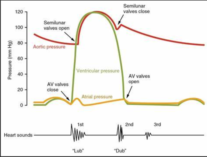
ventricular pressure is higher than atrial pressure so AV valves close
ventricular pressure is higher than aortic pressure so semilunar valves open
ventricular pressure falls below aortic pressure so semilunar valves close
ventricular pressure falls below atrial pressure so AV valves open