Skeletal System
Intro to the Skeleton
Joints provide movement for bones
Support, protection, movement, storage, manufacturing
Examples of protection: ribcage protects the heart & lungs from injury
Examples of storage: storage of minerals to be released into the bloodstream, storage of fat in yellow bone marrow
manufacturing: Production of red & white blood cells from red bone marrow
Bones are a solid matrix of living cells and fibers surrounded by calcium deposits
Bones are classified by their shape
Long bones have 2 basic regions: diaphysis (shaft) and epiphyses (ends of bone)
Epiphyseal plate (growth plate) is a thin layer of internal cartilage between the diaphysis and epiphyses
Articular cartilage is an external layer of cartilage that covers the ends of the epiphyses, providing smooth movement of joints and cushion from shock
In the diaphysis of the long bone, there is a hollow medullary cavity
Red bone marrow fills this cavity in young people, but age causes it to be replaced with fatty yellow bone marrow
New blood cells are produced in the bone marrow (hematopoiesis)
Summary: The skeletal system provides many functions for the body. Bones are classified by shape and have a specific structure with bone marrow in the center of the diaphysis and articular cartilage surrounding the epiphyses.
Microscopic Anatomy of Bone
The outer layer of bone is called periosteum, which is tough connective tissue
Periosteum is the location of muscle attachment and bone repair
Beneath the periosteum is a thick layer of compact bone
At the ends of long bones, the spongy bone layer is beneath the compact bone
Spongy bone is a lattice of trabeculae that are found along lines of stress for perfect resistance from compression
Spaces between the trabeculae are filled with marrow or blood vessels
Compact bone is arranged in cylinders called osteons
Osteons are arranged in concentric circles called lamellae
Lamellae surround a central (or Haversian) canal that contains blood vessels and nerves
Central canals are connected by perforating (Volkmann's) canals running perpendicularly
Osteocytes are mature bone cells that make up the majority of the bone structure
Osteocytes are connected with tentacle-like canaliculi, which allow them to receive nutrients and remove wastes
Osteoclasts and osteoblasts line the Haversian canals and the surfaces of the compact and spongy bone
Osteoclasts break down bone, while osteoblasts produce new bone
Summary: Spongy bone contains large spaces while compact bone is made of column-shaped osteons. Specialized bone cells build and destroy bone, while canaliculi keep the bone cells connected to nutrients.
Bone Formation and Remodeling
An embryo's skeleton is made of cartilage
Osteoblasts begin to secrete mineral deposits that replace the cartilage during embryo development
This process is known as ossification
As a child grows, columns of chondrocytes at the epiphyseal plate divide and deteriorate as the matrix around them calcifies
These cells become osteoblasts, which form spongy bone
Osteoclasts secrete acid to enlarge the medullary cavity as the bone grows
Inorganic components of bone: 65% mineral salts made of hydroxyapatites, which provide bone strength and hardness
Organic components of bone: 35% osteoid made of ground substance and collagen, which provides flexibility and tensile strength
Lack of hydroxyapatite causes "Rickets," lack of collagen causes "Brittle Bone Disease"
Calcium needs to be maintained at a certain level in the blood
Bones act as a storage tank for calcium
Two hormones, calcitonin and parathyroid hormone (PTH), trigger the processes of bone creation or dissolution
Bone homeostasis: when Ca2+ level is too high, calcitonin is produced to decrease it; when Ca2+ level is too low, PTH is produced to increase it
Osteoclasts break down bone to release Ca2+ into the blood, osteoblasts absorb Ca2+ from the blood to build bone
Steps of bone repair: hematoma forms, callus forms, callus ossifies, compact bone forms
Summary: Bone is formed as cartilage calcifies. The osteoid portion of the bone provides flexibility while mineral salts give bones strength. Hormones direct the constant remodeling of bone. When a bone is broken, a hematoma forms, followed by a callus to repair the fracture.
Axial skeleton
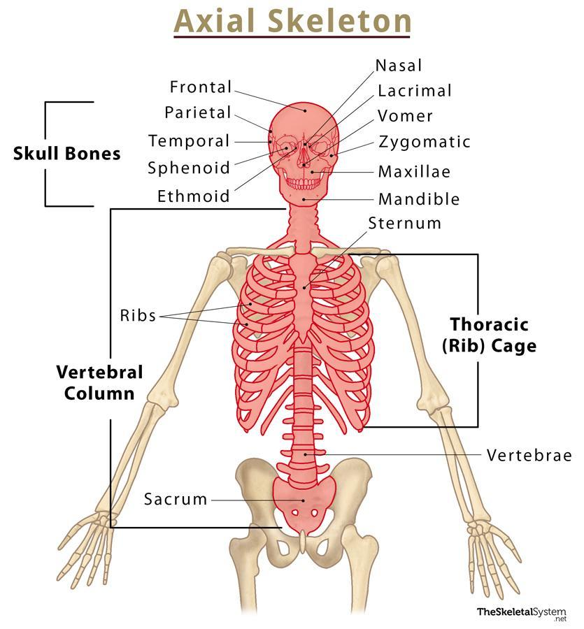
Axial skeleton: central axis of the body, includes the skull, ribs, sternum, and vertebrae (80 bones)
Appendicular skeleton: pectoral and pelvic girdles, bones of the arms, legs, pelvis, and shoulders (126 bones)
Bones found within the axial skeleton: cranium, mandible, cervical vertebrae, ribs, thoracic vertebrae, manubrium, lumbar vertebrae, sternum, sacrum, coccyx
Bones of the cranium are circled in yellow, other bones are major facial bones
Vertebral column extends from the skull to the pelvis, provides support and protects the spinal cord
Consists of 33 vertebrae at birth, but some fuse in adolescence
Intervertebral discs separate the remaining vertebrae and provide cushioning and shock absorption
Cervical vertebrae (7), thoracic vertebrae (12), lumbar vertebrae (5), sacral vertebrae (5 fused), coccyx (4 fused)
The spine has convex and concave curves
Primary (convex) and secondary (concave) curvatures allow for better balance and weight distribution
Summary: The skeleton is divided into axial and appendicular portions. Within the axial skeleton are the skull, vertebral column, and ribs. The vertebral column has 5 sections. The sections that aren't fused together have intervertebral discs for cushioning.
Appendicular Skeleton
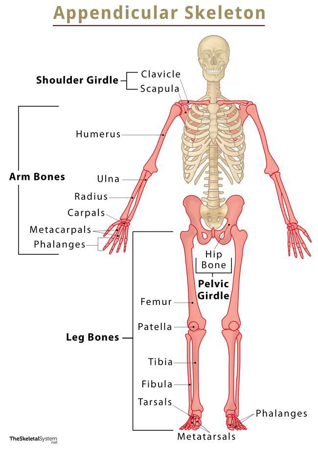
Bones found within the appendicular skeleton: clavicle, scapula, humerus, radius, ulna, carpals, metacarpals, phalanges
Bones found within the appendicular skeleton: pelvic girdle (pelvis), femur, patella, tibia, fibula, tarsals, metatarsals, phalanges
3 structural types of joints:
Fibrous joints:
Immovable or slightly movable
Held together by fibrous connective tissue
Cartilaginous Joints:
Immovable or slightly movable
Held together by cartilage
Synovial joints:
Highly movable
Contain synovial fluid for frictionless movement
Components of a synovial joint:
Joint capsule filled with synovial fluid surrounds the end of the bones.
Synovial membrane and articular cartilage line the joint cavity.
Ligaments attach the bones of the joint.
Ligaments:
Connect bone to bone
Tendons:
Connect muscle to bone
Types of synovial joints:
Pivot joint:
Between C1 and C2 vertebrae
Hinge joint:
Elbow
Saddle joint:
Between trapezium carpal bone and first metacarpal bone
Ball & socket joint:
Hip
Condylar joint:
Between radius and carpal bones of wrist
Gliding joint or plane joint:
Between tarsal bones
Summary:
The appendicular skeleton consists of the pelvis, legs, and arms.
The bones of the skeleton are connected by joints, which vary in structure and function.
Synovial joints are the most movable and can provide a range of movements based on the shape of the bones involved.
Bone Fractures
Bone fractures are breaks or cracks in the bone.
Fractures can occur due to trauma, such as falls or accidents, or due to underlying conditions like osteoporosis.
Common types of fractures include:
Closed fracture: the bone breaks but does not penetrate the skin.
Open fracture: the bone breaks and penetrates the skin, increasing the risk of infection.
Greenstick fracture: a partial break in the bone, commonly seen in children.
Comminuted fracture: the bone breaks into multiple fragments.
Symptoms of a fracture may include pain, swelling, bruising, deformity, and difficulty moving the affected area.
Treatment options for fractures include immobilization with casts or splints, realignment of the bone (reduction), and in some cases, surgery.
Recovery time for fractures varies depending on the type and severity of the fracture, as well as individual factors like age and overall health.
Complications of fractures can include infection, delayed healing, malunion (improper alignment during healing), and nonunion (failure of the bone to heal).
Rehabilitation exercises and physical therapy may be necessary to regain strength and mobility after a fracture.
Study Plan: Intro to the Skeleton
Day 1: Introduction to the Skeleton and Bone Structure
Read and understand the functions of the skeletal system.
Memorize the examples of protection, storage, and manufacturing provided by the skeleton.
Study the classification of bones based on shape.
Learn the structure of long bones, including the diaphysis, epiphyses, epiphyseal plate, and articular cartilage.
Understand the concept of the medullary cavity and the difference between red and yellow bone marrow.
Review the process of hematopoiesis in the bone marrow.
Day 2: Microscopic Anatomy of Bone
Familiarize yourself with the periosteum and its functions.
Study the composition and arrangement of compact bone and spongy bone.
Understand the structure of osteons and lamellae in compact bone.
Learn about the canals present in compact bone, including the central (Haversian) canal and perforating (Volkmann's) canals.
Memorize the functions of osteocytes, osteoclasts, and osteoblasts.
Understand the role of canaliculi in connecting osteocytes and providing nutrients.
Day 3: Bone Formation and Remodeling
Review the formation of bone from cartilage during embryo development.
Understand the process of ossification
Study the growth of bones during childhood and the role of osteoclasts in enlarging the medullary cavity.
Memorize the inorganic and organic components of bone and their functions.
Learn about the importance of calcium in bone health and the role of hormones (calcitonin and parathyroid hormone) in maintaining calcium levels.
Understand the steps of bone repair and the formation of callus and compact bone.
Day 4: Axial Skeleton
Study the components of the axial skeleton, including the skull, ribs, sternum, and vertebrae.
Memorize the different types of vertebrae and their numbers.
Understand the structure and functions
Learn about the primary and secondary curvatures of the spine and their significance.
Review the role of intervertebral discs in cushioning and shock absorption.
Day 5: Appendicular Skeleton and Joints
Read and understand the bones of the appendicular skeleton:
Clavicle, scapula, humerus, radius, ulna, carpals, metacarpals, phalanges
Pelvic girdle (pelvis), femur, patella, tibia, fibula, tarsals, metatarsals, phalanges
Learn the three structural types of joints:
Fibrous joints: immovable or slightly movable, held together by fibrous connective tissue
Cartilaginous joints: immovable or slightly movable, held together by cartilage
Synovial joints: highly movable, contain synovial fluid for frictionless movement
Understand the components of a synovial joint:
Joint capsule, synovial fluid, synovial membrane, articular cartilage, ligaments, and tendons
Day 6: Types of Synovial Joints
Focus on the types of synovial joints:
Pivot joint: between C1 and C2 vertebrae
Hinge joint: elbow
Saddle joint: between trapezium carpal bone and first metacarpal bone
Ball & socket joint: hip
Condylar joint: between radius and carpal bones of the wrist
Gliding joint or plane joint: between tarsal bones
Study the characteristics and movements associated with each type of synovial joint
Day 7: Bone Fractures
Learn about bone fractures:
Causes of fractures: trauma and underlying conditions like osteoporosis
Common types of fractures: closed, open, greenstick, comminuted
Recognize symptoms of fractures: pain, swelling, bruising, deformity, difficulty moving
Understand treatment options for fractures:
Immobilization with casts or splints
Realignment of the bone (reduction)
Surgery in severe cases