Week 7: Presynaptic Processes and Neurotransmitter Release
Presynaptic Processes: Neurotransmitters, Ca2+ channels, vesicular release, and presynaptic proteins
Purpose of notes: Provide a comprehensive, exam-ready synthesis of presynaptic neurotransmission from the provided transcript, with emphasis on definitions, mechanisms, components, and practical implications.
What is a neurotransmitter and why?
Neurotransmitters are chemical signals released from presynaptic nerve terminals into the synaptic cleft to affect a postsynaptic cell.
Historical framing: the “Soup vs Spark” controversy highlighted the chemical nature of signaling at synapses (as opposed to purely electrical transfer).
Key historical milestones:
John Eccles vs Henry Dale debate and Otto Loewi’s frog heart experiments (1921) demonstrated chemical transmission by vagal stimulation releasing acetylcholine (ACh) that slowed heart rate.
Loewi and Dale awarded the 1936 Nobel Prize for chemical transmission of nerve impulses.
Eccles, Hodgkin, Huxley awarded the 1963 Nobel Prize for ionic mechanisms in synaptic transmission.
Practical takeaway: neurotransmitters are chemical messengers released from presynaptic terminals to influence postsynaptic physiology.
Neurotransmitter criteria
A transmitter must satisfy a set of criteria to be defined as such, with acknowledged exceptions:
Synthesis: produced within the neuron.
Storage: stored within the neuron.
Release: released upon depolarization of the neuron.
Receptor: causes a biological effect on a postsynaptic receptor when released.
Inactivation: there is a mechanism for inactivation (uptake or degradation).
Postsynaptic mimicry: if applied to the postsynaptic membrane, should mimic the effect of release.
Note: some substances do not fit all criteria in every context, leading to nuanced classifications.
Classical (small molecule) vs Non-classical (large peptides) neurotransmitters
Classical (small molecules):
Amino acids: Glutamate, γ-aminobutyric acid (GABA), Glycine
Monoamines: Noradrenaline (norepinephrine), Dopamine, Adrenaline (epinephrine), 5-hydroxytryptamine (5-HT, serotonin), Acetylcholine (ACh)
Non-classical (large peptides):
Peptides: Substance P, Vasoactive intestinal peptide (VIP), Somatostatin, Neuropeptide Y (NPY), Enkephalin (Enk), Kisspeptin
Gases: Nitric oxide (NO), Carbon monoxide (CO)
Lipids: Anandamide
Conceptual distinction: Classical transmitters tend to act rapidly and locally; non-classical transmitters (peptides, gases, lipids) can have slower, longer-lasting, or broader signaling roles.
Comparison: Classical vs Non-classical neurotransmitters
Classical vs Non-classical (summary):
Size: Small molecules vs Neuropeptides (4–100 amino acids) for non-classical.
Synthesis: Uptake/enzymes for classical vs Protein synthesis for neuropeptides.
Vesicles: Small vesicles for classical; large dense-core vesicles for neuropeptides.
Duration of action: Fast and short for classical vs slow and long for non-classical.
See detailed comparison figure in transcript for specifics.
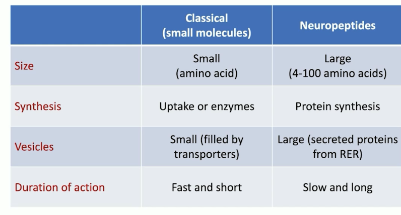
Summary of neurotransmitter concepts
Neurotransmitter: a substance released at a synapse by one neuron that modulates a postsynaptic cell in a specific manner.
Primary classes: classical (small molecules) vs non-classical (peptides, gases, lipids).
Exceptions exist; the classification helps predict release mechanisms and postsynaptic effects.
Presynaptic processes: basic process and nerve terminal organization
Goal: Describe how neurotransmitter release is organized at the presynaptic terminal.
Sequence of events (neurotransmission):
Transmitter synthesized and stored.
Action potential arrives.
Depolarization opens voltage-gated Ca2+ channels (VGCCs).
Ca2+ influx through channels.
Ca2+ triggers vesicle fusion with presynaptic membrane.
Transmitter released into the synaptic cleft by exocytosis.
Transmitter binds postsynaptic receptors.
Opening of postsynaptic channels.
Post-synaptic current alters excitability (EPSP/IPSP).
Reuptake and/or degradation terminate the signal.
Organization of the nerve terminal includes a reservoir of vesicles, the active zone, and a dense network of proteins coordinating release.
Voltage-gated Ca2+ channels (VGCCs) and roles in release
VGCCs couple membrane depolarization to transmitter release.
Major VGCCs involved in fast synaptic transmission are high-voltage-activated channels:
N-type (Cav2.2): classic blocker: conotoxin (from cone snails)
P/Q-type (Cav2.1): classic blocker: agatoxin (from spiders)
Calcium entering through VGCCs binds to vesicle-release proteins to trigger vesicle fusion and transmitter release.
Reference: Gambardella (2014) Prog Neurobiol, 213:87-96.
Transmitter release in packets: quantal release
Concept: neurotransmitter release occurs in packets called quanta.
Quantum content: the amount of transmitter in one vesicle; at the neuromuscular junction, ~ molecules of acetylcholine per vesicle.
Postsynaptic response to a single quantum is the miniature end-plate potential, mEPP.
mEPP size is about 1/100 of the end-plate potential (EPP) produced by whole-nerve stimulation:
Consequence: normal neurotransmission largely results from the release of many vesicles simultaneously.
Historical note: Bernard Katz and colleagues contributed foundational work on quantal release; Katz received the Nobel Prize in 1970.
Postsynaptic receptor interactions in quantal events: ~ receptors interact with the ACh in the neuromuscular junction example.
This quantal framework links vesicle content, release probability, and postsynaptic response.
Co-transmitters: co-release vs co-transmission
Co-transmitters: some terminals release more than one transmitter type.
Low-frequency stimulation:
Preferentially raises near-membrane Ca2+ leading to release from small clear-core vesicles (classical transmitters).
High-frequency stimulation:
Increases global Ca2+ and can trigger release of neuropeptides from large dense-core vesicles in addition to small-molecule transmitters.
Definitions:
Co-release: two transmitters packaged in the same vesicles and released together.
Co-transmission: transmitters packaged in distinct vesicles with differential Ca2+ sensitivity or spatial segregation, allowing selective release depending on activity patterns.
Reference: Varga et al., 2014 Curr Opin Neurobiol. 29:25-32.
Co-release and co-transmission: practical distinctions
Co-release scenario (same vesicles): both neurotransmitters are released together when the terminal depolarizes.
Co-transmission scenario (distinct vesicles): differential release from vesicle populations based on Ca2+ sensitivity; possible spatial segregation to different boutons or targets.
Implications: enables nuanced signaling, targeting different postsynaptic receptors, and allowing complex modulation of neural circuits.
Relationship between synaptic vesicle exocytosis and quantal transmitter release
Visualization and evidence:
Electron micrograph showing the arrangement of Ca2+ channels near vesicle fusion sites and vesicle fusion events (freeze-fracture EM).
Manipulating AP duration (e.g., with 4-AP) changes the number of vesicle fusions and quanta released.
Schematic of frog presynaptic terminals shows vesicles arranged in rows and connected to each other.
Practical takeaway: the probability and pattern of vesicle fusion govern the quantal output of a synapse.
Co-transmitters and vesicle pools: a mechanistic view
At low activity, selective Ca2+ microdomains near the membrane favor release from small clear-core vesicles (classical transmitters).
At higher activity, global Ca2+ elevation triggers release from large dense-core vesicles carrying neuropeptides and other modulators.
This dynamic allows a single neuron to switch signaling modes based on activity patterns.
Presynaptic vesicles and their roles
Vesicle membrane surface is densely populated with proteins (only a subset shown in illustrations).
Roles of vesicle-associated proteins include tethering, docking, priming, fusion, recycling, and maintenance of vesicle pools.
Example: Synapsin proteins help tether vesicles to the cytoskeleton, maintaining a reserve pool by crosslinking vesicles to each other and to actin.
Proteins in vesicle recycling: docking, priming, fusion, and beyond
Key steps and protein players:
Docking: involves GTP-binding proteins and SNAREs (to position vesicles at the membrane).
Priming: SNAREs, NSF, and SNAPs prepare vesicles for fusion.
Fusion: Synaptotagmin 1 acts as the Ca2+ sensor; SNAREs mediate fusion.
Coating: Clathrin, synaptotagmins, synaptobrevin, NSF, SNAPs regulate vesicle coat formation.
Budding: Dynamin, clathrin, actin drive vesicle formation from membranes.
Uncoating: Clathrin, Hsc-70, auxilin, synaptojanin remove coats after vesicle formation.
Structure and function of the SNARE complex
The SNARE complex is the core fusion machinery (“SNAP receptor”) and is essential for vesicle fusion.
Core components:
Synaptobrevin (vesicular SNARE)
Syntaxin (plasma membrane SNARE)
Snap25 (plasma membrane SNARE)
Synaptogamin (vesicular Ca2+-binding protein) acts as the Ca2+ sensor (in many contexts) and interacts with the SNARE complex to promote fusion.
The SNARE complex brings vesicle and plasma membranes into close apposition to enable fusion pore formation and transmitter release.
Synaptic vesicle recycling: models
Three main models of vesicle recycling discussed:
Kiss-and-run: vesicles transiently fuse, release transmitter through a transient pore, then reseal and are reused without full collapse into the membrane.
Clathrin-mediated endocytosis: vesicles fuse and collapse into the membrane; new vesicles are reformed at a distance via clathrin coats.
Ultrafast endocytosis (new model, Science 2015): rapid internalization of membrane, vesicle membrane delivered to an endosome, followed by clathrin-mediated regeneration of synaptic vesicles from the endosome.
Key takeaway: vesicle recycling can occur via multiple pathways, potentially coexisting and operating at different timescales depending on activity.
Summary of vesicle biology and neurotransmission (recap)
The surface of synaptic vesicles is densely populated with proteins, most of which contribute to vesicle recycling and cycling.
Many vesicle-associated proteins function as tethering or anchoring components; exact roles for some remain incompletely understood.
The SNARE complex is central to vesicle fusion.
There are three recognized models of vesicle recycling—clathrin-mediated, kiss-and-run, and ultrafast endocytosis with endosomal regeneration.
Connections to broader concepts and implications
Electrical signals are transduced into chemical signals at the synapse, enabling synaptic density and network-level computation through transmitter diversity, vesicle pools, and receptor subtype distribution.
Activity-dependent release patterns (frequency, duration) shape which transmitters are released (classical vs peptide) via distinct vesicle populations and Ca2+ microdomains.
Understanding vesicle recycling pathways informs pharmacology and potential therapeutic targets for synaptic dysfunction.
The historical Nobel Prize-context anchors the significance of chemical neurotransmission and the discovery of ion-channel mechanisms in neural signaling.
Key terms to remember
Neurotransmitter, postsynaptic receptor, EPSP, IPSP, mEPP, EPP, quantal release, quantum, vesicle, dock, priming, fusion, SNARE, Synaptotagmin, SNARE complex, VGCC, Cav2.2 (N-type), Cav2.1 (P/Q-type), conotoxin, agatoxin, Kiss-and-run, clathrin-mediated endocytosis, ultrafast endocytosis, endosome, dense-core vesicles, small-clear-core vesicles, co-release, co-transmission, TAC (transmitter-associated cortex placeholders if needed).
References and notes
Loewi, Otto (1921) demonstration of chemical transmission; Nobel Prize 1936.
Nobel Prize context: Eccles, Hodgkin, Huxley (1963) for ionic mechanisms of synaptic transmission.
Katz, Bernard (Nobel Prize 1970) on quantal release at the neuromuscular junction.
Varga et al. (2014) Curr Opin Neurobiol. Co-release and co-transmission explored.
Gambardella (2014) Prog Neurobiol, 213:87-96 on VGCC roles.
Science (2015) on ultrafast endocytosis and vesicle recycling models.