2: Auto-enzymatic & Allo-enzymatic Digestion & Nutrient Absorption
Learning objectives
Categorize livestock as auto-enzymatic or allo-enzymatic digester
Classify animals/avian species as omnivores or herbivores and identify their unique modifications
Identify the anatomy, functions and secretions of each organ in the digestive tract of important livestock species
Explain how absorption occurs through the phospholipid bilayer
General anatomy, digestion, & absorption
In general, everything inside the Kingdom Mammalia shares the same basic common digestive anatomy, aside from a few evolutionary modifications. Some have evolved exaggerated organs and some have even lost function or an organ completely. However, humans and livestock all have the following:
Principle parts of the digestive system (across all species):
Mouth
Esophagus
Stomach
Small intestine
Large intestine
Caecum/Colon
Anus
Major accessory organs: secrete digestive enzymes and bile into the intestinal tract which aid in digestion.
salivary glands
liver
gall bladder – exception* horses
pancreas
Types of Digestion
Mechanical Digestion: Breaking down of food into smaller particles to be processed by digestive system
Example: mastication (chewing)
Teeth chop food into smaller pieces which then pass through the digestive system
In most species, the majority of the mechanical digestion occurs in the mouth, but the stomach performs a little mechanical digestion as well through the action of churning.
The gizzard is the only form of mechanical digestion in avian species.
Chemical Digestion: Breaking down of food particles through chemical reactions
Example: stomach acid
Hydrochloric acid (pH ~ 2) denatures protein
Aids in solubilizing macronutrients through hydrolysis
Chemical reactions are usually associated with enzymatic digestion.
Enzymatic Digestion
What’s an enzyme?: An enzyme is a protein secreted by an organism that serves to ‘break down’ a substrate into two smaller products. Enzymes are secreted throughout the digestive tract with the greatest quantity coming from the pancreas.
Ex: Sucrase is the enzyme that breaks down Sucrose. The sucrase enzyme plus water breaks Sucrose down into the smaller products of glucose and fructose.
Characteristics
Enzymes are substrate specific; they work on only one type of substrate/nutrient type
Regulates from a state of low activity to high activity by attaching to a highly specific active site
Increases the rate of a reaction for a chemical reaction
The nutrient and body interface:
Now, lets look through our digestive tract tube like a telescope with a body around it. We are inside the donut hole; this is called
the lumen. We have mechanically, chemically and enzymatically broken down our food... now what?
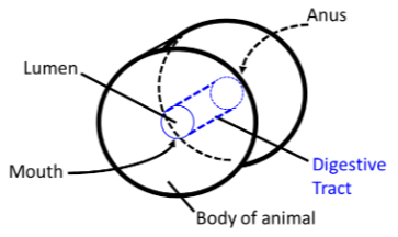
The Lumen: (the inside of the telescope):
Lumen is defined as the inside space of a tubular structure
The lumen contains digested food
Food remains inside the lumen, and is of no use, until it is absorbed into the body
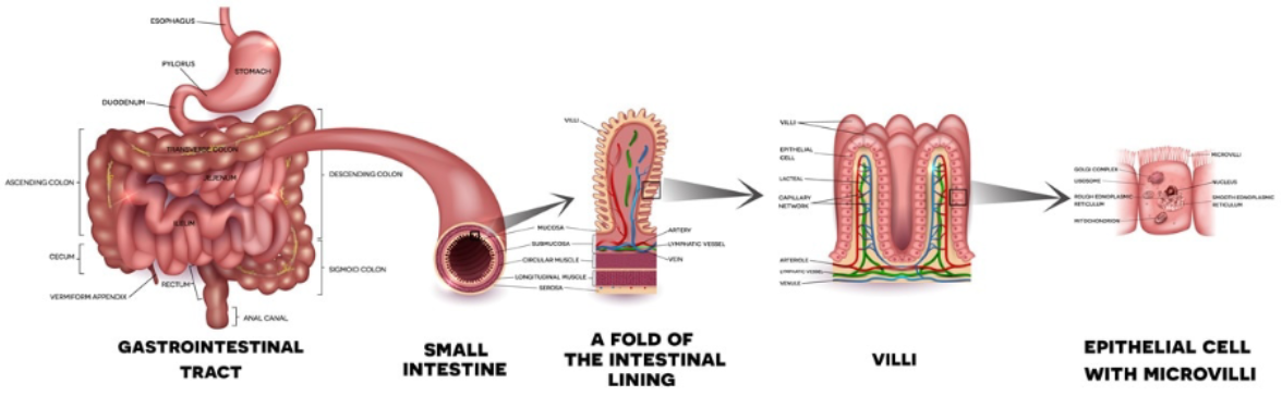
Absorption of nutrients predominantly occurs from the lumen of the small intestine through the layers of the digestive tract into general blood or lymphatic circulation
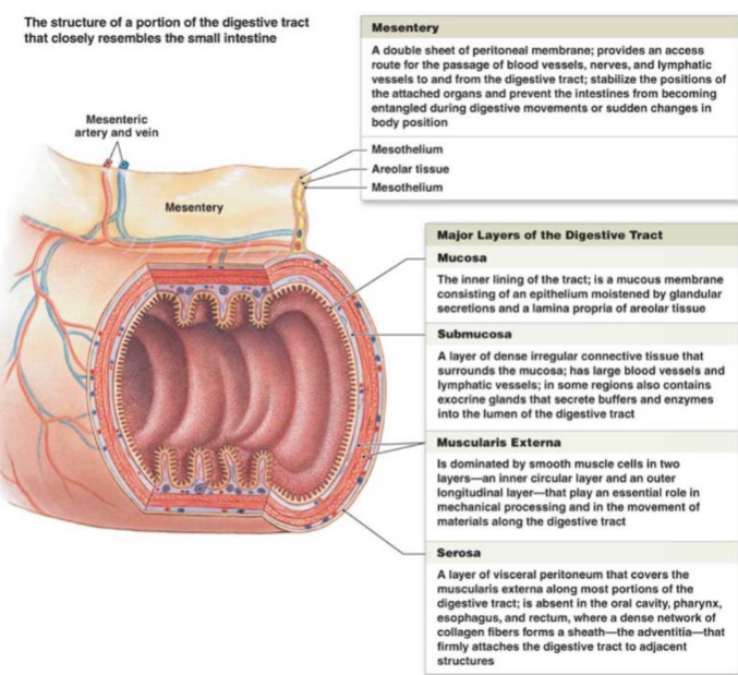
The Layers of the Small intestine
The two most active sites of digestion and absorption occur in the small intestine. The small intestine is made of layers. From inner most layer to outer most layer:
Mucosa
Submucosa
Muscularis
Serosa
Mesentery
The most active layers are the mucosa and the submucosa where nutrients are absorbed and transported. The other layers are
primarily concerned with the structure and organization of the organ within the body cavity.
MUCOSA – innermost mucous membrane layer of the digestive tract to which digested feed comes into contact; site of absorption through the epithelial cell’s phospholipid bilayer
The mucosal layer has crypts and folds and is lined with villi.
Villi are small finger like projections that contain portions of the mucosal and submucosal components.
Functions:
Increase absorptive surface area
Slow down passage rate
House absorptive cells
Transport absorbed materials to the blood or lymph system. Each villus is lined with a single layer of cells enterocytes (AKA epithelial cells): These are the primary absorptive cells for all nutrients. Each villus contains an arteriole, venule and lacteal providing access to the circulatory and lymphatic system
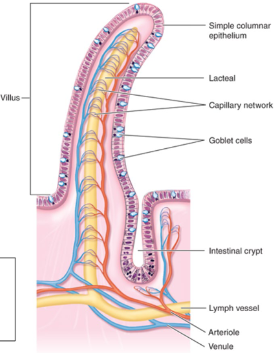
Enterocytes
1. Cells are continually formed at the base of the villus in crypts.
2. As they mature the “move up” to the tip of the villus
3. After they are “worn out” by chemical and physical attrition
they are extruded into the lumen.
4. ~ 17 billion cells/day are lost in this manner in humans!
(Moog, 1981)
5. These sloughed cells make up a significant portion of the
metabolic fecal nitrogen (aka endogenous nitrogen).
6. Diets high in fiber (or those with an abrasive effect) tend to
increase this loss.
(e) The surface area of a villus is further enhanced by microvilli lining the
lumen side of the enterocyte. The microvilli also have a fuzzy
projection called the glycocalyx.
(i) Glycocalyx: glycoprotein appendage of the microvilli which
houses enzymes (sticky mucus layer)
(ii) Microvilli + Glycocalyx = “Brush border”
(iii) The brush border helps to trap nutrient substrates and further digest them into small sub-units that can be absorbed by