Diagnostic Tests
Arriving at a clinical diagnosis
Using information about the current illness or problem of the patient (pt.) (Most important: is it an emergency or life-threatening), pt. history, physical exams, diagnostic tests
Current Illness → when did the symptoms/signs start? Under what circumstances? Have you had this before? Are they getting worse, better, or stable? What steps did the pt. take? Did anything help or change? If the pt. has had this before are there any significant changes?
Patient History: current or past medical history, family history, concurrent illnesses, alcohol, drugs, smoking, social issues, employment status, and any other important information.
Physical exam: an art of observation that is systematic but with a routine, got to know your tools
Noninvasive Procedures: ENT/eye inspection, a neurological test of reflexes, urine test, throat swab, x-ray, ultrasound, MRI, CT, and ECG (does NOT break the skin)
Invasive Procedures: blood draws, spinal tap, biopsy, endoscopy (BREAKS the skin)
Common Labs
- CBC - complete blood count + differential
- CMP - (comprehensive metabolic panel) K, Na, Cl, Ca, blood urea, glucose, N (BUN), Liver enzymes (enz) (AST, ALT)
- Plasma Proteins - albumin, enz, immunoglobulins (Igs)
- Serological test - antibodies
- Hormone - thyroid hormones
- Disease Markers - heart attack, pancreatitis, liver disease, renal disease
- Erythrocyte sedimentation rate - only useful in myeloma, temporal arteritis, polymyalgia rheumatica
- drugs
- Urine - Protein, microorganisms, blood
- Stool - blood, parasites, fats
- CSF - cells, proteins, microorganisms
Basic Lab Vernacular
- High Red Blood Cells (RBCs) - erythrocytosis or polycythemia
- Low RBCs - anemia or erythroblastopenia
- High White blood cells (WBCs) - leukocytosis (a sign of infection)
- Low WBCs - Leukopenia
- High lymphocytes - lymphocytosis
- Low lymphocytes - lymphocytopenia (a sign of cancer or HIV)
- Cytosis - high
- Penia - low
Measuring Electrical Activity (Diagnostic)
EEG → electrical activity of the brain used to detect seizures and brain activity after trauma
EMG → electrical activity of the muscles used to detect neurological conditions
Electrocardiogram (ECG, EKG) electrical activity of the heart
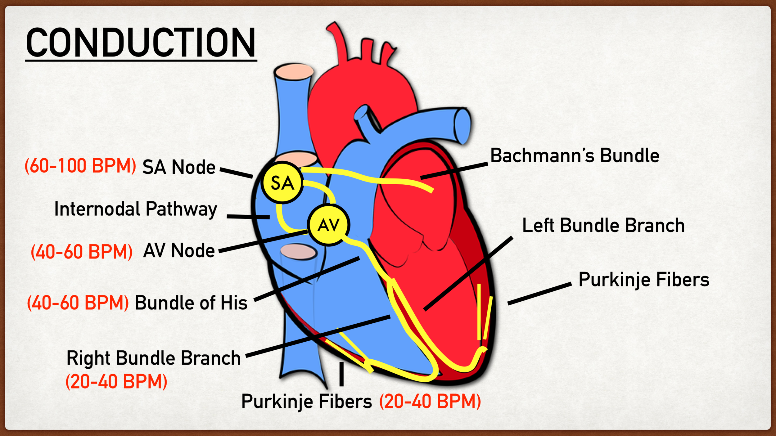
Sinoatrial Node to the Atriaventricular Node.
Fast Heart rates (tachycardia) leave not time for proper refill of the heart.
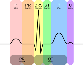 P wave: atrial depolarization (SA node)
QRS complex: Ventricular depolarization (V-contracts)
T wave: Ventricular Repolarization
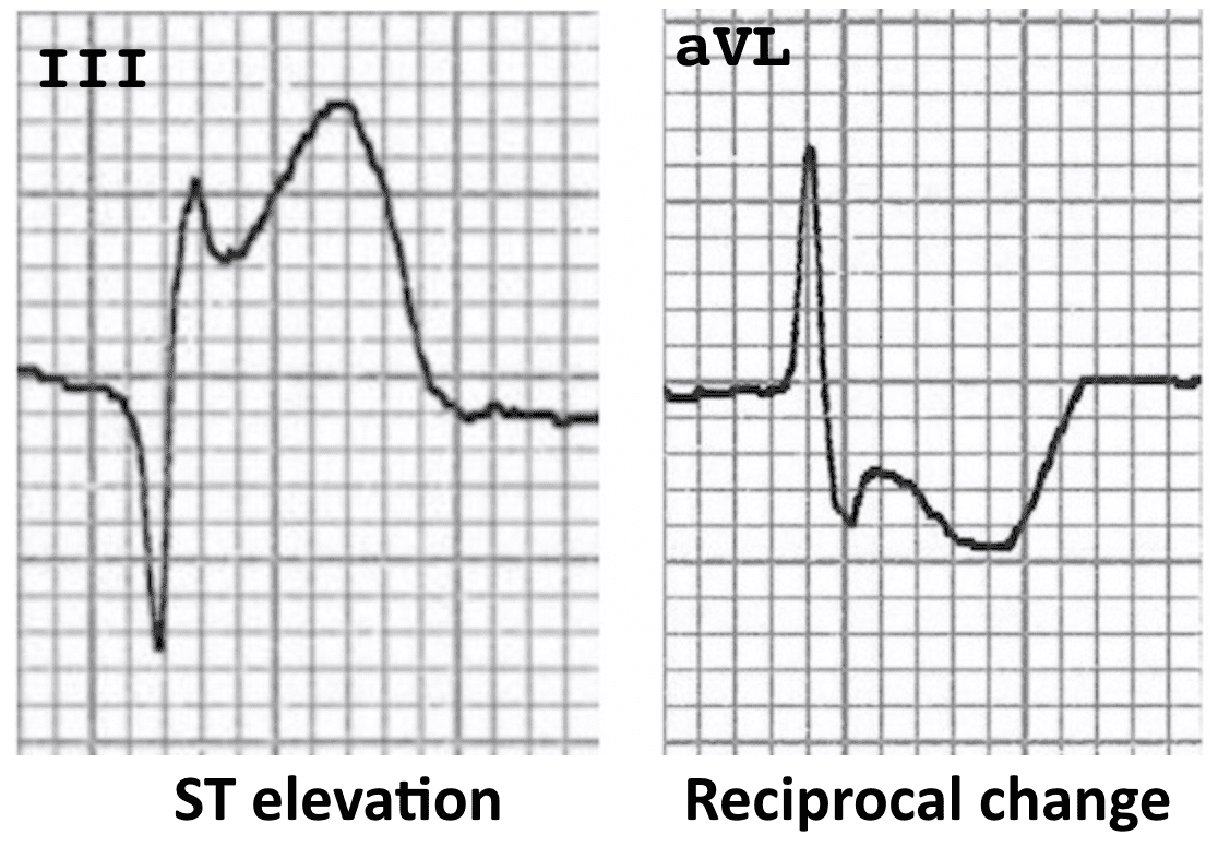
ST elevations occur in myocardial infraction
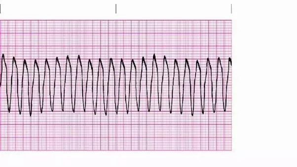
Ventricular Flutter - 250-300 bpm, sinusoidal waves
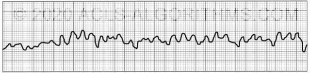
Ventricular flutter can lead to V-fibrillation where there’s no clear electrical activity.
Radiological Studies
X-rays (noninvasive)
Discovered by Wilhelm Roentgen in 1895 who worked with Electrical beams and discovered that a fluorescent screen nearby started glowing.
X-rays go through tissues, but not bones and metal
X-rays (and gamma rays) are electromagnetic radiation identical to light but have a much shorter wavelength. Where light has a wavelength of 6000 angstroms, X-rays are 1 angstrom (0.1 nm), and Gamma rays are 0.0001 angstroms.
X-rays and radioactivity used to be measured in rem (Roentgen Equivalent in Man). Now it is measured in Sievert (Sv). 1 Sv = 100 rem.
CT scan (Usually noninvasive)
CT (computerized tomographic) uses a motorized scanner that circles around patients and obtains many serial x-ray images that are then compiled into a 3-D image.
Reveals much more than an X-ray
Individual CT scans use lower intensity X-rays but have higher radioactivity (more X-ray exposure).
Can be enhanced with contrast (this is invasive)
Nuclear Medicine (invasive)
In nuclear medicine, patients are injected with material (usually iodine) that is enriched in areas of highest activity
Can be therapeutic in cancer treatment
MRI (Noninvasive)
Magnetic Resonance Imaging uses strong magnetic and radiowave fields (no metal in the room) and is very noisy (patients are provided with ear plugs/headphones).
MRI is superior for imaging soft tissues which can be “weighted” based on the structure that they could enhance based on RF pulse.
T1: one tissue is bright (the fat), used for anatomy
T2: two tissues are bright (fat and water), used for pathology
PET scan
Position Emission Tomography - injection of radioactive materials and uses gamma rays (Invasive)
Ultrasound
Ultrasound - uses a transducer to generate high-frequency sound waves (>20,000 Hz) to detect tissues in the body, humans cannot hear these sound waves which generate an echo that is reflected back differently to the transducer because tissues in the body have different densities.
Ultrasound is used for heart conditions, blood vessels, internal organs, etc
Painless and works through the skin.
Biopsies, histology, and cytology oh, my!
Biopsy - performed to obtain tissue for histologic examination (breast, bone-marrow)
Histology - examination of tissue with the microscope, the tissue is intact with its morphology preserved.
Cytology - cells recovered by various means, SINGLE cells are usually recovered tissue is not preserved, less invasive than a biopsy (pap smear, etc)
Tissue Processing
Clinicians (or pathologists) remove tissue by needle or surgery and preserve it in a fixative or frozen for examination by a pathologist. Upon receiving the tissue, the pathologist inspects the tissue, describes the tissue, marks the tissue, and dissects the tissue. Tissues must be processed before sections can be obtained for histological examination. They are frequently embedded in paraffin. Another way to preserve it is the “frozen section,” which is frequently done in surgery to give the surgeon information needed for his work. The frozen section is quickly freezing the tissue in butanol/liquid Nitrogen.
Sections must be stained before they can be examined by a pathologist. Most commonly it is an H and E stain. Special stains help discriminate tissues. PAS (Periodic acid schiff) for detection of glycogen in liver and muscle; fungi also have a positive PAS positive. Trichome stains collagen in the live, fibrosis, and cirrhosis.
Immunofluorescence staining identifies cell types, tissues, and molecules. Examples include immunoglobulins and complement proteins in a post-infectious state.
Hemolysin (H) is a basic dye that stains the nucleus blue.
Eosin (E) is an acidic dye that stains the cytoplasm pink.