Female Anatomy
Anatomy
Externally
breasts - provide nutrition for infants
vulva - opening to the vagina and various functions for intercourse
Internally
vagina - provides entry point for sperm and ext canal for babies
uterus - provides home for developing embryo/fetus during pregnancy
ovaries - produce ova that are carried to the uterus via the uterine tubes
Breasts
feature a body with nipple at apex
nipple surrounded by areola
feature sensory nerve fibers that trigger the milk ejection (let-down) reflex
capillaries and nerves close to skin surface here
areolar gland secretions protect nipple from chapping/cracking during nursing
axillary tail (tail of spence) extends towards armpit
suspensory ligaments hold in place
non-lactating feature adipose and collagen
mammary glands develop in pregnancy
15-20 lobes with alveoli (acini) lead to lactiferous duct → open into lactiferous sinuses → into nipple
glands atrophy after nursing ceases
myoepithelial cells are contractile cells found around ducts/acini that function in milk expression (stimulated y oxytocin)
breast milk is comprised of fats, carbs, proteins, vitamins, minerals, and water
also contain bioactive factors that augment the immature immune and digestive systems
Lactation
Initial stage (3-5 days postpartum) produces a nutrient-rich colostrum
milk “matures” after initial stage and shifts to a supply-demand system
breats emptying is crucial to maintaining production
prolactin stimulates milk production
let-down reflex invloves oxytocin
nipple nerve endings signal the hyothalamus to release oxytocin
milk forced from alveoli into ducts
Vulva
external genitalia = vulva
mons pubis → fat over pubic symphysis
labia majora and minora surround pudendal cleft
laia minora form the prepuce (clitoral hood)
pudendal cleft contains clitoris and vestibule
clitoris → erectile tissues that swell if aroused
more nerve endings than any other body part
vulvar vestibule → surrounds vaginal and urethral openings and contains the vestibular glands
lesser vestibular (skene’s/paraurethral) glands
secrete antimicrobial and ejaculate proteins
similar to prostate secretions
greater vestibular (bartholin’s) glands
secrete vaginal lubricants
highly variable morphology
variations based on amount of estrogen influence during development
more estrogen = larger, thicker structures
decrease in estrogen cn cause atrophy
most variation occurs within the size, shape, and color of the labia minora
2018 study measured the labia of 657 participants (age range 15-84) and found:
average labia majora= 3.1 inches long
average labia minora = 1.65 inches long
BMI correlated with labia majora size
vaginal delivery correlated with labia majora length
Clitoris
clitoris = glans clitoris + body + crura + clitoral hood/prepuce + vestibular/clitoral bulbs
glans clitoris varies in size and highly innervated
~8,000 nerve endings
prepuce protects the glans clitoris
body formed by the joinging of the crura
vestibular bulbs expand the vulva
Blood Supply
internal pudendal a. supplies the dorsal aa.
dorsal aa. gives rise to the deep dorsal aa., which supplies the corpora cavernosa that fill eith blood during erections
tunica albuginea
surrounds and separates the corpora cavernosa
forms the body and crura
helps sustain erection
dorsal vv. drain to the vesical plexus → internal iliac vv. → common iliac vv. → IVC
Musculature
various muscles act on the vulva
bulbospongiosus - closes the vagina and contributes to clitoral erections and orgasm contractions
ischiocavernosus - assists with clitoral erection
perinal mm. (deep transverse and superficial transverse)
deep transverse fixes the central tendon of the perineum (erineal body) and supports pelvic floor
levator ani mm. (illiococcygeus, pubococcygeus, and puborectalis)
supports pelvic structures, urination, childbirth, and defecation
sphincters (external anal and external urethral)
Internal Anatomy
internal organs include the ovaries, uterine tubes, uterus, and vagina
Vagina
located between bladder and rectum
8-10 cm distensible muscular canal
able to expand and priduces its own lubricant
vaginal membrane (“hymen”) around the vaginal opening
typically presents as a semi-circula ring of tissue that may be a fetal remnant
rarely cover entire opening (imperforate hymen)
many myths surround this structure
adventitia, muscularis, and mucosa layers
aglandular, but lubricated by serous fluid from cervical glands and Bartholin’s glands’ secretions
involved on sexual pleasure, menstruation, pregnancy, and childbirth
walls contain many sensory nerve endings
allows for passage of mentrual fluid
allows for entry of sperm into the uterus
referred to as the “birth canal” during childbirth
acidic environment (pH 3.8-5.0) impacted by age (4.0-4.5 in reporductive years), hydration status, diet, and safe interncourse
pH too low = reduces fertility
pH too high = increased risk of infection
microorganisms stabilize vaginal ecosystem
lactobacillus acidophilus ferments glycogen from decay of vaginal mucosa into lactic acid and releases H+
Uterus
thick, muscular chamber that opens into the roof of the vagina and tilts forwards over the urinary bladder
harbors and expels a fetus
three main parts:
fundus
body (corpus)
cervix → connects to vagina
glands secrete cervical mucus (20-60mg/day or up to 600 mg during ovulation)
protects from bacteria and can prevent sperm from entering uterus
uterine wall made of three layers
perimetrium → external serosa layer
myometrium → middle muscular layer
thickest layer made mostly of smooth muscle
produces labor contractions
endometrium → inner mucosa layer
simple columnar epithelium
multiple glands and cell types
functional layer superficially (menstrual shed)
basal layer (deep) that regenerates a new functional layer each cycle
site of attachement during pregnancy
Uterine Tubes
a.k.a “oviducts” or “fallopian tubes”
~10 cm long from ovary to uterus
muscular tube lined with ciliated cells
fimbriae, infundibulum, ampullam, isthumus
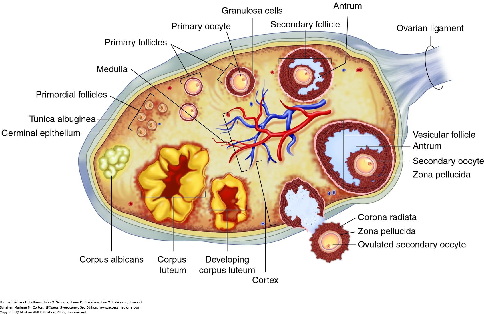
Ovaries
production of oocytes and sex hormones
oogenesis is the formation of oocytes
become ova (sing.ovum) once fertilized
each egg develops in a fluid-filled follicle
ovulation → follicle bursts and releases egg
follicles mature to house oocytes
primordial → primary → secondary → tertiary → vesticular (graafian/mature)
secrete estrogen, progesterone, and androgens
after ovulation, follicles become the corpus luteum; atrophy to corpus albicans