Midterm1 Review
MOD 1
Control Panel
25kV to 150kV is typically used in diagnostic work → impacts quality
25 to 1200mA is the range of tube current → impacts quantity
line compensation: prevents voltage fluctuations (can reduce IQ and risk pt dose) and adjusts it to keep it constant.
AEC: device that measures the quantity of radiation that reaches the image receptor, which will automatically terminate the exposure when the image receptor has received the required radiation intensity
APR: a set of pre-programmed exposure factors that allows for quick access to average/recommended exposure factors
Generator
main role of generator is to supply a steady high power source with little voltage ripple to the x-ray tube → consistent beam of electron energies striking the anode → consistent x-ray beam energy
high frequency power with near constant potential voltage
X-ray Tube
filaments are made of thoriated tungsten
Large Filament – for larger body parts – more heat dissipation - less spatial resolution (vice versa for small)
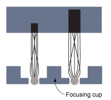
rotating anode = rotating disc anodes which allows a new part of the focal spot to be hit by the electrons for every exposure → longer anode life
three functions:
to conduct the electrons back to the generator through the cables
provide a base to support the target ring/focal spot and also to dissipate heat
produce xrays! (NOTE kinetic electron energy makes 99% heat and only 1% x-ray photons)
thermal infrared energy
electromagnetic energy
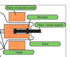
target/ focal spot = made of tungsten for its high atomic number and rhenium for strength
glass/vacuum envelope: not vacuum = the tube becomes inefficient, producing fewer photons (gassy)
protective tube housing: the part we move and adjust
filtration:
inherent - found within the xray tube
added - Al sheets between the tube and collimator exit glass mirrors and plastic windows within the collimator
MOD 2
characteristics:
-e only ionizes/ejects only if the photon energy is greater than the binding energy → -e from higher shell will drop down to fill vacancy (characteristic cascade) → the difference between the two binding energy is released as characteristic radiation
only -e at K-shell (inner) produce diagnostic photons, high binding energy = lower energy state -e
brems:
resulting photon energy reach to the same kVp energy selected (80kVp = 0-80keV)
Half Value Layer
represents the penetration ability the x-ray beam has through differing thicknesses of different materials (objects)
MOD 3
inverse square law: to see the intensity change as SID changes
doubled SID = quartered intensity
direct square law: to counteract the intensity lost
diverging beam contributes to shape & size distortions
OID increases = mag increases, sharpness reduced
SID increases = divergence reduces (with collimation) = reduced mag
*mag increases = OID increase, SID decrease * (think regarding diverging beam)
shape distortion occurs of the beam divergence and the position of the part (should be perpen to beam)
anode effects
line focus principle
smaller anode surface angle = smaller effective focal spot = better IQ
anode heel effect
focal spot blurs
can be reduced by minimizing OID
photon scatter
increased by increased kV and field size
coherent: low energy photons interact with atom → release scattered xray → increase noise very slightly
compton scatter: outer shell -e with incident xray → scattered xray and ionization of atom → may reach the receptor but doesn’t contribute to IQ = radiation fog → use grid to absorb scatter
photelectric effect: inner shell -e with incident xray → photoelectron emitted instead of scatter xray (cascade effect) → therefore ionization occurs but not scatter → this photoelectron gets absorbed in tissues
MOD 4
CR
elimination of expensive film and chemical-heavy wet processing equipment
latent image: incident x-ray photon that excites the photostimulable phosphors (PSP)
PMT converts light signals to analog light signals
ADC converts analog signal to digital
MOD 5
mag mode in fluoro
II system uses more mA when using mag mode than FPD fluoro