Muscles and Muscle Tissue Study Notes
Muscles and Muscle Tissue: Anatomy and Physiology
Introduction
Content modified by Iryna McGuire
Text derived from Human Anatomy & Physiology by Elaine N. Marieb and Katja Hoehn, Tenth Edition.
Muscle Functions
Movement of bones or fluids (e.g., blood).
Maintaining posture and body position.
Stabilizing joints.
Heat generation (especially prominent in skeletal muscle).
Types of Muscle Tissue
Skeletal Muscle Tissue
Characteristics:
Attached to bones and skin.
Striated appearance.
Voluntary control (conscious control).
Considered powerful.
Primary focus of this chapter.
Cardiac Muscle Tissue
Characteristics:
Located only in the heart.
Striated appearance.
Involuntary control.
Smooth Muscle Tissue
Characteristics:
Found in the walls of hollow organs (e.g., stomach, urinary bladder, airways).
Not striated.
Involuntary control.
Comparison of Muscle Types (Table 9.3)
Skeletal Muscle:
Location: Attached to bones or skin.
Cell shape: Single, long, cylindrical, multinucleate with obvious striations.
Cardiac Muscle:
Location: Walls of the heart.
Cell shape: Branching chains of cells; uni- or binucleate; striations present.
Smooth Muscle:
Location: In walls of hollow visceral organs; multiunit muscle in intrinsic eye muscles, airways, and large arteries.
Cell shape: Single, fusiform, uninucleate; no striations.
Special Characteristics of Muscle Tissue
Excitability (Responsiveness or Irritability): Ability to receive and respond to stimuli.
Contractility: Ability to shorten when stimulated.
Extensibility: Ability to be stretched.
Elasticity: Ability to recoil to resting length.
Skeletal Muscle Overview
Each muscle is supplied by:
One artery
One nerve
One or more veins.
Connective Tissue Sheaths of Skeletal Muscle
Epimysium: Dense, regular connective tissue surrounding the entire muscle.
Perimysium: Fibrous connective tissue surrounding muscle fascicles (groups of muscle fibers).
Endomysium: Fine areolar connective tissue surrounding each muscle fiber.
Skeletal Muscle Attachments
Direct Attachment:
Epimysium of muscle fuses to the periosteum of bone or the perichondrium of cartilage.
Indirect Attachment:
Connective tissue wrappings extend beyond muscle, forming a ropelike tendon or sheetlike aponeurosis.
Microscopic Anatomy of a Skeletal Muscle Fiber
Cylindrical cell up to 30 cm (approximately 12 inches) in length.
Contains:
Multiple peripheral nuclei.
Many mitochondria.
Glycosomes for glycogen storage and myoglobin for O2 storage.
Myofibrils, sarcoplasmic reticulum, and T tubules.
Sarcolemma
The sarcolemma is a specialized cell membrane surrounding striated muscle fiber cells.
Functions similarly to a typical plasma membrane, but adapted for muscle cell activities.
Myofibrils
Densely packed, rodlike elements accounting for about 80% of cell volume.
Exhibit striations: perfectly aligned repeating series of dark A bands and light I bands.
Sarcoplasmic Reticulum (SR)
A network of smooth endoplasmic reticulum surrounds each myofibril.
Functions in the regulation of intracellular levels.
T Tubules and Triads
T Tubules are continuous with the sarcolemma, facilitating the transmission of action potentials into the interior of the muscle fibers.
A triad consists of a transverse tubule and two terminal cisternae of the sarcoplasmic reticulum, which stores Ca²+ ions for muscle contraction.
Sarcomere
Defined as the smallest contractile unit (functional unit) of a muscle fiber.
The region between two successive Z discs, composed of thick and thin myofilaments made of contractile proteins.
Features of a Sarcomere
Thick filaments: Run the entire length of an A band.
Thin filaments: Extend the length of the I band and partway into the A band.
Z-disc: A protein sheet anchoring the thin filaments and connecting myofibrils, coin-shaped sheet of proteins.
M line: A protein line made of myomesin that holds adjacent thick filaments together.
Sarcomere: The functional unit of a muscle fiber, defined as the segment between two Z-discs and responsible for muscle contraction.
Neuromuscular Junction
The site where a motor neuron meets a muscle fiber.
Excitation by the neuron is the only functional way to activate contraction.
It represents a chemical synapse formed by the contact between a motor neuron and a muscle fiber.
The same principles apply to both the contraction of a single fiber and a whole muscle.
Neuromuscular Junction Implications
Auto-Antibodies (Myasthenia Gravis)
In conditions such as Myasthenia gravis, auto-antibodies target acetylcholine receptors (AChR), leading to impaired muscle activation due to the prevention of acetylcholine binding and consequent muscle fiber activation.
Muscle Development and Regeneration
Satellite Cells: These cells can be incorporated into muscle cells, facilitating protein synthesis and aiding in the repair and growth of muscle tissues.
Fibrosis: Involves the replacement of muscle fibers with scar tissue.
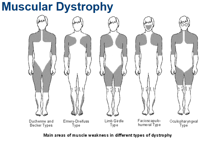
Muscular Dystrophy
A group of inherited diseases characterized by muscle destruction.
Muscles may enlarge due to fat and connective tissue deposits, while the muscle fibers undergo atrophy.
Types include:
Duchenne and Becker Types
Emery-Dreifuss Type
Limb Girdle Type
Facioscapulohumeral Type
Oculopharyngeal Type
Duchenne Muscular Dystrophy (DMD)
Recognized as the most common and severe type of muscular dystrophy.
Inheritance: Sex-linked, carried by females and expressed in males (occurring at a rate of 1 in 3500).
Symptoms include clumsiness and frequent falls; this typically leads to respiratory failure in the 20s.
Treatment: Currently no cure, but advancements such as viral gene therapy or infusion of stem cells with the correct dystrophin genes show promise.
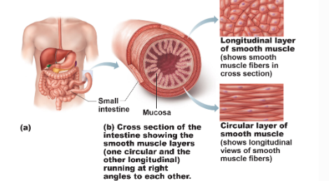
Smooth Muscle
Found in the walls of most hollow organs except the heart.
Typically organized in two layers:
Longitudinal layer
Circular layer.
Special Features of Smooth Muscle
Hyperplasia: Smooth muscle cells can divide and increase in number.
Example: Effects of estrogen on the uterus at puberty and during pregnancy can induce hyperplasia in smooth muscle cells.
Summary Notes
What are the three types of muscle tissue?
The three types of muscle tissue are skeletal, cardiac, and smooth muscle.
What types of muscle do not have striations?
The types of muscles that do not have striations are smooth muscle, which is found in the walls of hollow organs and is involuntary in nature.
What type of muscles are voluntary?
The voluntary muscles are skeletal muscles, which are attached to bones and facilitate movement through conscious control.
Learn the structural organization of skeletal muscles. (from a muscle fiber to a whole muscle)
The structural organization of skeletal muscles comprises several levels, beginning with individual muscle fibers (myofibrils) that group into fascicles, which are bundled to form the entire muscle.
What is the repeating structural and functional unit of skeletal muscles?
The repeating structural and functional unit of a skeletal muscle is the sarcomere, which contains myofilaments arranged in a highly organized manner, allowing for contraction.
Learn the structure of a skeletal muscle cell. How is it different from the majority of other cells in your body?
The structure of a skeletal muscle cell differs from most other cells in that it is multinuclear, meaning it contains multiple nuclei within a single cell membrane, which is essential for muscle function and repair. It is different from the majority of the cells in your body by having a unique striated appearance due to the precise alignment of actin and myosin filaments, which is critical for its ability to contract efficiently.
What is a triad? What is the structure of a triad?
A triad is a specialized arrangement of muscle cell structures consisting of one T-tubule and two terminal cisternae of the sarcoplasmic reticulum, which play a crucial role in facilitating the rapid transmission of electrical signals and the release of calcium ions necessary for muscle contraction. (acts as a rapid signaling unit).
Learn the structure of a sarcomere. What is the Z line? What is the M line?
The structures of a sarcomere are essential for muscle contraction and consist of various components, including the Z line, which marks the boundaries between adjacent sarcomeres, and the M line, located in the center of the sarcomere and serving as an anchoring site for the thick filaments.
Learn the structure of a neuromuscular junction.
The structures of a neuromuscular junction include a chemical synapse formed between the motor neuron and the muscle fiber, where neurotransmitters such as acetylcholine are released to stimulate muscle contractions.
Learn the mechanism of muscle development and regeneration.
The mechanisms of muscle development and regeneration are complex processes involving satellite cells, which are activated in response to muscle injury or stress, allowing for muscle repair and growth. Fibrosis, which is the process where connective tissue replaces muscle tissue, can hinder muscle regeneration and flexibility if excessive.
What is muscular dystrophy?
Muscular dystrophy is a group of genetic disorders characterized by progressive weakness and degeneration of the skeletal muscles, ultimately leading to a decrease in muscle function and mobility.
What protein is affected in muscular dystrophy?
Dystrophin is the protein that is primarily affected in muscular dystrophy, particularly in Duchenne Muscular Dystrophy (DMD), where mutations prevent its production, resulting in muscle degeneration.
Learn the signs and symptoms of muscular dystrophy and available treatments.
The signs and symptoms of muscular dystrophy are clumsiness, frequent falling, difficulty running or jumping, muscle weakness, and changes in posture. As the condition progresses, individuals may also experience difficulty with breathing and heart problems. Treatment options include physical therapy to maintain mobility, the use of corticosteroids to help improve muscle strength, and potential interventions such as cardiac care and respiratory support as needed. As well as gene therapy, or infusion of stem cells, which are being investigated as potential treatments to restore dystrophin production and improve muscle function.