Unit 12A Child Bone Dev Disorders
DISORDERS OF MUSCULOSKELETAL FUNCTION: DEVELOPMENTAL AND METABOLIC DISORDERS
Overview of Musculoskeletal Function
The development of skeletal structures begins in utero and continues to change throughout lifespan.
Throughout childhood, skeletal structures grow in length and diameter, sustaining a large bone mass.
Modeling: refers to the formation of the macroscopic skeleton, ceasing at physical maturity (usually 18-20 years of age).
Once skeletal growth attains adult size, the process of bone remodeling is responsible for skeletal maintenance and repair:
- Remodeling entails bone resorption and formation.
- With aging, bone resorption and formation become uncoupled, leading to loss of bone
Causes of Skeletal Disorders
Skeletal disorders may develop secondarily (2º) due to:
- Abnormal growth and developmental processes due to hereditary or congenital influences.
- Other disorders developing later in life due to nutritional deficiencies, metabolic disorders, hormonal influences, or aging
Alterations in Skeletal Growth and Development
Bone Growth and Remodeling
Embryonic and Fetal Development
- The skeletal system originates from mesodermal and neural crest cells in the developing embryo.
- Development of vertebrae and axial skeleton begins in the embryo at approximately the 4th week:
- Paddle-shaped limb buds of lower extremities appear late in the 4th week.
- Hand pads develop by days 33-36, and finger rays are evident by days 41-43.
- Ossification begins by the 9th week
Abnormalities of Skeletal Development
Abnormalities that start in embryonic development are relatively uncommon, often limited to defined embryonic structures:
- Examples include congenital absence of a phalanx, formation of extra bones (supernumerary digits), or fusion of adjacent digits (syndactylism).In utero positioning during fetal development may lead to more common problems:
- Newborns may exhibit imprints of in-utero positioning; however, these are often physiologic (not anatomic) in origin
Bone Growth in Childhood
Bone growth occurs during the first two decades of life:
- The skeleton undergoes overall growth with long bones growing at a rapid rate, regulated by the epiphyseal growth plate:
- Chondrocytes synthesize cartilage in the growth plate.
- As long bones grow in length, cartilage cells multiply and enlarge, eventually calcifying and dying.
- This attracts osteoblasts which migrate into the area to secrete osteoid, assisting in mature bone formationContinuous processes within the epiphyseal growth plates include cartilage synthesis, calcification, erosion, and osteoblast invasion, allowing for bone growth without altering bone shape.
The growth plate cells cease division at puberty, and the epiphysis fuses with the metaphysis

Factors influencing growth include:
- Trauma that may lead to epiphyseal separation, typically occurring at the weakest part of the growth plate.
- Nutritional and metabolic influences, like scurvy (vitamin C deficiency) which impairs the formation of the bone's organic matrix, slowing growth at the epiphyseal plate, and rickets (vitamin D deficiency) which affects calcification → soft bonesHormones such as thyroid hormone, insulin-like growth factor, and insulin are critical for physiologic growth:
- Alterations in hormone levels can impact growth.
- After puberty, the responsiveness of the epiphyseal plate to hormones decreases, leading most individuals to stop growing by age 20, although some bones (skull, jaw, fingers, and feet) may continue to grow
Alterations During Normal Growth Periods
Infants and children experience changes in muscle tone and joint motion during physiological growth:
- Toeing-in, toeing-out, bowlegs, and knock-knees are common occurrences during infancy and childhood, usually corrected during normal growthPhysiologic flexion contractures of the hips may be evident in the infant
During the first year of life, the lower extremities straighten in preparation for walking
Musculoskeletal assessments of newborns are crucial:
- Identify abnormalities for early intervention.
- Establish baselines for reference.
- Educate and counsel parents and caregivers.
- Facilitate treatment
Torsional Deformities
Infants and toddlers have lax ligaments that tighten with age:
- Hypermobility combined with torsional forces during growth can lead to deformities.
- Common torsional deformities include toeing-in (metatarsus adductus), toeing-out (femoral retroversion), and tibial torsion:
- Toeing-in (metatarsus adductus) → Pigeon-toed: Most common congenital foot deformity (1:1000-2000 live births), characterized by adducted forefoot (kidney-shaped appearance).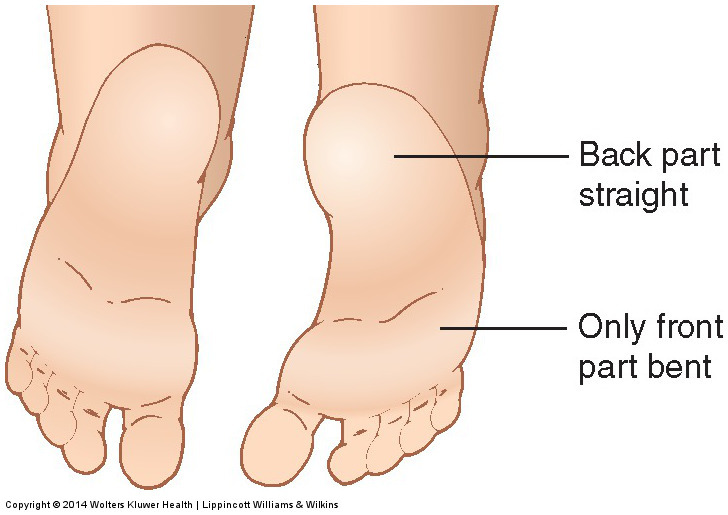
- Usually, results from fetal positioning in utero, can occur unilaterally or bilaterally
- Toeing-out (femoral retroversion): Less common than toeing-in, often linked to external femoral torsion, usually self-corrects when the child walks proficiently
- Tibial torsion: Internal tibial torsion is the most common cause of toeing-in in children under 2 years, often improves with growth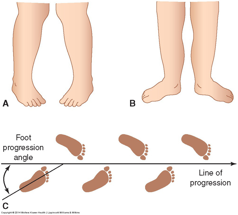
Genu Valgum (knock-knees): Clinically marked by decreased space between knees, typically resolves spontaneously:
The medial malleoli in the ankles cannot be brought in contact with each other when the knees are touching
- Most apparent in children aged 3-4 years, can be ignored up to age 7.Usually resolves spontaneously
- Associated with obesity in older children and adults
Genu Varum (bowlegs): Outward bowing of the knees, prevalent in infants and toddlers:
Outward bowing of the knees > one (1) inch when the medial malleoli of the ankles are touching
Majority of infants and children have some bowing of the legs up to 18 months of age
- Commonly resolves as limbs grow, but should be evaluated if persisting past 2 years of age to rule out rickets or tibia vara


vIf genu varum or genu valgum persist and are not corrected, osteoarthritis may develop in adulthood 2º abnormal intra-articular stress
§Persistent genu varum can → gait awkwardness and ↑ risk for sprains and fx
§Uncorrected genu valgum may → subluxation and recurrent dislocation of the patella with a predisposition to chondromalacia, joint pain, and fatigue
Flatfoot
Flatfoot (pes planus): Characterized by the absence of the longitudinal arch of the foot:
- Infants typically have wider, fatter feet, mistaken for flatfoot until arch development at approximately 2 years of age.§Flatfoot criterion: talus points medially and downward so that the heel is everted and the forefoot is inverted
- Types of flatfoot:
- Rigid flatfoot: Fixed, with no apparent arch, linked to neuromuscular diseases (e.g., cerebral palsy).
- Flexible flatfoot: More common, arch disappears only during weight-bearing, less prone to pain
Hereditary and Congenital Deformities
Congenital deformities: Osseous abnormalities present at birth, ranging from mild to severe:
- Examples include polydactyly (extra digit) → most common anomalymacrodactyly (hypertrophic digit)
syndactyly (webbing)
absence of a bone
Common etiologies remain often unknown, but can include hereditary influences and external agents impacting fetal development
Osteogenesis Imperfecta
Osteogenesis Imperfecta (OI): A common hereditary disorder characterized by defective synthesis of type I collagen:
- Generally inherited as autosomal dominant trait, with four major subtypes identified.
- Clinical manifestations include fragile bones, short limbs, soft/thin cranium → triangular face, and abnormal connective tissue featuresMutations in the genes connected with type I collagen synthesis impact the development of bones, joints, ears, ligaments, teeth, sclerae, and skin
- Most severe defects occur in OI type II, often resulting in stillbirth or death during infancy
In adults affected by (OI), hearing loss 2⁰ otosclerosis of the tiny bones in the middle ear is common
§Parent/caregiver teaching:
oAffected child’s bones are fragile
oSteps can be implemented to prevent fractures
Developmental Dysplasia of the Hip
Developmental Dysplasia of the Hip (DDH): Abnormal hip development leading to instability, malformation, subluxation, or dislocation:
- More common in first-born children and x6 in females.§Left hip is more often involved than the right 2⁰ the more common left occipital intrauterine positioning of most infants
- Etiology includes:hereditary → positive family hx
environmental → fetal position, tight uterus, breeched
Female
Developmental dysplasia of the hip is associated with the presence of other congenital anomalies
Dislocated hips, the head of the femur is located outside of the acetabulum
vDx:
§Early diagnosis is important because tx is easiest and most effective if begun during the first six (6) months of life
§Repeated dislocations → damage to the femoral head and the acetabulum

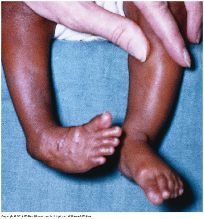
Congenital Clubfoot (talipes): Very common pediatric orthopedic condition affecting 1-2 per 1000 live births:
Occurrence follows a multifactorial inheritance pattern:
- Often idiopathic, with abduction → foot is plantar flexed and inverted, accounting for most idiopathic casesFeet can be passively overcorrected or brought back into the opposite position
- Treatment may involve braces, casts, or surgery
Scoliosis
Scoliosis: Lateral deviation of the spinal column often resulting in a 3-dimensional spinal deformity:
- Affects the coronal or frontal plane and may include vertebral rotation.
- Classified by the age of onset and etiologyFemales are most affected
Idiopathic Scoliosis: Spinal curvature with no established etiology, most common in healthy children:
- Divided into iinfantile (<3 yr/old)
juvenile (4-9)
adolescent (>10) types = most common
vEtiology: most likely complex and multifactorial
§Heredity appears to be involved
neurofibromatosis T1
vClinical manifestations:
§Although the scoliotic curve may be present in any area of the spine, the most common curve is a right thoracic curve → a rib prominence on the convex side and hypokyphosis from rotation of the vertebral column around its long axis as the spine begins to curve
Congenital Scoliosis
vCongenital scoliosis: 2º disturbances in vertebral development during 6th-8th weeks of embryonic development
§Divided into two (2) categories: failures of formation and failures of segmentation
1)Failures of formation: absence of a portion of the vertebra, such as hemivertebra (absence of a whole side of the vertebra) and wedge vertebra (missing only a portion of the vertebra)
2)Failures of segmentation: absence of the normal separation between the vertebra
§Affected child may have addional anomalies and neurological complications
Neuromuscular scoliosis
vdevelops 2° neuropathic or myopathic dz
§There is often a C-shaped curve from the cervical to the sacral regions
§Noted with cerebral palsy, muscular dystrophy, myelodysplasia, and poliomyelitis
§Myopathic neuromuscular scoliosis develops with Duchenne muscular dystrophy, and is usually not severe
Scoliosis: Clinical Manifestations
§Usually first noted because of the deformity
oHigh shoulder, prominent hip, or projecting scapula may be noted
§Idiopathic scoliosis is usually a painless process
oPain may be present in severe cases, usually in the lumbar region
•Pain etiology: pressure on the ribs or on the crest of the ilium
oAdults with less severe deformity may experience mild backache
§Dyspnea may be evident 2º diminished chest expansion and/or GI disturbances from crowding of the abdominal organs
§If scoliosis is left untreated, the deformity may progress to compromise cardiopulmonary function → ↑ risk for neurologic complications
Diagnosis: Identified via physical examination for cardinal signs, crucial for early intervention to prevent severe deformity
vCardinal signs:
§Uneven shoulders or iliac crest
§Prominent scapula on the convex side of the curve
§Malalignment of the spinous processes
§Asymmetry of the flanks
§Asymmetry of the thoracic cage
§Rib hump or paraspinal muscle prominence when bending forward