dental radiographs
Collimator
Definition: A collimator is a device that narrows a beam of particles or waves. In dental x-ray procedures, it is a tube that helps control the size and shape of the x-ray beam.
Types:
Round Collimator: Circular tube form.
Rectangular Collimator: Reduces patient exposure by approximately 40%.
Function: Placed on the area of interest on the x-ray film. Reduces patient radiation exposure by filtering out unnecessary x rays.
Intensifying Screen
Definition: An intensifying screen is used in conjunction with x-ray films to reduce patient exposure.
Usage: Typically utilized during extraoral x-rays, especially with Digital Panoramic Tomography (DPT).
Functionality: Enhances the effect of the x-ray beam and allows for lower exposures.
Types of Dental X-rays
Intraoral X-rays:
IOP (Intraoral Periapical): Focuses on specific teeth, useful for root canal treatment and diagnostic purposes.
Bitewing: Shows interproximal caries and helps visualize bone levels between teeth.
Anterior Occlusal X-rays: Used to detect supernumerary teeth and evaluate the anterior region of the mouth.
Extraoral X-rays:
OPG (Orthopantomogram) or Panoramic X-ray: Shows a broad view of the jaws and teeth. Commonly used in orthodontics.
Lateral Cephalogram: Orthodontic x-ray used to evaluate the jaw relationship and facial growth patterns.
Lateral Oblique: Provides an image of the mandible from an oblique angle.
Dental X-ray Machines
OPG Machine: Used to create panoramic images.
CT Scanning: Produces cross-sectional images, useful for detailed evaluation.
CBCT (Cone Beam Computed Tomography): A specialized CT that provides 3D images with less radiation, suitable for implants and orthodontics.
Regulations and Safety in Radiology
IRMER: Ionizing Radiation (Medical Exposure) Regulations. Laws governing x-ray facilities to protect staff and patients.
IRR: Ionizing Radiation Regulations. Focus on safe practices in handling radiation.
ALARA Principle: "As Low As Reasonably Achievable". Aim to minimize radiation exposure.
Radiographic Errors
Types of Errors:
Foreign Body Artifact: Caused by forgetting to remove dental appliances, jewelry, etc., before imaging.
Blurred Image: Occurs from patient movement or incorrect collimation during the exposure.
Reverse Film Error: Occurs when the film is placed backward in the holder; results in low-density images with lead foil markings.
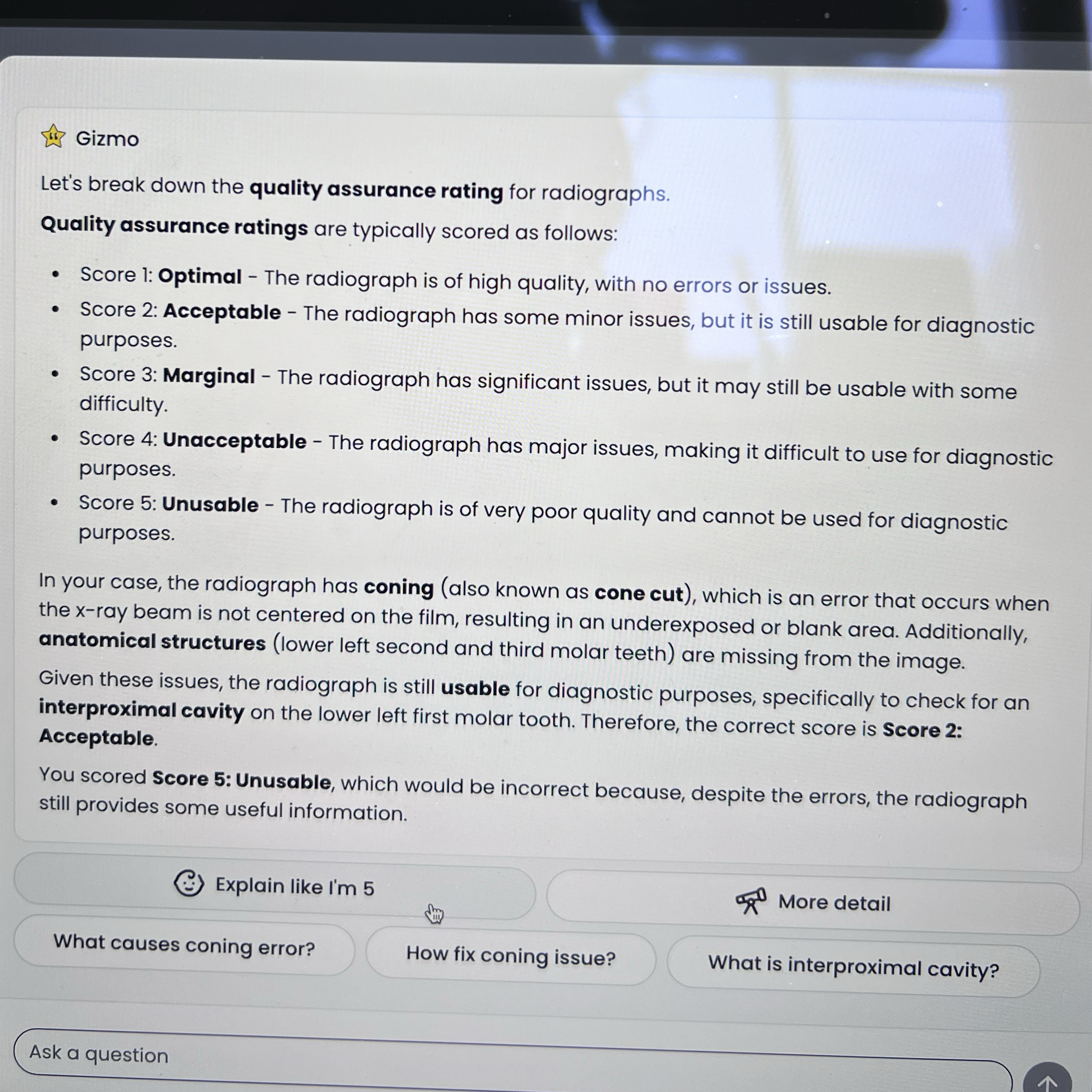
Cone Cut: Happens when the collimator isn't aligned with the x-ray film.
Incorrect Vertical/Horizontal Angulation: Can lead to overly long or short images and overlapping due to improper positioning of the x-ray beam.
Film Processing Steps
Developer Solution:
Temperature: 18-22°C.
Time: 4-5 minutes.
Reduces silver halide crystals to form a dark image.
Water Bath: 8-10 seconds to rinse excess developer.
Fixer Solution: 8-10 minutes to remove unsensitized crystals, resulting in a clear film.
Final Water Rinse: 10-15 minutes to remove excess chemicals.
Film Composition and Functionality
Outer Envelope: Protects the film.
Lead Foil: Prevents backscattered radiation which might otherwise create fog on the image.
Black Papers: Protect the film from light.
Image Formation: Created by the sensitive silver halide crystals inside the film.
Key Concepts in Radiology
Optimization: Refers to minimizing radiation exposure while achieving quality diagnostic images.
Controlled Zone: Area around the radiation source that should be approximately 2 meters to ensure safety.
Treatment of Radiographic Issues
Handling Radiation Contamination: Understanding contamination leads to safety protocols during procedures.
Genetic Mutation Risk: Explained due to unnecessary radiation exposure, highlighting patient care importance.
Summary of Important Regulations
RPA (Radiation Protection Advisor): Provides advice on safe use of radiography.
RPS (Radiation Protection Supervisor): Ensures compliance with safety measures in radiographic imaging.