ch 57 burns
Always anticipate pain & edema with burn pt’s
Priority: ABC, then fluid resuscitation
children & elderly are at high risk for burn injuries due to their skin
Initiates local changes to the integumentary system
Tissue destruction
Inflammatory cascade
In severe burns, can lead to a systemic response
etiology
Thermal burns include 80% all burns (most common)
Fire/flame injuries
Scald injuries
Non-thermal burns involve:
Electricity: possible widespread internal tissue damage, including tissue & bone destruction, which is not easily assessed
common to see cardiac & renal complications
Rhabdomyolysis → myoglobinuria
muscle breakdown releases myoglobin (oxygen-storing protein) which appears in the urine
Dysrhythmias, particularly fatal ones
Chemical: ammonia, lye
Radiation: time between exposure & symptoms may be immediately or days/weeks/months/years later
could occur as part of treatment or warfare
classification & severity
burns are classified by total body surface area (TBSA) & depth of injury
other contributing factors:
type & location of burn
pt age
Gender
past medical history (comorbidities)
Presence of inhalation (damage to resp system) and/or other injuries
3 phases of major burn injury (see later in outline):
emergent / resuscitative phase: from onset of injury to completion of fluid resuscitation
acute / intermediate phase: from start of diuresis to complete wound closure
starts within 2-3 days post burn, may lasts weeks to months
rehabilitation phase: from wound closure to return of optimal physical & psychosocial adjustment
assessing injury size
rule of 9s
used primarily for thermal burns
Not accurate for children or for extremes of height & weight (short, tall, obese)
Body is divided into five areas, each with increments of 9% TBSA
Head & neck: 9% (4.5% front & 4.5% back)
Arms: 18% (9% each arm; 4.5% front & 4.5% back)
Anterior & posterior trunk: 36% total (18% front & 18% back)
Anterior & posterior legs: 36% total (18% each leg; 9% front & 9% back)
Perineum: 1%

Lund & Browder
Preferred to determine TBSA in children as it accounts for changing body proportions of children (ex: larger head & smaller legs)
Chart determines the TBSA burned for each body part according to age of pt
Allows clinicians to document the TBSA according to burn depth
palmar method
used for small burns
the pt’s palm (including fingers) = 1% TBSA (adults) or 0.5% (children)
Specific to the pt since everyone’s palm size is different
less accurate
depth of injury/tissue destruction
influencing factors:
Etiology
Temperature
Duration of contact with the burning agent
skin thickness: depth of tissue/layers affected
thin skin (elderly) = deeper burn injury
burn depth assessment:
burn depth | tissue involvement | wound characteristics | pain assessment | healing |
superficial (1st degree) | minimal damage to epidermis | dry, no blisters; pink or red; blanches easily | hypersensitive | 3-7 days with no scarring |
superficial partial thickness (2nd degree) | entire epidermis but minimally to dermis | blisters may be closed or open and weeping; pink or red; mild edema; blanches easily | hypersensitive | 7-14 days with no scarring |
deep partial thickness (2nd degree) | entire epidermis & deep layers of dermis | blisters are flat & may be closed or open; waxy appearance; cherry red, white, mottled, or pale in center; edema; sluggish or no blanching | less pain; hypersensitive around wound edges but may only be sensitive to pressure in the center | possibly 3-6 weeks & possibly some scarring, or the wound may have to be surgically excised & grafted |
full thickness (3rd degree) | destruction of entire epidermis & dermis; may involve subcut fat, muscle, and/or bone (high morbidity) | dry, leathery, pale, white, brown, tan, black, charred; no blanching; possibly contracted if muscle is involved | no pain in the center of wound but may be sensitive to pressure | always scars; will not heal without surgical excision & grafting |
4th degree burn | see below |
superficial (1st degree)
slightly edematous
Commonly due to sunburn & minor flash burns
heals on its own
superficial partial thickness (2nd degree)
Hair follicles, sebaceous glands, sweat glands remain intact
near normal cap refill (pt is still being perfused)
Moist, fluid-filled blisters appear within minutes
Very painful
heals on its own
deep partial thickness (2nd degree)
decreased cap refill (compromised perfusion is a concern)
heals on its own
immersion injury
a partial thickness (2nd degree) burn injury (can be superficial or deep)
involves a clear line of demarcation
Usually done on purpose
ex: a hot bath during elderly abuse

full thickness (3rd degree)
includes destroyed hair follicles, sebaceous glands, sweat glands, & nerve endings
bright red to white; dry & leathery (brownish color)
Insensitive to touch

4th degree
Total tissue destruction down to bone with bone exposure
May be caused by prolonged exposure to a fire or flame, high voltage electrical injuries or exposure to highly concentrated chemicals
Extremities may appear charred & mummified
Amputation usually required
High morbidity & mortality

injury location
any injury to head & neck → edema → risk of acute airway compromise
scarring
Loss of function, even after healed
Joint contractures
age & gender
high mortality rates in elderly
Elderly skin is thinner & less elastic → deeper burn depth
Women are at less risk for burn injury but higher mortality when they are burned
pathophysiology
3 phases of major burn injury (see later in outline):
emergent / resuscitative phase: from onset of injury to completion of fluid resuscitation
acute / intermediate phase: from start of diuresis to complete wound closure
starts within 2-3 days post burn, may lasts weeks to months
rehabilitation phase: from wound closure to return of optimal physical & psychosocial adjustment
integumentary system
When microcirculation is damaged:
Skin cannot cool itself after the heat source is removed
Burning process & damage to skin continues
Results in damage to the dermis
Skin is sterile immediately after a burn injury
Eschar develops over the burn wound, creating an environment for bacteria growth
Burns under 20% TBSA = local response
Burns over 20% = local & systemic response
considered major burns
systemic response: damaged tissue (injury) → inflammatory process → cytokines & other chemical mediators release into systemic circulation → systemic changes & cellular permeability changes → fluid shifts from intravascular to interstitial spaces → compromised perfusion
Fluid shifts & shock → tissue hypo-perfusion & organ hypo-function
Each system is compromised due to less perfusion
Fluid shift from intravascular to interstitial means we are losing volume (to the interstitial space) → inadequate volume & perfusion
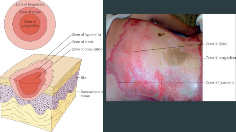
zones of injury
zone of hyperemia (outer)
warm or exposed area of skin
zone of stasis
compromised circulation
zone of coagulation (inner)
very compromised or absent circulation
most severe area of injury
edema stage (1st stage)
First 24 hours
Fluid leak from intravascular to interstitial spaces
rise in osmotic pressure
rise in capillary permeability
Vasoactive substances release
high interstitial edema
intravascular hypovolemia: drop in intravascular volume
capillary leakage:
Small wound: edema greatest 8-12 hrs post-injury
under 20% TBSA will not cause hypovolemia
Large wound: edema greatest 24 hrs post-injury
over 20% TBSA → hypovolemia due to tissue disruption, increased cap permeability, & fluid shift from intravascular to interstitial space on a much greater scale
pic below is an example of tissue disruption, increased cap permeability, & fluid shift from intravascular to interstitial area

cellular response / burn shock:
inadequate perfusion & fluid resuscitation → burn shock → eventual systemic shock
sodium & water shift into cells as potassium K+ moves out → intracellular swelling
fluid leaks out of blood vessels → low circulating volume, while swelling compresses capillaries → decreased perfusion to cells/tissues
possible cell death
high potassium levels intravascularly (extracellularly)
no cell perfusion → anaerobic metabolism begins → high lactic acid levels & metabolic acidosis
becomes a systemic issue
CV system changes
over 40% TBSA:
Significant myocardial dysfunction
heart can’t compensate from hypovolemia & reduced oxygen delivery → decreased myocardial contractility & CO within mins of injury
Generalized edema develops within first 24 hrs following severe burn → increases workload of heart
pt’s exposed to electrical currents may have myocardium necrosis & be predisposed to cardiac dysrhythmias
critical intervention for burn shock: restore intravascular volume by fluid resuscitation
first 24 hours
Intravascular volume loss & decreased venous return → decreased preload, cardiac filling pressure, CVP & PCWP
preload = amount of fluid coming back to the heart
Activation of SNS system & catecholamine release → tachycardia, vasoconstriction
early phase: classic s&s of compensated shock
fluid shift → intravascular volume depletion → shock (interstitial (tissue) edema, low BP, high pulse)
peripheral vascular effects
evaluate each extremity for color, temp, pulse, cap refill, sensation, pain, motor movement
Loss of perfusion → risk of necrosis & loss of limb
may not be able to feel for a pulse or temp due to the state of the burn
Eschar
Stiff (nonelastic), tough, dead tissue covering full-thickness burn or deep partial thickness
increasing edema acts as a restricting force → tissue can’t expand → compartment syndrome (see next heading)
Can compromise limb circulation, particularly in circumferential burns
burn-induced compartment syndrome
excess fluid in extravascular space → increasing edema acting as a restrictive force → tissue can’t expand → compartment syndrome (interruption in perfusion at cellular level)
important if ocurring around chest due to breathing compromise r/t chest restriction
Fasciotomy: surgical incision into fascia to relieve pressure or tension

resp system changes
AIRWAY IS THE #1 PRIORITY IN BURNS
Edema peaks ~24 hrs, so airway swelling can worsen over time
intubation may be needed later even if not on arrival
Signs of inhalation injury:
mucosal lining changes of oropharynx & larynx
Presence of soot / sooty sputum, hoarseness, singed nasal hairs, edema, or blisters
Symptoms of direct injury occur over first 24 hours
Stridor: screeching sound on inhalation
Hypoxia
Resp distress
Injury to lower airways is a result of noxious gas inhalation, not a direct injury
usually rapidly fatal
parenchymal injury (puh-REN-kih-muh)
Involves entire resp tract including alveolar membrane
profound bronchospasms & hypoxia are usually lethal within first few hours of injury
Restrictive injury r/t edema
Burns to neck and thorax
fluid shift from intravascular to interstitial → restricted chest wall movement
Escharotomy
GI system changes
Systemic vasoconstriction → paralytic ileus & mucosal edema
N/V, abdominal distention
Bowel ischemia → increased intestinal mucosal permeability & gut edema → bacteria translocate outside GI tract → increased risk of sepsis/septic shock
Stress ulcers (Curling’s) in stomach / duodenum are common
Severe burns → reduced blood flow to stomach / duodenum → damage to lining → Stress (Curling's) ulcers
Abdominal pain; hematemesis & melanic stools
renal system changes
Decreased renal blood flow & GFR
lowers urine output
raises creatinine & BUN
RBC destruction → hematuria
AKI can develop
Tubular necrosis
Myoglobin & hgb accumulate and occlude renal tubules
Treatment: flushing kidneys avoids myoglobin coagulation within the kidney itself & therefore avoids AKI
Electrical burns will require higher urine output (75-100 mL/hr)
neuro system
decreased cerebral perfusion → restlessness, confusion, anxiety, etc.
sodium shifts → cerebral edema
Carbon monoxide injury is under the umbrella of burn injury
carbon monoxide or associated head injury → neuro changes
carbon monoxide binds to Hgb stronger than O2
immune system changes
Prolonged humoral & cellular immunosuppression → possible immune dysfunction lasting over a month
Products released from activated inflammatory cells increase vascular permeability & local tissue ischemia
Activation of the inflammatory cascade occurs immediately after a burn injury
Systemic inflammatory response syndrome (SIRS)
Inflammatory response & release of chemicals increases risk of infection
metabolic changes
cellular metabolism decreases the first few days after a burn
Oxygen consumption decreases during burn shock
BMR may be double
Body weight drops quickly
Delayed wound healing, muscle wasting, & bone loss can occur
anxiety & psychiatric issues
Increased levels of anxiety
Delirium
Acute stress & PTSD symptoms
Sleep disturbances
Depression
assessment & care
First step in care: stop burning process by removing pt from burning source
burned area should be cooled with cool (not cold) water for at least 5 mins
Chemical burns should be lavaged with large amounts of water ASAP
Remove clothing & jewelry to prevent further burning
Clothing that adheres to the skin should be left in place
swelling with jewelry (ex: rings) can further decrease perfusion
Cover burned areas with dry bandages
Wrap pt in blankets
Do not interrupt blisters
primary assessment
A B Cs
Airway
Humidified oxygen is administered
Intubate to secure airway if necessary
Automatically assume that pt will have edema
Breathing
Circulation
2 large bore IV lines
Place on a cardiac monitor
neuro status
pt should be awake & able to follow commands
Decreased neuro status may indicate anoxic injury or additional neurologic injury
Anxiety & restlessness may indicate decreased cerebral perfusion
trauma assessment
done after initial assessment
AMPLE
A = allergies
M = medications
P = past medical history
L = last meal
E = events surrounding the injury (what happened?)
suspicious burn patterns
Suspicious burn patterns inconsistent with history may indicate self-inflicted, assault-related, or illegal activity.
Suspicious patterns of injury in children and the elderly include:
Scalds with uniform depth (from a hot bath)
Bilateral symmetry
Absence of splash marks
Multiple, superficial, well-defined circular burns (from cigarettes)
Small point burns
minor burns
Wound dressings
2 goals:
Maintain a moist environment to facilitate wound healing
Prevent infection
Common topical burn wound creams: Bacitracin ointment (OTC) & silver sulfadiazine (Silvadene; prescription)
Topical agents applied directly to wound can be left open to air or covered with a semi-open or closed dressing
pain management
assess & treat prior to beginning wound care using developmentally appropriate pain scales
pt undergoing initial wound evaluation & debridement may require an IV narcotic
pt transfer to a burn center
best treatment for pt’s with major burns
Criteria evaluated to determine if referral is warranted:
Burn size (usually over 20% TBSA)
Burn etiology
Pre-existing medical conditions (can be under 20% TBSA but with severe comorbidities)
Concomitant traumas
a pediatric patient needing a pediatric unit
Individuals with specific psychosocial needs
emergent / resuscitative phase
from onset of injury to completion of fluid resuscitation
fluid movement from intravascular to interstitial space = loss of fluid
Burns over 20% TBSA require fluid resuscitation to counteract intravascular fluid loss from increased capillary permeability
Goal: perfuse organs & maintain adequate urine output on maintenance IV fluids
given through a central line
formula in a heading below
monitoring & therapeutic devices:
Large bore IV
Urinary catheter* check policy
pt is high risk for infection so this may not be done
NG tube * check policy
pt is high risk for paralytic ileus so this may not be done
Continuous cardiac monitoring
ICU/Burn Unit setting
airway management
Begins with resp assessment
Elevate HOB
Supplemental oxygen
Place pt on 100% humidified oxygen if needed and/or until CO level determined
Assess for s/s of hypoxemia
Early intubation is encouraged since it’s hard to intubate once max edema has set in
continual resp monitoring
Signs of inhalation injury
Initial chest x-ray to assess for signs of atelectasis, pneumonia, and edema
Monitor ABGs & vitals
Evaluate pt’s with burns to the neck, chest, and abdomen for development of compartment syndrome & edema that may compromise breathing
nursing interventions (respiratory)
Immediately report difficulty breathing and/or swallowing
Instruct patient to cough & deep breathe
Explain procedures to pt
Importance of maintaining warm environment
Risk factors for infection
ABA’s burn consensus formula
Most common formula used during the first 24 hours post burn
formula: 2-4 mL Ringers Lactate X weight (kg) X %TBSA = mLs in first 24 hrs
answer = mL’s infused for fluid resuscitation within first 24 hrs post burn
first half is infused the first 8 hrs, the 2nd half is infused over the following 16 hrs
ex: 70 kg patient, 50% TBSA, 2 mL = 7,000 mL in first 24 hrs
first 8 hrs = 3,500 mL
following 16 hours (after the initial 8 hrs) = 3,500 mL
2 mL for thermal & chemical burns (Brooks formula)
4 mL for electrical burns (Parkland formula)
monitoring
nursing roles: giving fluids & continually assess pt status
Hourly urine output is the gold standard for monitoring burn resuscitation
We want kidneys to be perfused to produce urine
monitor lung sounds & JVD
successful resuscitation = adequate urine output after 2 hours of maintenance-rate fluids
indicates adequate kidney perfusion & restoration of fluid loss
dextrose-containing crystalloid solutions may be used once resuscitation is successful
Fluid type & rates are adjusted hourly based on urine output
Acceptable vital sign parameters:
HR under 120 bpm
systolic BP over 100 mmHg
clear lung sounds
monitor hemodynamics:
CVP measured hourly
PAWP under 18 mmHg indicates adequate resuscitation
low value = dehydration; high value = fluid overload
electrolyte status
Hyperkalemia occurs early as potassium leaks from damaged cells into the bloodstream (vasculature)
Correcting metabolic acidosis temporarily resolves hyperkalemia
Hyperkalemia can cause:
Heart dysrhythmias & ventricular failure
pt is on a cardiac monitor
Muscle weakness
EKG changes
Hyponatremia may occur with hypo- or hyperkalemia
hypo- & hypernatremia can cause change in neuro status
adjust fluid administration rates if there’s fluid excess
Caution with elderly, cardiac or renal impairment
compartment syndrome
Excessive fluid resuscitation → fluid accumulation in abdominal cavity → compartment syndrome
those at risk: circumferential burn wounds to neck, chest, abdomen, & extremities
Burn eschar constricts the burned area → impaired circulation
edema → subcut fluid expansion → impaired circulation
Prevention: escharotomy
Possible switch to colloid fluids
Neurovascular assessment: pulses, color, temp, paralysis
wound debridement
clean with warm water / sterile saline & antimicrobial soap / wound cleanser
Assess location, size, depth of injury
coverage / dressing:
major burn is dressed similarly to a minor burn
Silver sulfadiazine: most common topical agent used
pain management
route & drug of choice: IV morphine or fentanyl
IV meds onset of action is rapid
Muscles may be destroyed
PO takes too long
Narcotics are titrated according to pt pain response
Boluses are given for anticipated procedures, such as wound care
Anxiolytics are often prescribed prior to procedures
acute / intermediate phase
from start of adequate urine output (for 2 hrs on IV fluid maintenance rates) to complete wound closure (starts 2-3 days post burn, lasts weeks to months)
pt enters into a hyper-metabolic state until after the wound is healed
Increased body temp
Tachycardia
Increased caloric needs
possibly as high as 5000 cals/day to prevent weight loss & a negative nitrogen balance
Weight loss
cardiac
Capillary leak stops (regains integrity after 48 hrs)
Fluid re-enters vasculature from interstitial space
Hemodilution ; decreased HCT
Increased urinary output
Hemodynamic monitoring for fluid balance (prevents fluid overload)
Central venous line
Pulmonary artery catheter
VTE prophylaxis: low molecular weight heparin
fluid & electrolytes
sodium loss due to diuresis & fluid shift into vasculature → hyponatremia
s&s: neuro changes, weakness, dizziness, muscle cramps, fatigue, headache, tachycardia, confusion
Potassium shifts into cells → hypokalemia
s&s: fatigue, muscle weakness, leg cramps, dysrhythmias (U wave development on EKG), paresthesias, decreased reflexes
resp system
Monitor those with upper airway injury carefully
Signs of respiratory compromise:
low o2 sat & RR, accessory muscles
Pneumonia
Acute respiratory failure / ARDS
Chest x-rays
ABG
Fever
Pulmonary secretions
WBC count
GI system
Monitor for stress ulcer and paralytic ileus
Bowel sounds q4h
Turn & position if allowed (NOT on burn site)
Check gastric pH
musculoskeletal system
healing burns form less flexible scar tissue → muscle contractures & decreased ROM
Pain leads patients to prefer a flexed position → encourage regular stretching & movement of burned areas
Splinting can be beneficial to prevent / reduce contracture formation
neurologic system
Usually no physical symptoms unless severe hypoxia or electrical injury complications occur
Disorientation
Combative
Hallucinations
Delirium: more acute at night & occurs more often in elderly
ABCDEF bundle may be implemented to reduce risk
nursing care
pain management:
Analgesics & sedatives
Morphine
Hydromorphone (Dilaudid)
Lorazepam (Ativan)
Midazolam
Prevent / manage infection
Sterile technique for all dressing changes
Culture & sensitivities are performed
Antibiotics ordered as warranted (not done prophylactically)
VAP
Systemic infection
nutrition
pt is in a hyper-metabolic state
oral diet (if applicable): high in calories, carbs, protein diet with supplements
daily calorie counts to monitor dietary intake
pt with large burns usually require enteral nutrition or TPN within first 24 hrs after injury
enteral nutrition:
lowers catabolic hormone production
improves nitrogen balance
lowers wound infection rate
lowers the incidence of diarrhea
decreases duration of hospital stay
Tight glucose control; target glucose 80-110 mg/dL
Total energy needs can be as much as 100 times normal
wound management
These pt’s are in a warm room
Prevention & infection control
Preservation of viable tissue
Promotion of wound closure with minimal complications
Wound cleansing
Topical antimicrobial therapy
most common: silver sulfadiazine, bacitracin, mafenide acetate cream (sulfamylon cream)
mafenide acetate cream (sulfamylon cream): monitor for metabolic acidosis
Debridement
Wound closure
wound cleaning
done via hydrotherapy
Place pt in hydrotherapy tub for 20-30 min, twice a day
Attendants wear gowns & gloves until wounds are healed
everything done around the pt is sterile
keep room at 80-90 degrees to prevent chilling
wound care
Multiple dressing changes or closed method
Sterile gauze dressings are laid over topical antibiotic
Dressing changes anywhere from every 12 - 24 hrs to once every 14 days
Staff PPE
open exposure method of healing
Burn is covered with topical antibiotic but there’s no dressing over wound
Usually limited to the care of facial burns
Isolation technique
Sterile linen
Room temp = 85 degrees
surgical management
débridement of eschar
Purpose: excision & coverage
Remove eschar & other debris
Leave viable tissue exposed.
Excision can result in considerable blood loss
Blood transfusions may be necessary
Cover with skin graft, (autograft), or other biological covering
methods:
Mechanical: hydrotherapy and wound irrigation
Biodebridement: biosurgical or maggot debridement therapy (MDT)
Chemical: enzymatic/fibrinolytic agents applied to eschar → digest necrotic tissue → speed eschar separation
Surgical: accomplished under anesthesia in OR
skin grafts
a small skin sample can be stretched/meshed or lab-grown to cover a larger area
Gold standard for temporary wound closures: allograft (from a DONOR; not the pt’s own tissue)
functions:
Decrease bacterial proliferation
Control heat loss
Decrease protein loss in wound exudate
Increase patient comfort
Protect underlying structures
Stimulate healing
Prepare & test wound bed for autografting
Gold standard for permanent wound closures: autograft (the pt’s own tissue)
nursing care
Success of grafts
Avoid infection
ensure perfusion
elevate extremity
ex: if graft is on arm, assess cap refill, temp, etc.
Maintenance of contact (positioning)
immobilize graft after surgery; exercise begins 5-7 days after
Change 1st dressing in 3-5 days
Donor site care
Monitor for infection, perfusion, bleeding (donor sites tend to bleed easily)
Cleaned in sterile fashion
Thrombostatic agent applied
Split thickness wound remains
Painful
average healing time: 10-14 days
negative pressure wound therapy
Consists of a sponge, suction tubing, & occlusive dressing
Creates negative pressure to:
Remove edema
Stimulate perfusion
Provide a closed system for wound healing
May be used on wounds or grafts
rehabilitation phase
from wound closure to return of optimal physical & psychosocial adjustment (can last several years with large burns)
Begins in hospital
nursing actions
help pt to restore quality of life & appearance
support the patient’s physical & emotional healing
Physical healing focuses primarily on wound healing
goal: limit the amount of scar tissue
wound & scar management
Assess for signs of infection
goal: limit the amount of scar tissue
scarring continues to develop for up to 2 years post injury
pt may have surgeries throughout this time
Active exercise is encouraged.
Maintain or rebuild muscle mass
Decrease edema
Reduce the risk of DVT
Promotes psychosocial well-being
hypertrophic scarring
a thickened, wide, often raised scar that develops where skin is injured
Laser therapy improves scar color, reduces height and tension, and relieves pain and itching
Injection of a corticosteroid solution
Can be prevented or greatly reduced by applying pressure garments

wound contractures
scar formation over joints → limited joint movement → wound contractures
Prevention begins during acute phase:
Exercise
Body positioning
Joint splinting
Pressure dressings
neuropathies
scar formation, edema, improper positioning of splints / dressings → nerve damage → neuropathies
prevention: position splits & dressings correctly
treatment: gabapentin, neurontin, SSRI / SNRI (cymbalta)
Symptoms:
Severe burning pain
Increased or decreased sensation
Cold intolerance
pain management
pain is described as aching
Oral opioids, acetaminophen, NSAID’s provide adequate pain relief for most pt’s
Evaluate effectiveness