Year 8 biology :(
Levels of Biological Organisation
The human body is structured into five distinct levels of biological organisation, ordered from the smallest basic unit to the most complex integrated system:
Cell: The smallest basic unit of a living thing.
Tissue: A group of similar cells working together to perform a particular function.
Organ: A structure made of different tissues working together to perform a particular function.
Organ system: A group of organs working together to carry out a broad, essential function.
Organism: A complete living thing made up of interacting cells, tissues, organs, and organ systems.
Biological Hierarchy Flowchart:
The Circulatory System
Main Function: The circulatory system acts as a transport network that moves substances around the body, including oxygen ( ), nutrients, hormones, and waste products.
The Heart: A muscular organ responsible for pumping blood throughout the body.
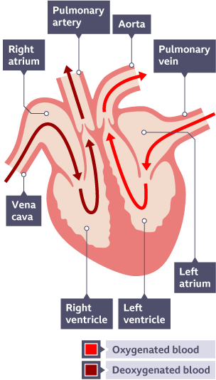
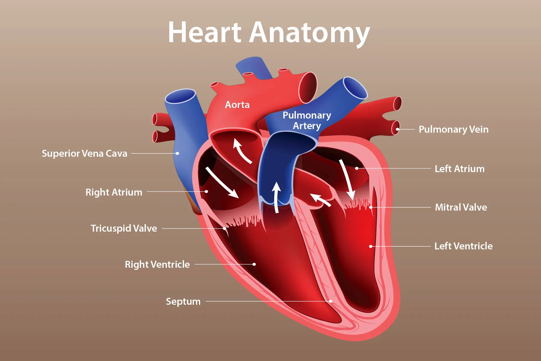
Structure: The heart consists of four internal chambers:
Right Atrium: An upper chamber that receives deoxygenated blood from the body.
Right Ventricle: A lower chamber that pumps deoxygenated blood to the lungs.
Left Atrium: An upper chamber that receives oxygenated blood from the lungs.
Left Ventricle: A lower chamber that pumps oxygenated blood to the rest of the body. It is more muscular than the right ventricle because it must produce greater pressure to propel blood throughout the entire organism.
Directional Blood Flow:
Right Side: Receives deoxygenated blood from the body and pumps it to the lungs.
Left Side: Receives oxygenated blood from the lungs and pumps it around the body.
Major Blood Vessels:
Aorta: The largest artery in the body; it carries oxygenated blood from the left ventricle to the rest of the body.
Vena Cava: The main veins(Superior and Inferior) that carries deoxygenated blood from the body back to the right atrium.
Pulmonary Artery: Carries deoxygenated blood from the right ventricle to the lungs.
Pulmonary Veins: Carry oxygenated blood from the lungs to the left atrium.
The Artery/Vein Exception:
Usually, arteries carry oxygenated blood and veins carry deoxygenated blood.
The exception involves the pulmonary(lung) system: the pulmonary artery carries deoxygenated blood, and the pulmonary veins carry oxygenated blood.
Defining strictly by flow: Arteries carry blood away from the heart; Veins carry blood towards the heart.
Heart Valves: These structures prevent blood from flowing backwards, ensuring it moves in the correct one-way direction through the heart.
Systemic Path of Blood Flow:
Blood Vessels and Blood Composition
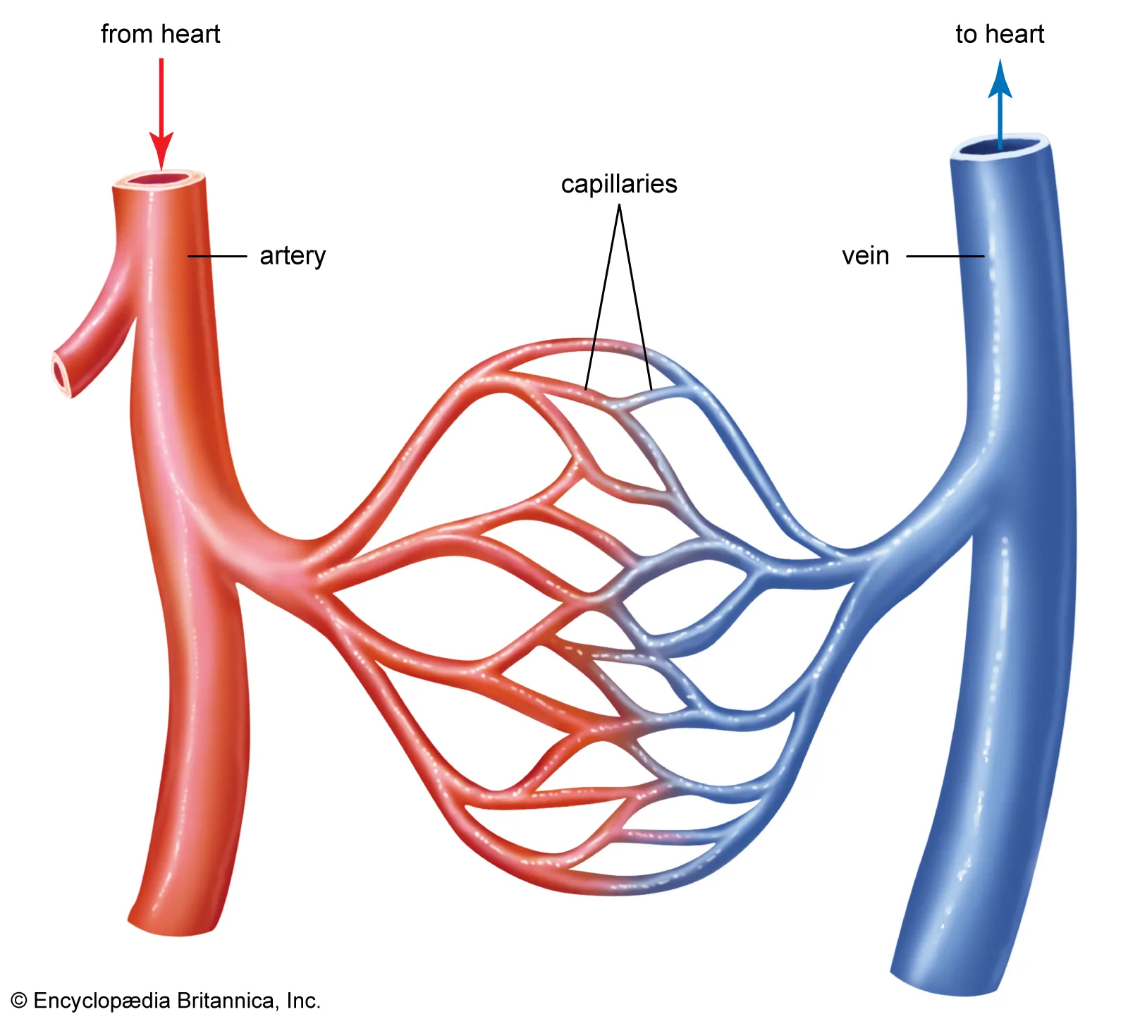
Arteries:
Carry blood away from the heart.
Feature thick, muscular walls to withstand relatively high pressure.
Usually carry oxygenated blood.
Veins:
Carry blood towards the heart.
Feature thinner walls compared to arteries.
Often contain valves to prevent backflow.
Usually carry deoxygenated blood.
Capillaries:
Tiny, extremely small blood vessels with very thin walls.
Connect arteries (via arterioles) and veins (via venules).
Allow for the exchange of substances (oxygen, nutrients, carbon dioxide) between the blood and body tissues.
Components of Blood:
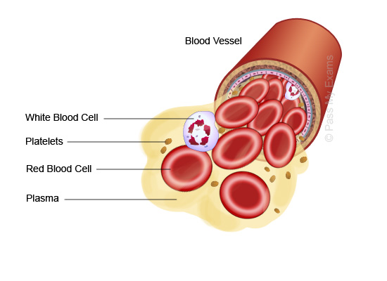
Red Blood Cells: Transport oxygen using a protein called haemoglobin, which binds to oxygen molecules.
White Blood Cells: Defend the body against pathogens (microorganisms like bacteria or viruses that cause disease).
Platelets: Essential for blood clotting to stop bleeding from injuries.
Plasma: The liquid component of blood that transports dissolved substances including nutrients, hormones, carbon dioxide, and wastes.
The Respiratory System
Main Function: Facilitates gas exchange by bringing oxygen into the body and removing carbon dioxide ().
Key Structures:
Trachea: The windpipe that transports air to and from the lungs.
Bronchi: Two main tubes branching from the trachea, one leading into each lung.
Bronchioles: Smaller tubes branching from the bronchi that carry air deep into the lungs.
Alveoli: Tiny air sacs at the end of bronchioles where gas exchange occurs.
Gas Exchange Process:
Oxygen moves from the air inside the alveoli into the surrounding blood capillaries.
Carbon dioxide moves from the blood into the alveoli to be exhaled.
Efficiency of Alveoli: Gas exchange is efficient because alveoli have a large surface area, very thin walls, and a robust blood supply.
Mechanics of Breathing:
The Diaphragm is a sheet of muscle under the lungs that controls chest cavity volume.
Inspiration (Breathing In): Diaphragm contracts moves down chest space increases air enters lungs.
Expiration (Breathing Out): Diaphragm relaxes moves up chest space decreases air leaves lungs.
The Digestive System
Main Function: To break down food into smaller substances for nutrient absorption and body utilization.
Key Processes:
Digestion: The act of breaking food into smaller substances.
Absorption: Moving digested nutrients into the bloodstream.
Enzymes: Substances that speed up chemical reactions, specifically those involved in digestion.
Pathway of Digestion:
Mouth: Mechanical breakdown occurs through chewing; saliva begins chemical digestion.
Oesophagus: A muscular tube transporting swallowed food to the stomach.
Stomach: Stores and churns food; uses acid and enzymes to continue digestion.
Small Intestine: Completes digestion and absorbs nutrients into the blood for transport to cells.
Large Intestine: Absorbs water from undigested material and facilitates faeces formation.
Rectum: Stores faeces before excretion.
Anus: The opening through which faeces exit the body.
The Nervous System
Main Function: Controls and coordinates body activities, allowing response to environmental changes.
Primary Components:
Brain: Processes information and controls body activities.
Spinal Cord: Transmits nerve signals between the brain and body; manages reflex actions.
Nerves: Carry electrical signals throughout the body to muscles, organs, and sensors.
Stimulus-Response Pathway:
Stimulus: A detectable change in the environment.
Receptor: Detects the stimulus.
Process:
Response: The resulting action or change in the body.
Skeletal and Muscular Systems
Main Functions of the Skeleton: Provides support, protection for internal organs, and a framework for movement.
Major Bones: Includes the Skull, Spine, arm bones, leg bones, and the Femur (the thigh bone and the longest bone in the body).
Joints: Locations where two or more bones meet.
Muscle Function: Muscles contract to produce movement.
Tendons: Connective tissues that attach muscles to bones. When muscles contract, they pull on the bone.
Opposing Pairs: Because muscles can only pull and cannot push, they work in pairs to move joints in different directions.
Movement Coordination:
The Excretory System
Main Function: Removes metabolic waste products and excess substances to prevent harmful buildup.
The Urinary System:
Kidneys: Filter the blood to remove wastes and excess substances, resulting in the production of urine.
Ureters: Tubes that carry urine from the kidneys to the bladder.
Bladder: Stores urine until it is excreted.
Urethra: The tube that carries urine from the bladder out of the body.
Excretion Pathway:
System Interdependence
Organ systems are interdependent, meaning they rely on each other to keep the organism alive and functioning.
Respiratory + Circulatory: The respiratory system brings into the blood and removes ; the circulatory system transports these gases between the lungs and the body's cells.
Digestive + Circulatory: The digestive system breaks down food and absorbs nutrients; the circulatory system transports these nutrients to all cells.
Circulatory + Excretory: The circulatory system transports metabolic wastes from cells to the kidneys; the kidneys filter these wastes for removal via urine.
Nervous + Muscular: The brain sends electrical signals via nerves to muscles, causing them to contract and produce movement.
Skeletal + Muscular: Muscles pull on the skeletal framework to produce movement, while the skeleton provides the necessary support and protection.