Week 6: Evolution of Colour Vision
Colour Vision and Opsins
Colours: promote contrast perception = enhance visibility of objects against surroundings
What is colour vision?
Visible light: electromagnetic waves that we can see
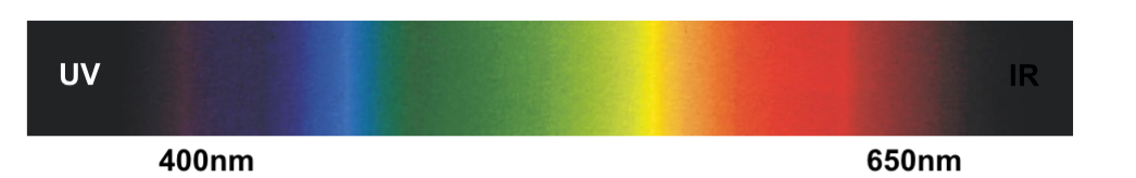
Colour vision is the ability to discriminate between different wavelengths of visible light, which ranges from about 400 to 650 nanometers.
An absence of light perception in this spectrum is interpreted as black.
Cone Photoreceptors: The cells responsible for detecting different wavelengths, contributing to color perception.
Color perception is subjective and interpreted by the brain based on signals received from photoreceptors.
Role of Opsins
Opsins: light-sensitive proteins in photoreceptors that determine spectral sensitivity. Variations in opsin structure lead to differences in wavelength sensitivity.
When light interacts with opsins, it triggers phototransduction, leading to a chemical signal responsible for reducing glutamate release in photoreceptors.
Structure of Opsins
Outer Segment of Cone Cells: Contains membranes (lamellae) wherein opsin proteins are embedded to maximize light sensitivity.
The membrane’s architecture increases the surface area for more opsins to be present.
Visual Pigment Composition: Opsins consist of transmembrane domains (seven helices) that enclose a chromophore—vitamin A (retinal).
Retinal's position within the opsin structure is critical for light sensitivity.
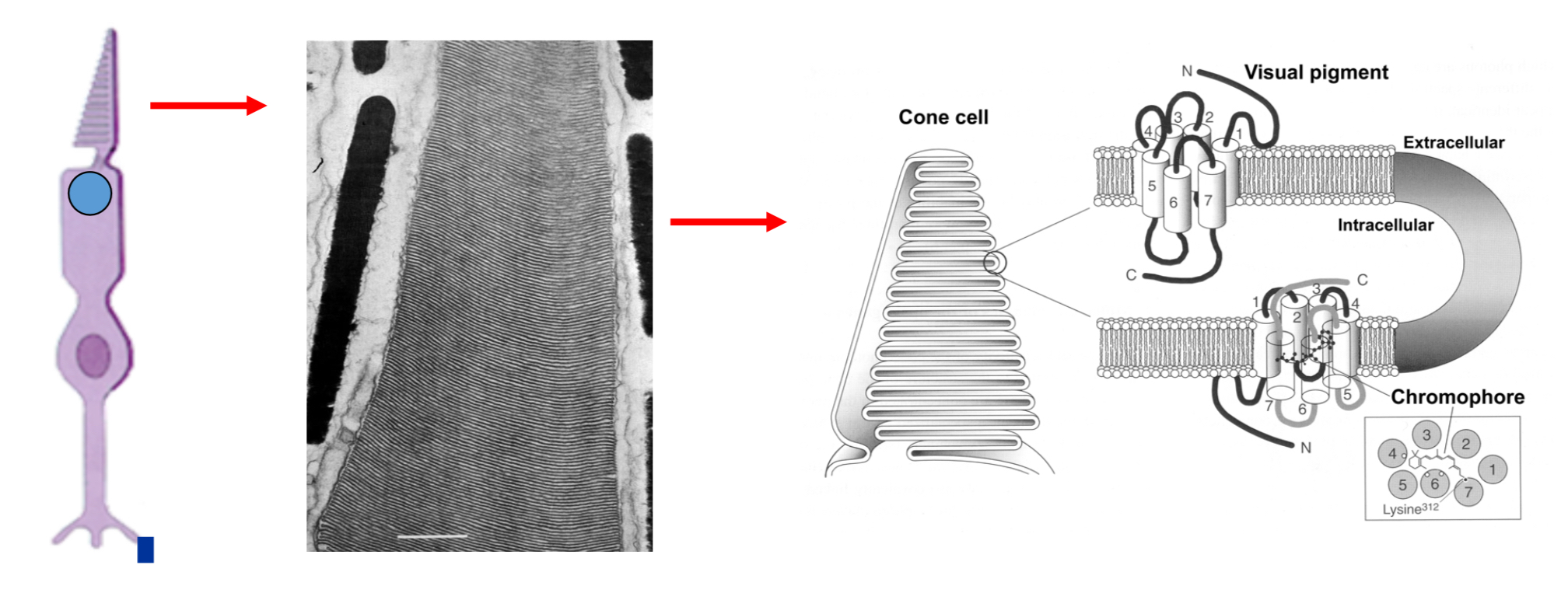
Visual pigment = opspin (proteins) + chromophore (vitamin A)
Opsin = 350 amino acids → opsin is a protein that sits in the membrane and binds retinal
Opsin has 7 transmembrane domains that are made up by a chain of amino acids
Phototransduction Process (study briefly)
When a photon hits the retinal, it changes shape (cis to trans), which alters bonds in the opsin.
This creates a cascade of chemical events leading to electrical signals interpreted by the brain.
Regeneration of Retinal: Once activated, retinal must revert to its original shape, which involves a series of biochemical reactions and transportation back to the receptor.
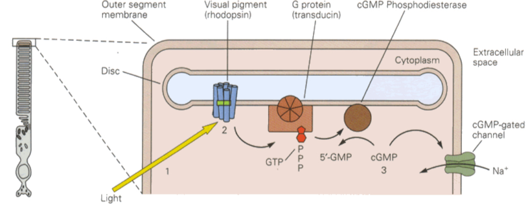
Light hits retinal
Retinal captures photons and changes shape
Opsin changes shape
Causes intracellular changes (domino effect) and change in membrane potential (=electricity)
How does opsin differentiate between different wavelengths?
Opsins differentiate wavelengths via specific molecular structures and positioning of retinal.
The amino acid sequence of opsins affects how well they can absorb different wavelengths.
Opsin binds retinal using two amino acids:
Key Amino Acids: Specifically, lysine (K) and glutamic acid (E) play crucial roles in anchoring and orienting the retinal.
Variations in the amino acid sequence lead to different sensitivities towards light wavelengths, effectively tuning the opsin for different light detection.
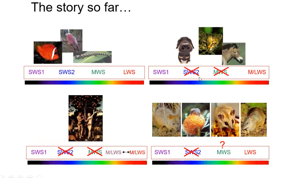
Cone opsins
Ancestral Opsin Gene: Likely a medium wavelength sensitive opsin that gave rise to other types through duplications.
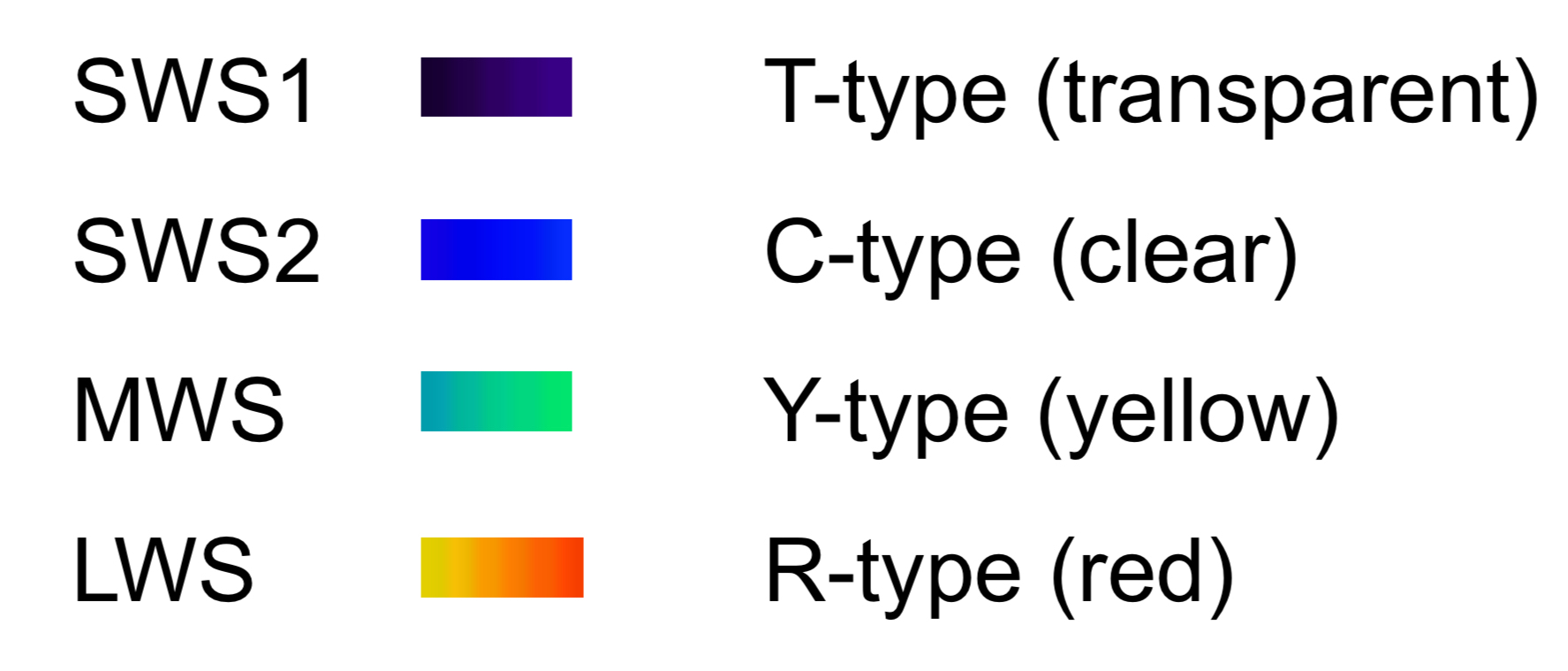
There are 4 types of cone opsins, each sensitive to different parts of the light spectrum:
SWS1 and SWS2 (short wavelength sensitive)
MWS (medium wavelength sensitive)
LWS (medium to long wavelength sensitive)

Natural selection acts on these mutations, resulting in evolutionary advantages based on the ability to detect specific colors, beneficial for survival in various environments.
Rod Opsin (used for night vision)
Only one rod opsin class: Rhodopsin (RH1)
It’s sensitive to low light, not colour
Not all vertebrates have all 5 opsin classes
Different species may lose or gain opsins depending on their environment and needs
Spectral tuning: Red vs. Green opsins
Mutations in the opsin gene can lead to small changes in light absorption sensitivity:
Position 180: Serine ↔ Alanine causes a shift of 3 nm.
Position 277: Tyrosine ↔ Phenylalanine causes a shift of 7 nm (made the wavelength 7nm longer).
Position 285: Threonine ↔ Alanine causes a shift of 14 nm.
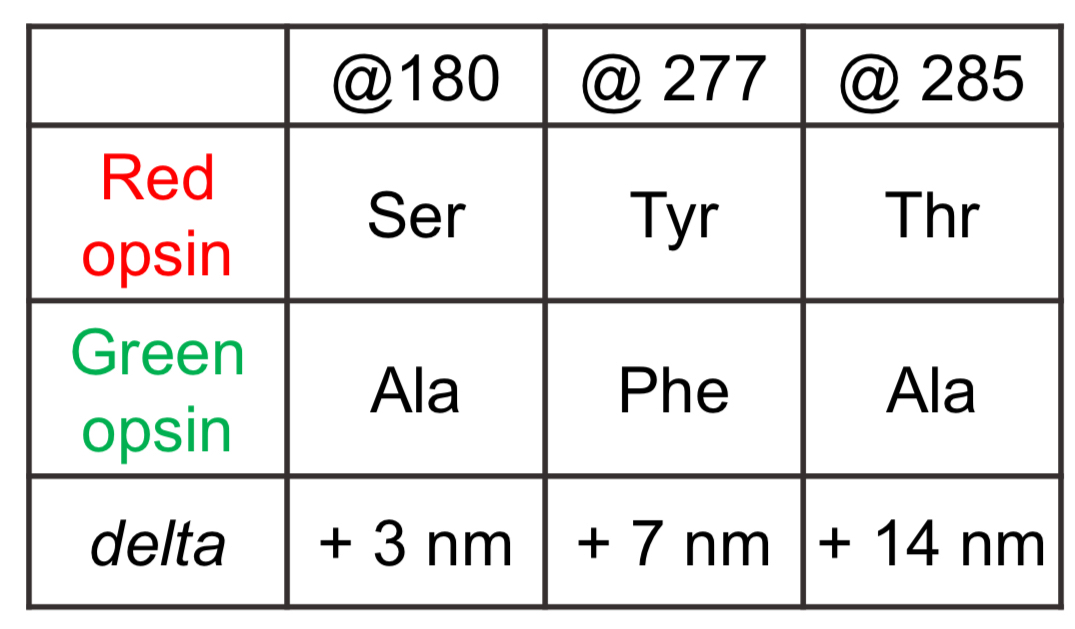
Fifteen total mutations
Three account for most of the spectral tuning
Total potential shift in colour perception due to mutations can accumulate up to 24 nm, highlighting how slight modifications can lead to significant evolutionary changes in visual perception.
_______________________________________________________________________________________________________________
Color Vision Overview
Color vision refers to the ability to perceive differences in wavelengths of light and is predominantly mediated by cone photoreceptors in the retina which contain optical proteins called opsins.
Mechanism of Color Discrimination
Wavelength Absorption by Cones:
Different cones in the retina are sensitive to different wavelengths of light (e.g., blue cones absorb light in the short wavelengths, and red cones in the longer wavelengths).
Color perception arises through the comparison of signals from different cones in terms of the relative activation based on the absorption of light.
Absorption Spectrum:
Cones do not directly transmit color information; instead, they send signals based on the amount of light absorbed at various wavelengths. This relative activation helps the brain to interpret color.
The activation of various cones linked to wavelengths allows us to perceive a specific color (e.g., green corresponds to a particular activation combination of cones).
Special Techniques in Color Vision Research
Spectrophotometry:
Synthetic Opsin Testing: Synthetic opsins can be created and tested in a spectrophotometer to measure their absorbance across different wavelengths.
Microspectrophotometry: This method allows testing of individual cone cells in a controlled environment to validate absorption characteristics.
Importance of Cone Types in Color Vision
Color Discrimination Necessity:
A minimum of two types of cones is necessary for color differentiation; having just one allows only for light detection.
Opsin Classes:
Vertebrates generally possess several types of opsins across the visible spectrum: ultraviolet, blue, green, and red.
Ancestral vertebrates likely had a tetrachromatic vision, which is advantageous in diurnal environments, allowing them to detect a wider range of colors.
Evolutionary Perspectives on Color Vision
Non-Mammalian Species:
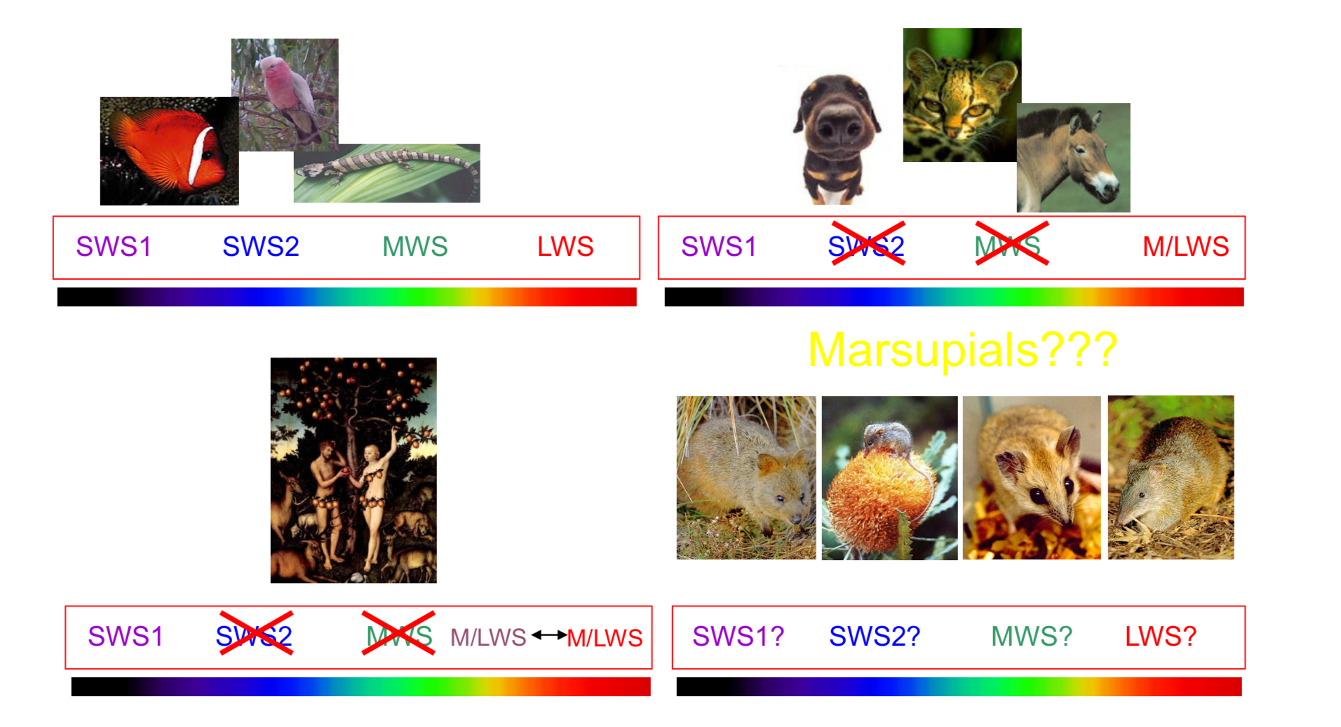
Many non-mammalian vertebrates (fish, birds, reptiles) retain all four classes of opsins supporting tetrachromatic vision, vital for survival (e.g., detecting food, escaping predators).
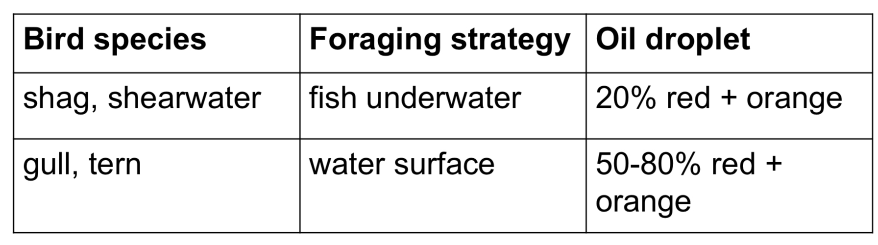
Oil Droplets: Present in some species (e.g., birds), oil droplets filter specific wavelengths, enhancing colour discrimination by providing acuity.
Four classes of cone opsins, present in ancestral vertebrates = tetrachromacy
Coloured oil droplets → oil droplets provide different specialisations and acuity
_______________________________________________________________________________________________________________
Evolutionary Perspectives on Color Vision
Different types of carotenoids contribute to the coloration of oil droplets found in avian and reptilian species.
Carotenoids are organic pigments that not only influence the colour of the oil droplets but also play a role in light filtering.
These pigments can enhance color discrimination by selectively absorbing certain wavelengths of light.
Types of Carotenoids:
Lutein: Commonly found in avian oil droplets, lutein is known for its yellow color and protective antioxidant properties.
Zeaxanthin: Another important carotenoid, zeaxanthin is found in many bird species and enhances the sensitivity to green wavelengths.
Astaxanthin: This reddish pigment can be found in some species and may aid in distinguishing between red and green hues.
Collectively, these carotenoids provide avian and reptilian species with enhanced visual capabilities, allowing them to navigate their environments more effectively by detecting a wider range of colours.
Different types of cones paired with different types of oil droplets.
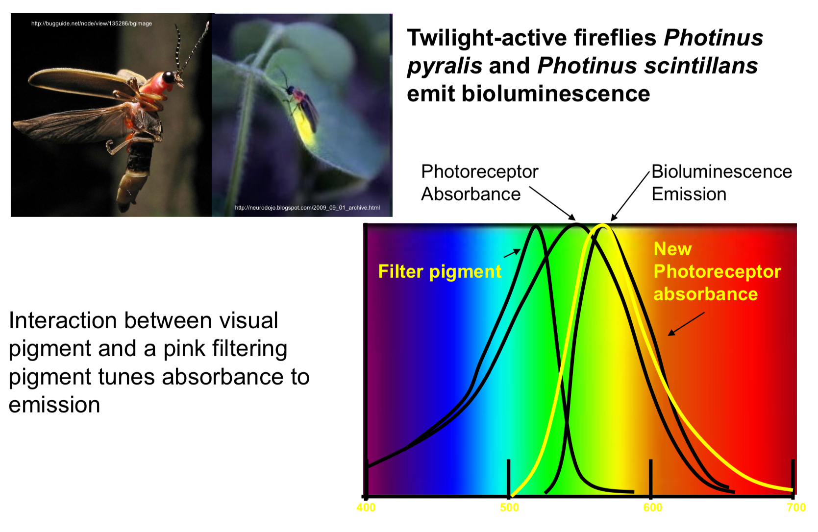
Filtering pigments in eye can tune photoreceptor sensitivity to better match the wavelengths of light emitted by bioluminescent organisms like fireflies.
Bioluminescent matching:
Fireflies like Photinus pyralis emit light at a specific wavelength (shown as the bioluminescence emission peak in the graph).
To best detect this light, their photoreceptors need to be sensitive to the same wavelength.
Photoreceptor Absorbance:
the original absorbance curve (black line) shows what wavelengths the unfiltered photoreceptor is most sensitive to
Role of filter pigment
a pink filtering pigments shifts the peak absorbance of the photoreceptor toward the emission wavelength
This is shown by the new absorbance curve (yellow line), which is now better aligned with the firefly’s emitted light
Summary
the firefly eye uses a filtering pigments shifts to shift the sensitivity of its photoreceptors so they can more effectively detect their own bioluminescent signals — an adaptation for improved signal detection in low-light environments
_______________________________________________________________________________________________________________
Avian and reptilian oil droplets
 _______________________________________________________________________________________________________________
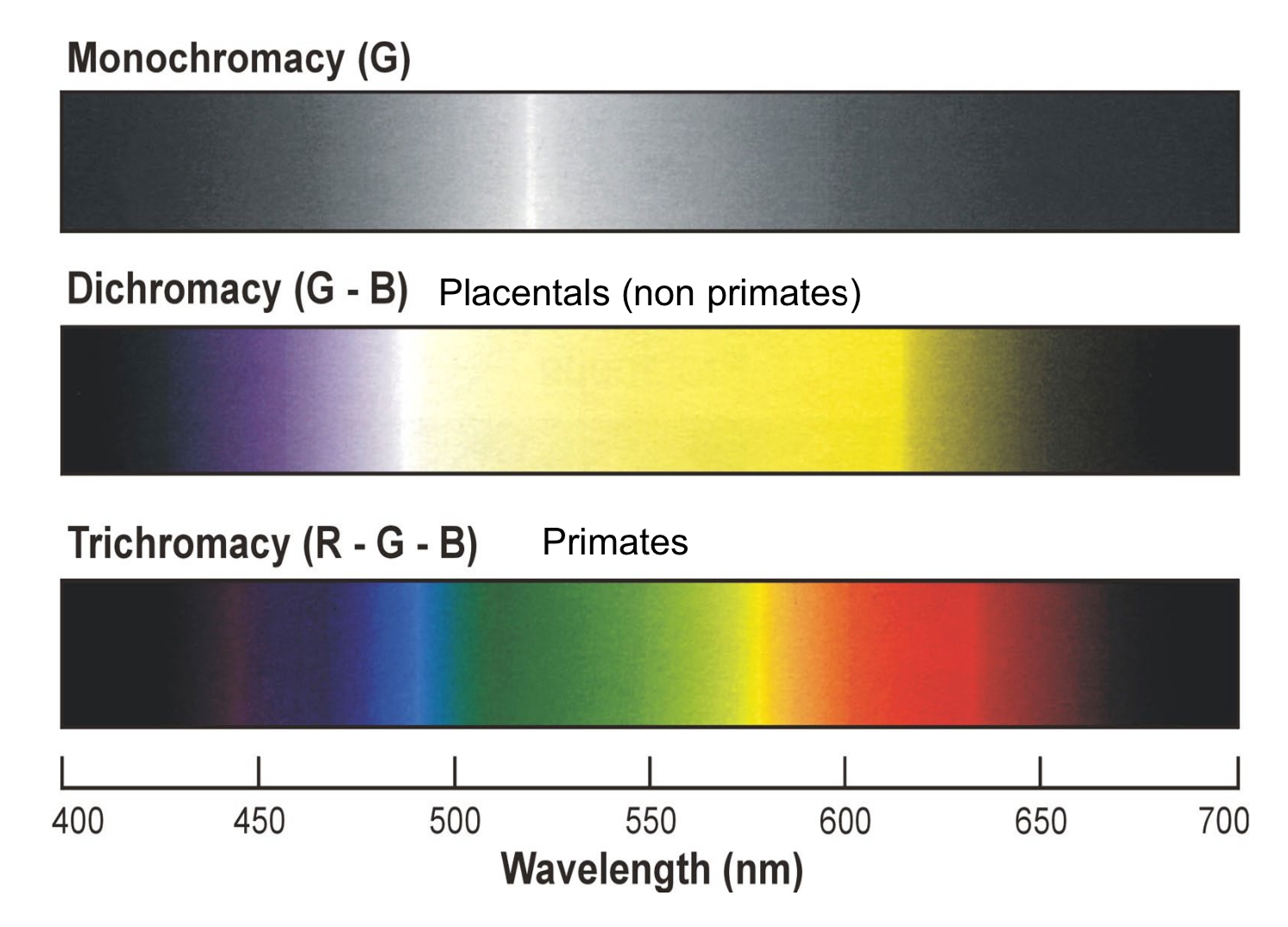
Adaptations in placental Mammals
Nocturnal Evolution:
Many mammals have evolved to be nocturnal, leading to a reduction in color vision capabilities and a predominance of rod cells over cones (less reliance on color).
^The common view is that mammals like cats and dogs are color blind due to the lack of specialized color opsins.
Two cone opsin genes LOST (nocturnality)
Two cone opsin genes retained = dichromacy
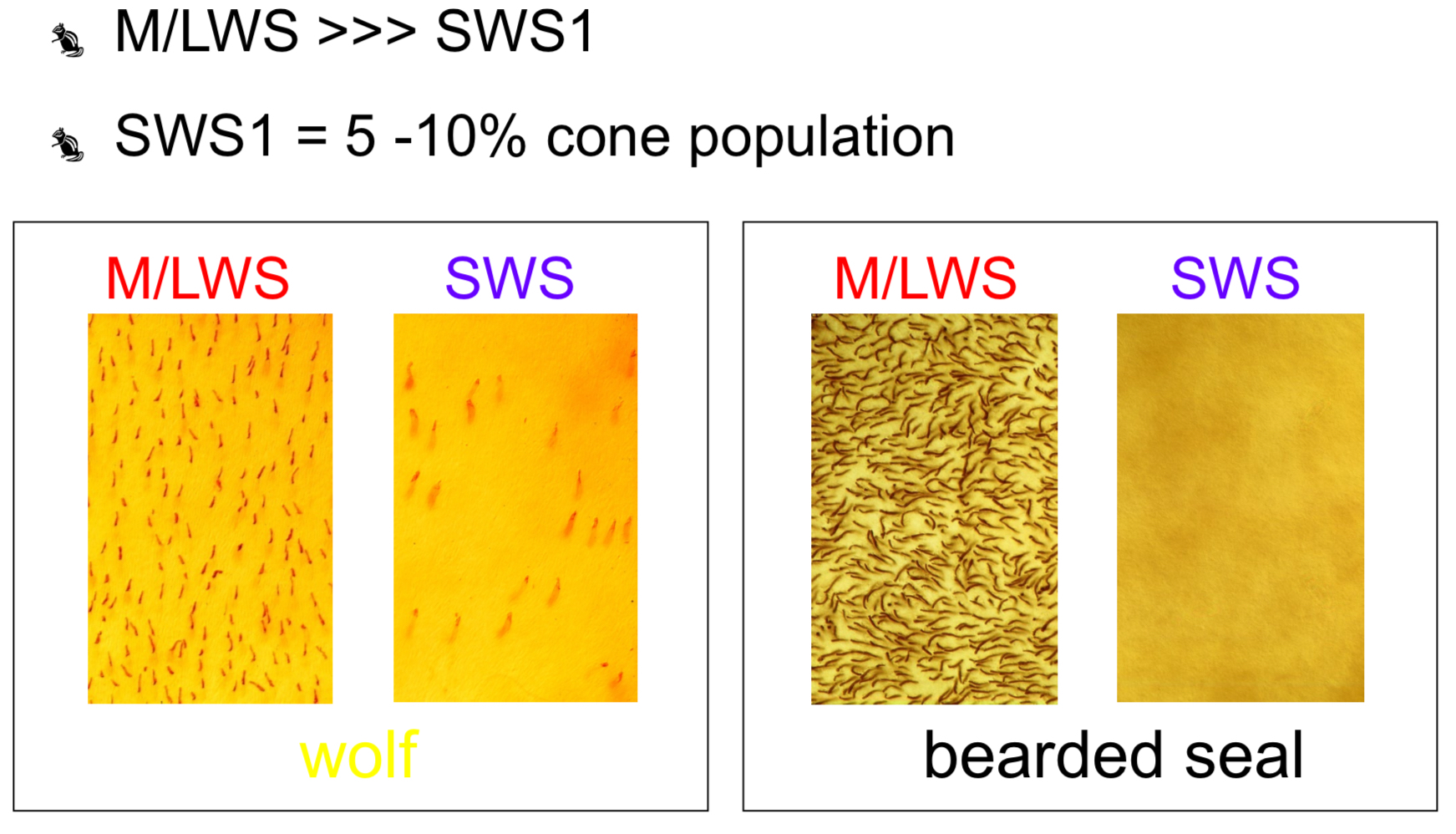
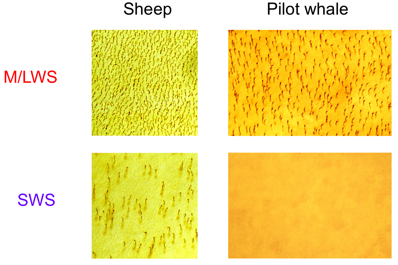
Placentals: terrestrial vs marine
Retinal Structure Comparisons:
Differences in cone distributions are noted between terrestrial animals (e.g., wolves) and marine mammals (e.g., bearded seals), highlighting adaptations to their respective environments.
Marine mammals often lack short wavelength sensitive cones, as in the case of the pilot whale, leading to monochromatic vision.
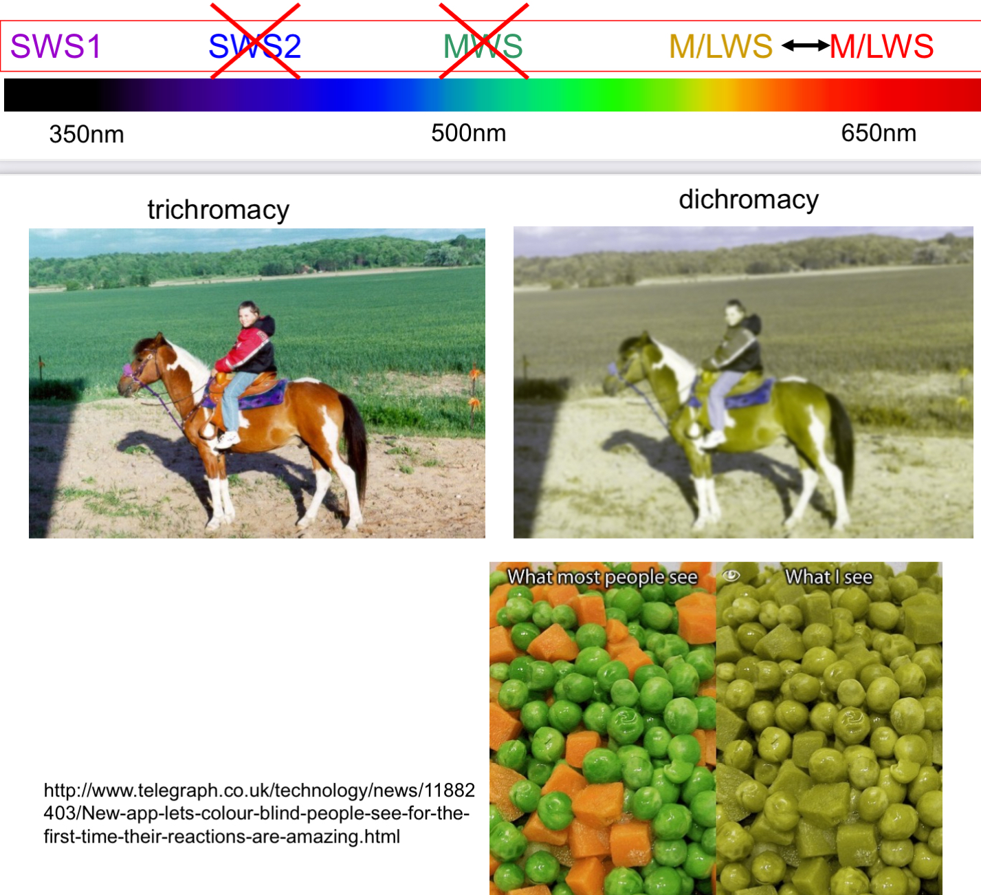
Primates and Enhanced Colour Vision
Trichromacy in Primates:
Unlike other mammals, some primates (e.g., monkeys) re-evolved a third opsin necessary for distinguishing between ripe fruits and green foliage, aiding their survival.
Three cone visual pigments = trichromacy
Third pigment re-evolved from duplication of M/LWS opsin gene - 45 mya
No oil droplets or double cones
Why are marine mammals monochromats?
Underwater environment is blue, so marine mammals should have retained the SWS opsin.
rod and cone interactions:
in low light, rods dominate
Other senses
may have taken priority, reducing the evolutionary pressure to maintain colour vision
Adaptation to marine environment
reflection of downwelling light: most light that penetrates water is from above (downwelling), and its properties change with depth
Shallow waters are red-shifted: in shallow water, the light has more red in it. But over time, early marine mammals lost their SWS opsin (blue-detecting), possibly due to early evolutionary conditions or because colour vision wasn’t that useful underwater
Summary
although it seems marine mammals should keep blue-sensitive vision (SWS opsin) for the blue ocean, low light, the dominance of rods, and early evolutionary adaptations (like red-shifted light in shallow waters) led to the loss of SWS opsin, making them monochromatic.
_______________________________________________________________________________________________________________
The Complexity of Colour Vision
Color Perception Differentiation:
Understanding the distribution of cones and their correlations with environmental factors is critical, as evidenced by the differing strategies among species.
Various strategies highlight the niche adaptations that arise from evolutionary processes.
Colour Vision in Marsupials
Introduction to Color Vision Controversy
Trichromats vs. Dichromats
The discussion starts with the concept of trichromacy, as evidenced by the ability to differentiate between green and red. The focus is on marsupials, where the classification between dichromats and trichromats has been debated.
Understanding Colour Vision in Different Species
Tetrachromats: Fish, birds, and reptiles are identified as tetrachromats, possessing four types of opsin proteins for color vision.
Marsupials: Generally identified as dichromats due to a loss of two opsin proteins, which is believed to be an adaptation to nocturnal life. However, some evidence suggests that certain marsupial species may possess trichromacy.
Species Overview
Dichromats:
Virginia opossum (American marsupial)
Tamar wallaby (Australian marsupial)
Trichromats:
Fat-tailed dunnart
Quenda (bandicoot)
Honey possum
Quokka
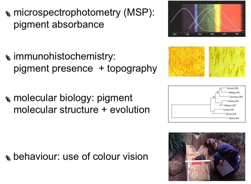
Techniques for Investigating Colour Vision
Microspectrophotometry (MSP)
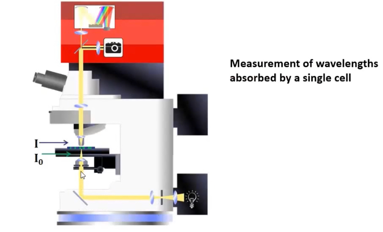
How MSP Works:
Measures wavelengths absorbed by individual cells.
Involves sending light of specific wavelengths onto samples to assess absorption.
Challenges:
Conducted in complete darkness to prevent bleaching of visual pigments, making isolation of single cells challenging.
Findings:
Tamar wallabies exhibit dichromacy, absorbing light at 420 nm and 528 nm.
Certain marsupial species (fat-tailed dunnart and honey possum) show three types of cone opsins, suggesting trichromacy.
Immunohistochemistry
Application: Utilized to identify opsin types in retinal hormones.
Limitations: Currently, antibodies primarily available for short and long wavelength sensitive opsins, limiting definitive conclusions.
Evidence from Studies:
In dunnarts, about 30% of cones remain unlabeled, indicating potential for a third opsin. Absence of unlabeled cones in Tamar wallaby confirms dichromacy.
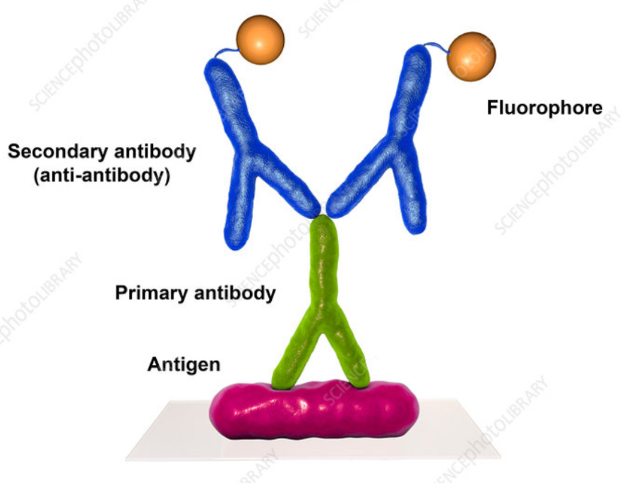
inconclusive: antibodies are available only to SWS and LWS cones
But ~30% unlabelled cones in dunnart retina - mysterious 3rd opsin?
No unlabelled cones in tammar wallaby retina - confirming dichromacy
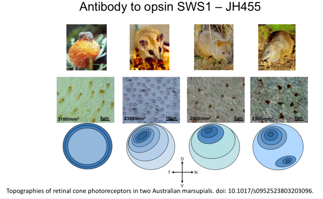
Understanding Opsins and Their Distribution
Short-wavelength sensitive opsin (JH455): Found in peripheral regions of the retina; associated with detecting light.
Medium/Long-wavelength opsin (JH492): More numerous in retinas, linked to acute vision and object recognition.
Dueling Perspectives on Opsins in Marsupials
The dichromat (Virginia opossum) has fewer cones (nocturnal) and mostly rod-dominated retina.
Long Wavelength Opsins: Evidence gathering through sectioning allows for a clearer understanding of different photoreceptor types.
Molecular Biology
Genetic Studies: Identifying cone opsin genes can determine dichromacy vs. trichromacy definitively.
The Tamar wallaby has been studied in detail, revealing only two visual pigments, affirming its dichromat status.
Still limited genomic studies on dunnarts regarding the third cone.
Functional Aspects of Colour Vision
Behavioral Studies
Does the retina send an electrical signal to the brain in response to specific wavelengths?
Electroretinogram Studies: Used for measuring neural responses of retinas to specific wavelengths.
Can the animal perform a behaviour that demonstrates discrimination between colours?
Colour Choice Tasks: Animals trained to distinguish between coloured chambers; success indicates effective colour vision.
Is the colour relevant in the animal’s environment?
Ecological Relevance: Determining if animals can perceive colours seen in their natural environment.
Electrophysiological Responses
The study shows that visual pigments in dunnarts and honey possums effectively respond to specific wavelengths, indicating functional color vision.
Behavioral Training Success Rates
Animals trained to choose colors showed varying success rates linked to their abilities to discern between them.
Dichromats often had a neutral point where a particular light wavelength appeared indistinguishable from white light, impacting their success in color discrimination.
Spectroradiometry and its Importance
This technique measures light wavelength reflections, showing how well-animal color vision aligns with environmental cues.
Evidence was presented using banksia flowers, a key food source for honey possums, showing that they could differentiate between matured and immature flowers given the wavelength sensitivities.
Conclusion - Diversity of Color Vision in Marsupials
The case of marsupials presents a complexity contrasting from their tetrachromatic fish and bird counterparts.
Diversity in Evolution: Some marsupials have likely evolved different strategies for color discrimination based on their ecological niches.
Future studies could further elucidate the diverse capabilities of color vision among marsupials and their evolutionary trajectories.
Dichromacy vs Trichromacy in marsupials or both?
seems that there is both?
Double cones, oil droplets, cone dimensions, rod:cone ratio
Ecological diversity diurnal, arrhythmic, crepuscular, nocturnal
this supports that some will be dichromats and some trichromats