Spinal Anesthesia
Spinal anesthesia (also known as spinal block, subarachnoid block, intradural block, and intrathecal block) is a form of regional anesthesia.
Typically administered via a fine needle (commonly 9 cm/3.5 inches long; 12.7 cm/5-inch needles for obese populations).
Can be performed via Midline or Paramedian approaches.
Pharmacological Agents
Choice of local anesthetic is primarily based on duration of action.
Lidocaine:
Duration: 45-90 minutes; SHORT duration
Provides excellent sensory and fairly profound motor block; not commonly used though d/t spinal complications
Bupivacaine:
Duration: 60-120 minutes; INTERMEDIATE duration
Provides a slightly more intense sensory block compared to Tetracaine.
Tetracaine:
Duration: 120-180 minutes (with epinephrine); LONG duration
addition of epi prolongs it the most compared to the others
Provides slightly more motor block compared to Bupivacaine.
Usually used for hip surgeries
VASOCONSTRICTOR Adjuvants Specifically for Spinal Anesthesia
Epinephrine: Dosage: 0.1 to 0.5 mg.
Minimal effects on increasing bupivacaine’s duration of action
Not usually added to ropivacaine at all or bupivacaine because no significant increase in duration
(less often) Phenylephrine: Dosage: 2 to 5 mg.
These drug’s vasoconstrictive properties decrease blood flow in the dura mater without affecting spinal cord blood flow = decreased drug clearance via dural vasculature
Risks:
Adding epinephrine to lidocaine may increase risk of neurotoxicity (but remember that lidocaine is not really used for spinals anymore)
Adding either epinephrine or phenylephrine to tetracaine may increase the risk for transient neurologic symptoms (TNS).
OPIOID Adjuvants Specifically for Spinal Anesthesia
Opioids and Analgesics are added to enhance surgical anesthesia and provide postoperative analgesia. Effects mediated at the dorsal horn of the spinal cord.
Intrathecal fentanyl: Dosage of 25 mcg for short outpatient procedures. Rapid onset of s/e, if will have them.
Intrathecal morphine: Dosage of 0.1 to 0.5 mg can provide postoperative pain relief for up to 24 hours but requires in-hospital monitoring for delayed respiratory depression.
Remember that everything going into the intrathecal space needs to be preservative free! The preservative free version of morphine is called Duramorph
Also remember that this prolonged resp depression is a result of the hydrophilicity of the solution
Smaller safe dose compared to epidural administration
Clonidine: Alpha-2 agonist that is less effective than opioids; augments sympathomimetic and hypotensive effects of LAs, so not commonly used. S/e: sedation, hypotension, bradycardia.
Level of Block
Major factors affecting spread:
Baricity of solution
This is the ratio of comparing the density of one solution to CSF
Density is the weight of 1 mL of solution in grams at a standard temperature
Specific gravity is density of a solution in a ratio compared to the density of water
**Summary: Baricity if CSF is 1, but the Specific gravity is 1.004-1.009. So hypobaric can mean <1 OR <1.003 and hyperbaric can mean >1 OR >1.010
Position of patient***
For the first 3 minutes after injection
If you do not do this, then it settles down into a saddle block; not effective for when dermatome was meant to be higher block
Contour of spinal canal
apex at L3 (more cephalad spread?)
most dependent area is T4 (more equal spread?)
Dose (mg) and volume (ml) injected.
Total injected dose (mg) = volume x concentration
Minor factors include:
Level of injection
speed of injection/barbotage (turbulence)
Think about there not really being a lot of volume for this to be a significant factor
size and angle of needle
physical status of patient
intra-abdominal pressure*** (results in reduced CSF volume) — need less of the actual volume/dose
Onset of block moves from caudad (tail/feet) to cephalad (head).
Notify the patient that they will feel the numbness spread form “the toes up”
Recovery of anesthesia recedes from the highest dermatome in a caudad direction.
Baricity and Patient Position
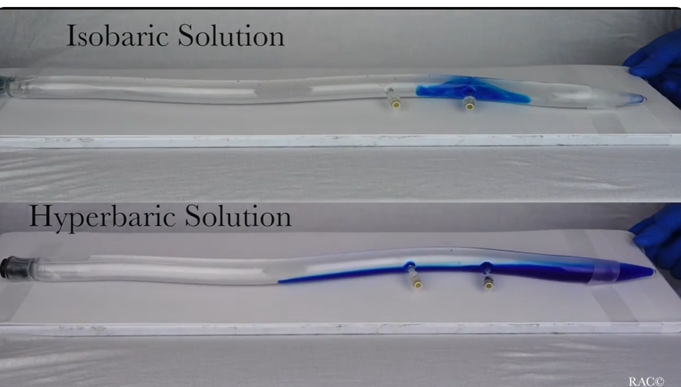
Baricity predicts the direction that LA will move upon injection into CSF. It is relative to CSF (1.004 to 1.009). Contour of vertebral canal is critical to distribution.
Hyperbaric:
Weighs more than CSF due to addition of dextrose; thus, it spreads more
It always travels down or towards gravity. “Sinks”. Greater cephalad spread when supine.
Most common for spinal anesthesia. EX: C-section, hernia repair (lower abdominal surgery), or saddle blocks (for peri/anal surgeries)
Example Solutions:
0.75% Bupivacaine with 8% glucose,
5% Lidocaine with 7.5% glucose,
0.5% Tetracaine mixed with 5% glucose.
Hypobaric:
Weighs less than CSF due to addition of sterile water
It will travel up the spinal cord or up towards the sky. “Float”
EX: when the surgical site is the "highest" point of the patient (such as prone jackknife for some perineal procedures or hip arthroplasty where anesthetic can float up up the nondependent side)
Example Solutions:
0.5% Lidocaine with sterile water
0.33% Tetracaine mixed in sterile water.
**lower concentrations than isobaric
Isobaric:
Weighs about the same as CSF, so will stay around the same level as injection (limited spread)
Typically provides profound motor block and prolonged duration. EX: Total Hip Arthroplasty, Knee surgery, or Procedures where position changes (as it is less likely to migrate unexpectedly)
Example Solutions (DIFFERENT FROM EACH OTHER!):
0.5% bupivacaine with water
2% Lidocaine with water
0.5% Tetracaine mixed in 50% CSF.
**hypo and hyper-baric are important when deciding how the patient should be position: hypobaric/hyperbaric depend on gravity to steer the location of the block. They are useful for a denser block rather than a more patchy one is isobaric. Although there is risk of cephalid spread once the LA reaches the thoracic kyphosis, the spine actually begins to slope "up" toward the cervical region and acts as a natural barrier; The heavy solution is unlikely to "climb" that hill unless you leave them in a steep Trendelenburg for too long. This is popular for abdominal surgeries; if the person’s fingers start getting numb (meaning in the cervical area now, a high spinal), this means just lay them flat or reverse trend to stop the flow up.
Spinal Anesthesia Math

**Review separate math paper
Physiological Changes Under Spinal Anesthesia
Hypotension and Bradycardia
Patients may experience dyspnea despite adequate ventilation. Proprioception, not reality. Spinal should have little effect on resting alveolar ventilation, they just cannot feel their muscles but they are working.
Quick tip: You can differentiate whether the patient is truly having a high spinal by having them squeeze your hand. If they can, that means C6-8 are still intact and so is diaphragm
Neuroendocrine Changes: Absence of adrenocortical response to painful stimuli.
Bladder: Atonic bladder; ureters contracted (urinary retention)
most common cause of prolonged PACU stay (since one of t he last things to return)
GI: contraction of small/large intestines, relaxation of sphincters (laxative effect)
Thermoregulation: Hypothermia due to loss of vasoconstriction. Inhibits shivering.
Decreased surgical bleeding: result of hypotension and reduction in peripheral venous pressure
Side Effects and Complications of Spinal Anesthesia
Neuro complications results from:
Direct trauma from needle/catheter.
Compression from hematoma, abscess, or LA injection within restricted areas.
Paresthesia: Transient sensation of tingling, burning, or numbness due to nerve brushing (often without long-term consequences/sequelae)
Important to stop needle advancement if paresthesia occurs. Wait until it resolves, then inject slowly; NEVER push against force. If they do not resolve, then you need to pick a different level.
Back pain: Commonly occurs due to ligament relaxation associated with anesthesia. Generally minor and short-lived. Frequent occurrence.
Spinal Hematoma:
Presents with severe back pain and neurologic deficits within 48 hours.
Diagnosis: MRI; immediate treatment involves evacuation of hematoma.
Post-Dural Puncture Headache
Loss of CSF exceeds the production rate, which then leads to downward displacement of the brain that stretches the meninges. Blood vessels in the skull distend to compensate for loss of CSF.
Children are at lower risk; risk increases substantially following puberty and slowly declines with age
Incidence also varies directly with size and type of needle; a 25 G pencil-point needle is the most appropriate to prevent this
Symptoms include:
Starts 12-24 hours post-procedure.
Postural component: dull/throbbing occipital —> frontal headache that is worse when sitting and better when lying down
N/v, malaise, anorexia, ocular disturbances (light sensitivity)
Treatment includes bed rest (lay flat), hydration (constant fluid intake), analgesics, caffeine (fiorecet), and possibly an epidural blood patch (if symptoms are severe, will provide immediate relief but risk of another dural puncture; considered definitive treatment).
15-20 ml sterile patient blood is injected into the epidural space near or below puncture site
You only inject until the patient verbalizes the pressure is too much/uncomfortable (do not push any more after the patient says this!)
Must remain supine for at least 1 hour following the placement of the blood patch (this is to prevent any pressure increases that could blow away the blood patch)
Holding the baby is still ok!
Cardiovascular Effects
Hypotension:
Occurs in 33% of spinal anesthesia patients due to sympathetic block (basically, the parasympathetic system takes over)
Symptoms include decreased venous return, cardiac output, and SVR.
Management:
Degree of hypotension parallels level of block and the intravascular fluid volume
Greatly exaggerated by coexisting hypovolemia
Treatment:
Restore venous return to increase cardiac output
5-10 degree head-down to improve venous return.
Not steep bc you do not want to risk cephalad spread of the LA
Fluid Loading: Administer 1 L fluid bolus prior to block (15 mL/kg)
Sympathomimetics: Ephedrine (10 mg IVP) and Phenylephrine (100 mcg).
Based on HR
Bradycardia and Asystole
Bradycardia occurs in 10-15% of patients, incidence increases with a higher level of block.
Mediated by blockage of cardioaccelerator fibers at T1-T4 and decreased venous return (Bezold-Jarisch reflex)
Treated by increasing preload and escalating drugs with (in order): atropine —> ephedrine —> epinephrine if necessary.
Based on level of bradycardia severity
Other effects:
High Spinal Anesthesia:
Excessive sensory/motor block leading to LOC shortly after injection. Manifests soon after injection.
This is different from the prioprioceptive loss of breathing sensation with a higher level spinal (T4). In that case, the patient is still having normal Spo2, be able to talk to you.
S/s:
weak grip when asked to follow commands
patient looks “floppy”
LOC
Tx:
maintain airway and ventilation
support circulation with sympathomimetics and IVFs
Place in head-down position and flex neck
**you do not want to sit them up, because this will worsen hypotension, at this point your goal is to maintain perfusion as much as possible while preventing further spread; but you are already getting spread, so most important thing is to prevent hypotension now
Tracheal intubation
Nausea: Although this can mean a good spinal block, it should also alert to possible systemic hypotension that could potentially lead to cerebral ischemia
Managed with sympathomimetics; nausea and vomiting incidence parallels the block level. Zofran will not help!
Urinary Retention: May necessitate catheter drainage.
Failed Spinal Options include:
Adding sedation or gas via mask (e.g., Propofol, Ketamine, Versed, Volatile).
Is it positional? Head down position to make it spread higher; if one sided, then rotate the patient to the side that is not numb (bed control)
Converting to general anesthesia
Repeating spinal, if there is time (not exceeding a reasonable single-shot dosage).
2.2 mL is the maximum volume that can be injected
If there was an additive to the first spinal attempt, do NOT add the additive to the second spinal attempt mixture
Spinal Kit Contents
Items in the Kit:
Povidone iodine packet.
Three swab sticks.
Fenestrated drape.
Towel.
Gauze pads (3" x 3").
5 mL syringe with 19 G x 1.5 filter needle
3 mL syringe with 25 G x 5/8” needle
1% lidocaine for SQ injection
Spinal agents including:
5% lidocaine, 0.75% bupivacaine, 1% tetracaine, 1:1000 epinephrine.
Introducer needle (20G x 1.5").
25g Whitacre spinal needle.
Sharps stick pad
adhesive bandage (don’t always have them, you can use a gauze pad to wipe away and hold short amount of pressure after procedure)
Spinal Needles
Two basic designs:
Open-ended cutting needle.
More potential for post dural puncture HA
The bevel must be parallel to the longitudinal dural fibers to minimize the tear
EX: Quincke, Pitkin
Closed tapered-tip pencil-point needle (noncutting).
Less potential for post dural puncture HA, as it separates dural fibers instead of cutting them
25g pencil-point is most common
EX: Whitacre, Pencan, Sprotte

Administration of Spinal Anesthetic
Position patient in a sitting or lateral position with lumbar back pushed out. Locate landmarks and identify target space.
Open trat, sterile glove, prep, and drape
Blot excess prep solution with gauze to prevent
Recommendation is that chloroprep is the superior cleaning supply, BUT both betadine and chloraprep are neurotoxic
Chloroprep is supposed to be scrubbed for friction. You only need to blot Betadine, not chloroprep.
Deliver local anesthetic (3 ml 1% lidocaine) with the smallest gauge needle to anesthetize skin and subcutaneous tissue using a good skin wheal.
Use a midline approach to insert the introducer needle into the interspinous ligament.
Bevel direction is parallel to longitudinal dural tissue fibers
Insert at top margin of the lower spinous process and advance with slight cephalad orientation
Once introducer is seated, insert the spinal needle ensuring to avoid early bone contact; redirect as needed.
Early encounter of bone (aka, spinous process) means you should withdraw the spinal needle back into the introduced and then redirect the introducer in small angular increments in a cephalad direction
Deep encounter of bone (aka, transverse process) means you should withdraw the spinal needle into the introducer and redirect the introducer in a caudad direction or lateral
**Paramedian approach: needle is inserted 1 cm lateral to the caudal aspect of the interspace. It is angled 10-15 degrees medially toward the spinal canal and angled slightly cephalad. . Primary use for elderly and arthritic patients (it bypasses the supra and interspinous ligaments, but you still feel the ligamentum flavum)
Upon entering the subarachnoid space (with a small pop), remove the style and give it a few seconds to confirm free flow of CSF (just let it come to the opening, don’t let it keep dripping)
CSF may initially be blood-tinged. Once CSF is clear, connect the LA syringe, aspirate again to confirm swirl of CSF in the fluid before inject the local anesthetic solution
If blood-tinged CSF continues to flow, the needle should be removed and inserted at a different level.
Inject in small increments; perform a second aspiration at the end of injection
If the patient has paresthesia, do not inject the LA and see if it goes away. If it does, try re-injecting again slowly but if paresthesia happens again, then remove the entire unit and re-try at a different level
Needle and syringe should be removed as a single unit; patient positioning according to target block level (e.g., saddle block vs T4 level; baricity of the LA)
Continuous Spinal
Involves inserting a catheter into the subarachnoid space for repeated drug administration.
Limit catheter insertion distance to 2-4 cm
Significant risk of post-dural puncture headache; therefore, it is not routinely employed.