Gross structure to fine structure
What’s in a muscle?
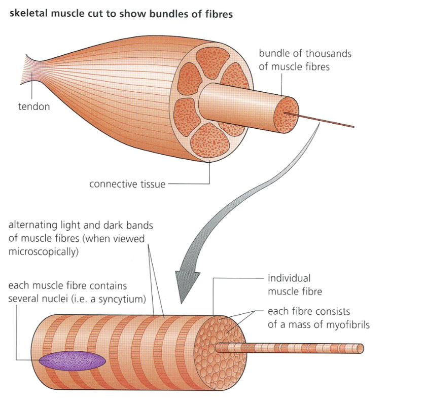
Muscle cells are called muscle fibres
Muscle → Muscle fibre → Myofibrils → Sarcomere → Actin and myosin
Skeletal muscle
Looks stripy due to protein filaments
Contain two protein called actin and myosin
Each muscle is made of muscle fibres
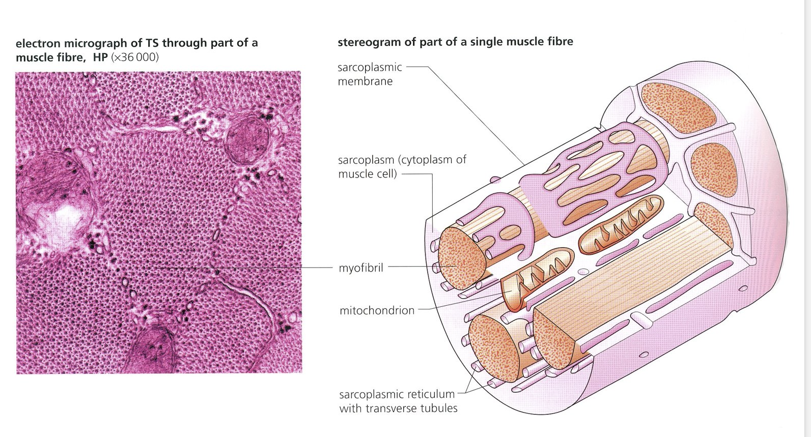
The muscle fibres are the same as most cells containing cytoplasm, mitochondria and endoplasmic reticulum
The muscle fibres contain many myofibrils, which are made up of sarcomeres
Very long, sometimes called acellular as they don’t seem to have distinct cell membranes and may have many nuclei (multinucleate)
Each cell is quite complex
Each cell has a surface membrane is called sarcolemma and cytoplasm is called sarcoplasm
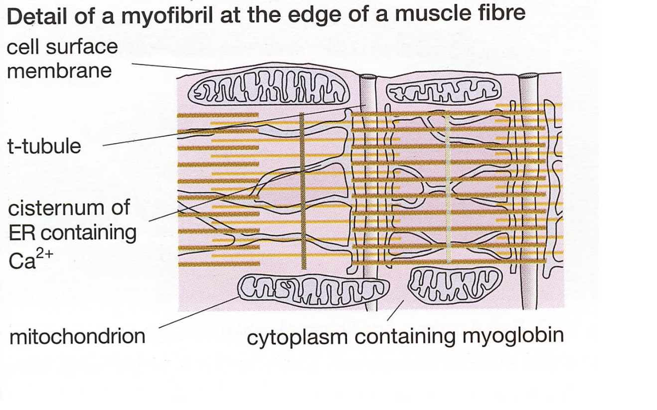
Large number of mitochondria = lots of ATP
Inside each fibre cell there are many longitudinally running fibres called myofibrils and it’s these that give the characteristic striations (stripes)
The sarcoplasm is interwoven with special sarcoplasmic reticulum (modified endoplasmic reticulum) and also infoldings of the sarcolemma called the T system or T tubules
These are important because they wrap around the myofibrils and are important in muscle contraction
Fine structure of muscles

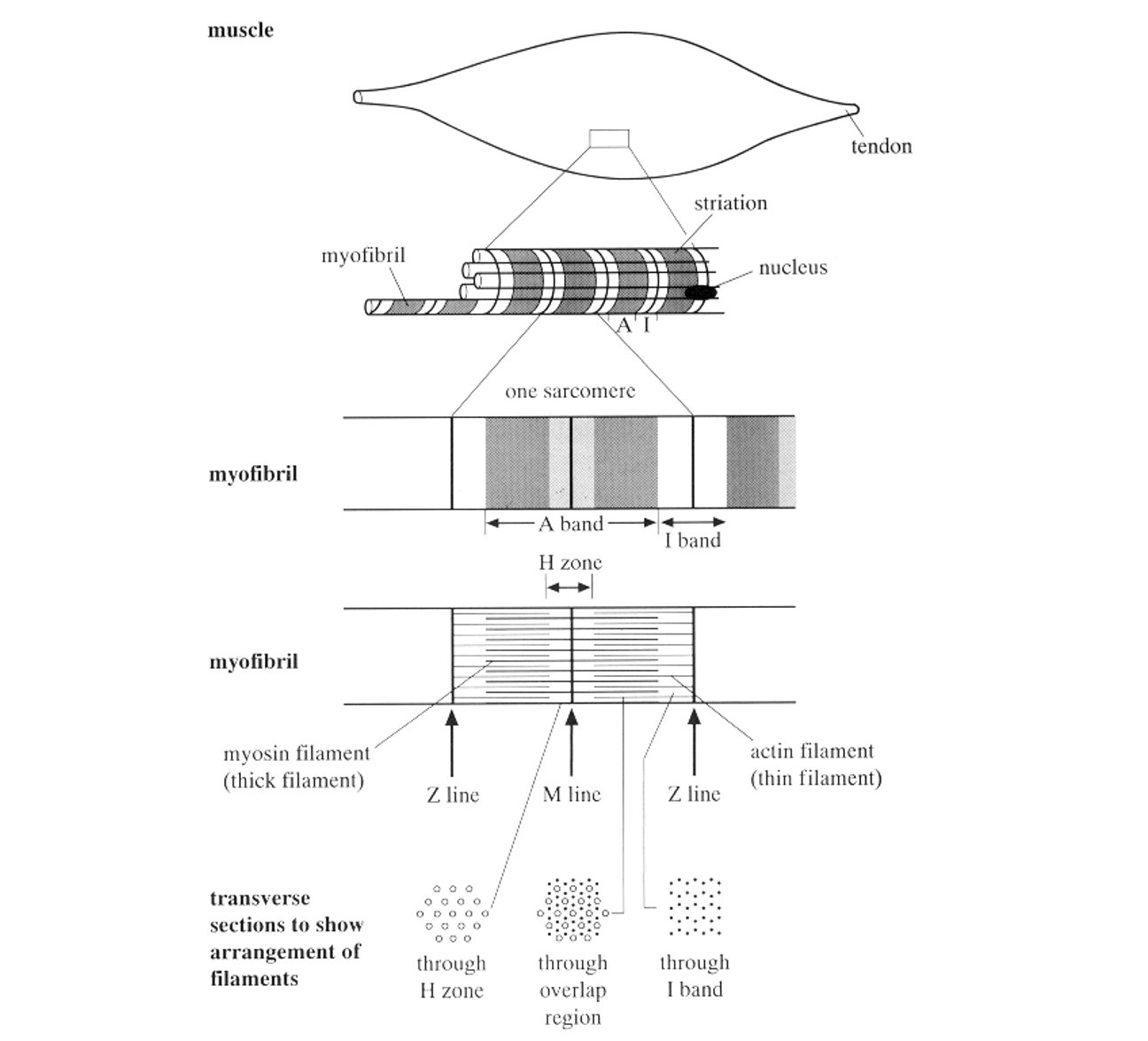
Sarcomeres
The markings of the myofibrils correspond to regular repeating units called sarcomeres
The sarcomere consists of different protein filaments, thin ones (actin) and thick ones (myosin)
The sarcomeres are joined end to end by thin plates called Z lines or Z plates
The filament form bands of differing shades
During contraction the filaments slide together to form a shorter sarcomere
Cross bridges are formed
Z line (Mark the end of sarcomere and are regions where actin fibres cross link)
A band or Dark band represents the length of the thicker myosin strands. (but the actin also overlaps here)
The I band or light band contains only actin
The H zone inside the A band is myosin only
The M line is a protein that holds the myosin together (like the Z line for actin)
Actin:
Thin filaments (8nm diameter)
Has myosin binding sites
Individual molecules are helical (2 of them)
2 regulatory proteins called Tropomyosin and Troponin are associated with it
Tropomyosin normally blocks the myosin binding site
Troponin can bind to calcium ions
Myosin:
Thich filaments (16nm)
Contain myosin heads that can bind to actin
Individual molecules have a common shaft and a protruding bulbous head
The heads can make cross bridges and contain ATP binding sites and an enzyme called ATPase.
Troponin and tropomyosin
Tropomyosin molecules coil around the actin
Troponin complex is attached to each tropomyosin