wk1: cytoplasm & organelles
Nucleus = stores genetic information
nucleolus = makes ribosomes
ribosome = makes protein
rough endoplasmic reticulum (RER) = folding/assembly of proteins
smooth endoplasmic reticulum (SER) = lipid and steroid synthesis and detoxification of drugs
Golgi apparatus = modifies, sorts and transports proteins
mitochondria = make energy (ATP)
peroxisome = regulates biochemical pathways that involve oxidation
lysosome = removes unwanted materials from the cell
vacuole = stores water and nutrients
vesicle = transport of materials
cytoskeleton = structure, support and transport
centrioles = cell division
The watery intracellular fluid surrounding the nucleus is called cytosol
Suspended within cytosol are structures called organelles (“little organs”) that perform specific functions to keep the cell healthy
Collectively, the cytosol and intracellular organelles make up the cytoplasm of the cell
Cell structure (cytoplasm & organelles)
The watery intracellular fluid surrounding the nucleus and fills the cell is called cytosol
Suspended within cytosol are structures called organelles (“little organs”) that perform specific functions to keep the cell healthy
Collectively, the cytosol and intracellular organelles make up the cytoplasm of the cell
Cytosol accounts for ~55% of the cell volume (varies in composition & consistency)
Cytosol is made up of ~75-90% water + 10-25% dissolved solutes (ions, glucose, amino acids, fatty acids, proteins, lipids, ATP, waste products, organic molecules
Organelles include ribosomes, endoplasmic reticulum (smooth and rough ER), Golgi apparatus, mitochondria, lysosomes, vesicles, centrioles & cytoskeleton
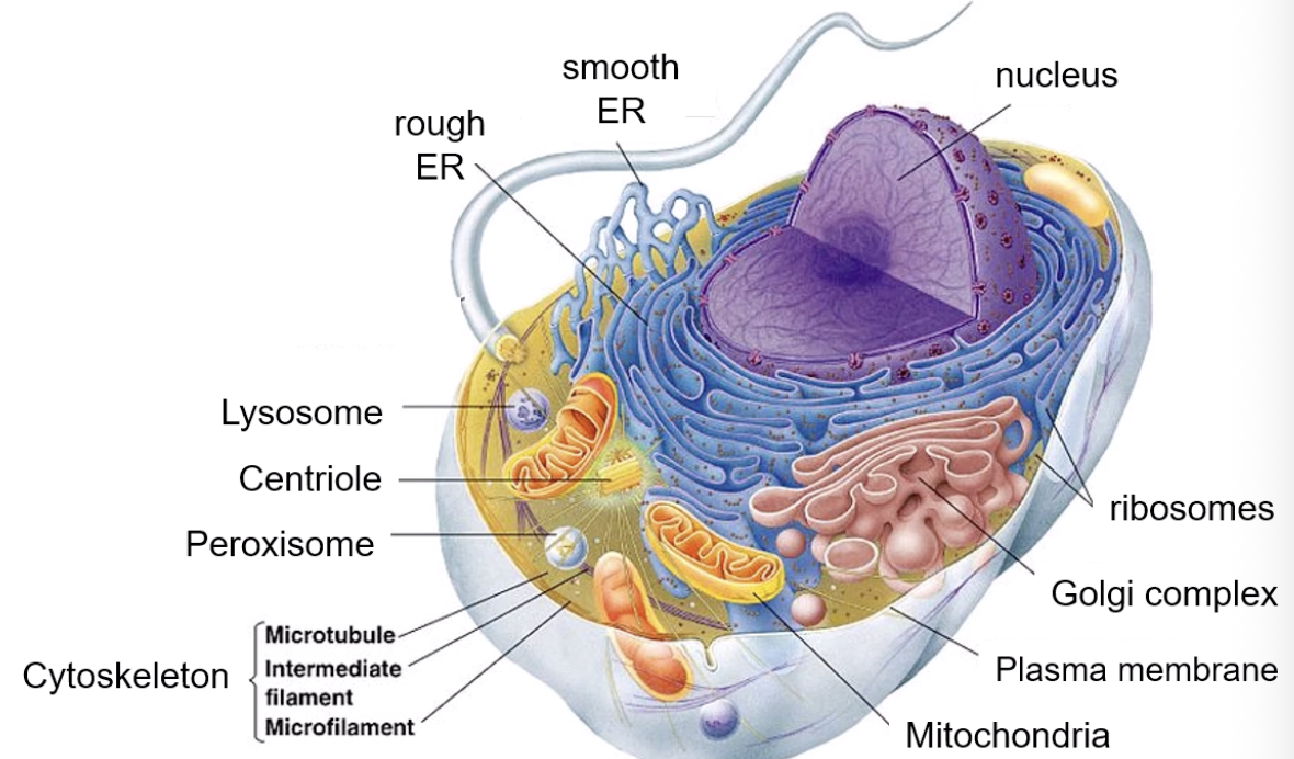
Ribosomes: function in protein synthesis, are composed of RNA and proteins, consist of two subunits (large & small), and are formed in the nucleolus.
Ribosomes can either be attached to the endoplasmic reticulum (where complex proteins that require modification are synthesised) or floating free in the cytosol (where short/simple proteins are synthesised)
Endoplasmic Reticulum: network of membranous sheets and channels extending throughout the cytoplasm, the ER is continuous with the nuclear envelope
Rough endoplasmic reticulum (RER): studded with ribosomes, functions to fold and assemble new proteins and give them three dimensional structure (proteins made by ribosomes are a chain of amino acids; they need to go through RER to fully develop)
Smooth endoplasmic reticulum (RER): lack ribosomes (does not synthesis proteins) - contains enzymes to synthesise lipids, steroids and carbohydrates
Golgi Apparatus: series of flattened, membranous sacs that modify (final tweaking), sort, and package proteins for secretion or delivery to other organelles.
Golgi Apparatus has two surfaces: entry - facing the RER (cis cisternae) & exit - facing the plasma membrane (trans cisternae)
Golgi Apparatus modifications include - addition/ removal of sugar molecules, addition of sulphate groups, addition of phosphate groups
Protein synthesised in ribosome → folded into ER → packaged into vesicle → shuttled to Golgi (cis) → vesicle fuses with Golgi complex → protein delivered into lumen → enzymes modify the protein → modified protein is shuttled into next cisterna via transfer vesicle → protein makes it way through Golgi complex →exits via trans cisterna in vesicle (time - 9:35)
Vesicles/vacuoles: transport substances - membrane bound sacs containing water, proteins, enzymes, hormones & waste. Vacuoles = vesicles that only contain water/waste
Mitochondria: ATP (energy) production (cellular respiration), double membrane (inner and outer).
Mitochondria inner membrane has numerous folds (cristae) extending into inner matrix - matrix contains enzymes, ribosomes & DNA (inside the folds cellular respiration occurs = glucose & oxygen are converted to ATP)
Red blood cells = 0 mitochondria
Muscle cells = 100s -1000s mitochondria per cell (the more mitochondria = the more energy needed to function)
Mitochondria has its own DNA; mitochondrial DNA encodes for proteins that the mitochondria needs to function and allows for communication to the nucleus
Mitochondrial DNA is inherited from maternal line
Lysosome: specialised vesicle that breakdown damaged organelles, pathogens & organic compounds (membrane-bound vesicle containing digestive enzymes & formed in Golgi complex)
Lysosome contain ~50 enzymes capable of breaking down proteins, nuclei acids, carbohydrates & lipids (break down materials no longer needed by the cell)
Lysosome optimal function in acidic conditions (pH ~5)
Cytoskeleton
Cytoskeleton supports plasma membrane & gives cell shape - supports positioning & movement of organelles - enables cell movement
Cytoskeleton structure: protein framework (filaments or slender tubes) throughout cell - made up of rigid filamentous protein chains, & highly labile for their continuous remodeling
Microtubule (microfilament): composed of actin, narrow diameter ~7nm, structure resembles double helix, highly dynamic; assemble/disassemble quickly
Microfilament functions as strength/structural support, facilitates cell movement/ changes in cell shape, involved in muscle contraction & cell division
Intermediate filament: protein composition varies among cell types, intermediate diameter ~8-11nm, multiple protein strands wound together, more permanent - not rapidly assembled/disassembled.
Intermediate filament functions as strength/structural support, movement of materials through cytoplasm, & anchoring organelles in place
Microtubule: composed of hollow tubulin tubes, large diameter ~25nm, highly dynamic - can grow/shrink quickly
microtubule function as strength/structural support (resist compression), movement of organelles, involved in cell division
Centrioles
Centrioles support the spatial arrangement of the cell & are involved in cell division
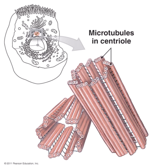
Structure: cylindrical structures, composed of cytoskeletal microtubules
Two centrioles form (at a right angle) a centrosome; the main microtubule organising centre of the cell, they’re surrounded by dense mass of proteins
cells that lack centrioles (red blood cells, neurons) cannot divide
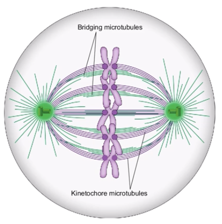
During cell division (mitosis), the centrioles are involved with movement of DNA strands
Centrosomes form the mitotic spindle - an apparatus that forms to separate duplicated chromosomes
Centrosomes move to opposite ends of the cell → pull on the duplicated chromosomes to ensure equal amounts of DNA are distributed to each of the cell daughters
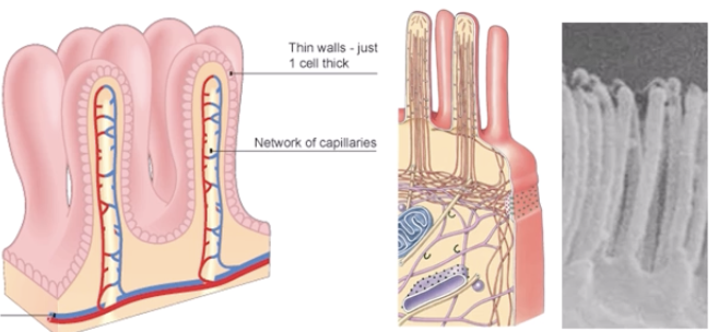
Projections that extend from cell surface maintained by cytoskeleton within cell
microvilli: finger-like extensions of plasma membrane, increase surface area thus aid absorption, located in digestive tract & kidneys - 100-1000 per cell, made up of actin filaments, ~0.1 - 1.0𝜇m length
Cilia: hair-like projections, aiding in the movement of substances across the cell surface (coordinated, sweeping movements), located in the respiratory tract & reproductive tracts, 100-1000 per cell, made up of microtubules, ~5-10𝜇m length
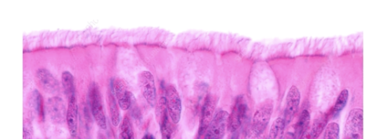
flagella: move entire call (locomotion), produces propellor-like movements & only located on sperm cells in humans, made from microtubules, ~10-200𝜇m length