Cerebellum
🧠 DETAILED MULTI-PARAGRAPH SUMMARY
The cerebellum (Latin for “little brain”) is a highly structured brain region located at the posterior aspect of the brain, beneath the occipital and temporal lobes. Although it accounts for only about 10% of total brain volume, it contains over 50% of the brain’s neurons, highlighting its dense computational capacity. Its primary function is not to initiate movement but to fine-tune motor activity, ensuring smooth, coordinated, and precise execution of voluntary movements. It operates through a rapid corrective feedback loop, continuously comparing intended movement with actual performance and making adjustments in real time.
Damage to the cerebellum leads to a range of motor deficits, including ataxia (loss of coordination), dysmetria (inability to judge distance or scale), and nystagmus (abnormal eye movements). Such damage disrupts balance, spatial accuracy, timing, and motor learning, underscoring the cerebellum’s essential role in refining movement and adapting motor skills through experience.
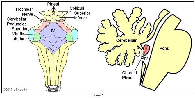
Structurally, the cerebellum follows a “rule of 3,” which helps organise its anatomy and function. It consists of two hemispheres separated by the vermis, and is divided into three lobes: anterior, posterior, and flocculonodular (vestibulocerebellum). It connects to the brainstem via three pairs of cerebellar peduncles: inferior (to medulla), middle (to pons), and superior (to midbrain). Functionally, it receives input (afferents) from three major systems: the vestibulocerebellum (balance and eye movements), spinocerebellum (body position and proprioception), and cerebrocerebellum (planning and coordination of voluntary movement). Outputs (efferents) are sent via deep cerebellar nuclei, primarily the fastigial, interposed, and dentate nuclei, to influence motor pathways.
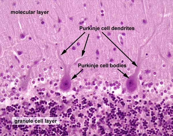
The cerebellum has a distinct internal organisation, consisting of an outer cerebellar cortex (grey matter) and an inner medulla (white matter). The cortex is arranged into three layers: the granule cell layer (input layer), the Purkinje cell layer (output layer), and the molecular layer (processing layer). Each layer contains specific neuron types that contribute to signal integration. Notably, Purkinje cells, which are inhibitory (GABAergic), serve as the sole output of the cerebellar cortex and project to deep cerebellar nuclei.
At the microcircuit level, cerebellar function depends on two major input pathways: mossy fibers and climbing fibers. Mossy fibers originate from the spinal cord and brainstem and synapse onto granule cells, whose axons (parallel fibres) activate Purkinje cells. Climbing fibers arise from the inferior olive and form powerful one-to-one synapses with Purkinje cells. The integration of excitatory and inhibitory signals within this circuitry ensures precise timing and coordination of motor outputs.
The balance between excitation and inhibition is critical for cerebellar function. Disruption of this balance—such as through alcohol’s enhancement of GABAergic inhibition—leads to impaired coordination and cerebellar dysfunction. Long-term potentiation (LTP) and depression (LTD) within cerebellar circuits contribute to motor learning, allowing the brain to refine movements over time.
📌 BULLET POINT SUMMARY
🔹 General Features
“Little brain,” located under occipital/temporal lobes
10% brain volume, >50% neurons
Coordinates movement via feedback loops
🔹 Functions
Motor coordination
Balance and posture
Timing and precision
Motor learning
🔹 Damage Effects
Ataxia
Dysmetria
Nystagmus
Impaired balance and coordination
🔹 Rule of 3
Peduncles: superior, middle, inferior
Hemispheres + vermis
Lobes: anterior, posterior, flocculonodular
Inputs: vestibulo-, spino-, cerebrocerebellum
Outputs: fastigial, interposed, dentate nuclei
Cortex layers: molecular, Purkinje, granule
🔹 Peduncles
Inferior → medulla
Middle → pons
Superior → midbrain
🔹 Functional Divisions
Vestibulocerebellum: balance, eye movement
Spinocerebellum: body position
Cerebrocerebellum: planning movement
🔹 Cellular Structure
Granule cells (excitatory)
Purkinje cells (inhibitory output)
Interneurons (stellate, basket, Golgi)
🔹 Inputs
Mossy fibers → granule cells
Climbing fibers → Purkinje cells
🔹 Outputs
Deep nuclei → motor pathways
🔹 Clinical Relevance
Alcohol disrupts GABA → impaired coordination
🧩 FILL-IN-THE-BLANK SUMMARY
Section 1: General Anatomy
The cerebellum means __little brain_____ in Latin.
It contains over __50%____ of the brain’s neurons.
It is located beneath the ___occip and temporalpe_____ lobes.
Section 2: Structure
The cerebellum has __2_______ hemispheres.
They are separated by the _vermis_________.
The three lobes are _anterior ________, __posterior________, and _floccc_________.
Section 3: Connections
The cerebellum connects to the brainstem via _peduncles_________.
The inferior peduncle connects to the __medulla_______.
The middle peduncle connects to the __pons_______.
Section 4: Functional Divisions
The _vestioccebellum____ controls balance and eye movement.
The _spinocebellum________ receives spinal input.
The _cerecebellum________ receives cortical input.
Section 5: Cellular Organization
The output cells of the cerebellar cortex are ___purkije_______ cells.
These cells are __inhi______ (excitatory/inhibitory).
Granule cells are ___exitatory______ (excitatory/inhibitory).
Section 6: Circuitry
_mossy________ fibers synapse with granule cells.
__climbing________ fibers synapse directly with Purkinje cells.
Purkinje cells inhibit the ___dentate ____ nuclei.
✅ ANSWERS (Fill in the blanks)
little brain
50%
occipital and temporal
two
vermis
anterior, posterior, flocculonodular
peduncles
medulla
pons
vestibulocerebellum
spinocerebellum
cerebrocerebellum
Purkinje
inhibitory
excitatory
mossy
climbing
deep cerebellar
📝 40 EXAM-STYLE MCQs
Questions
The cerebellum is located:
A. Frontal lobe
B. Brainstem
C. Posterior brain
D. Spinal cordCerebellum contains:
A. 10% neurons
B. 25% neurons
C. >50% neurons
D. 5% neuronsMain function:
A. Sensation
B. Movement coordination
C. Vision
D. HearingDamage causes:
A. Paralysis
B. Ataxia
C. Blindness
D. AphasiaVermis separates:
A. Lobes
B. Hemispheres
C. Layers
D. CellsInferior peduncle connects to:
A. Pons
B. Midbrain
C. Medulla
D. CortexMiddle peduncle connects to:
A. Medulla
B. Pons
C. Midbrain
D. CortexSuperior peduncle connects to:
A. Medulla
B. Pons
C. Midbrain
D. Spinal cordFlocculonodular lobe function:
A. Vision
B. Balance
C. Hearing
D. SmellSpinocerebellum receives:
A. Visual input
B. Spinal input
C. Auditory input
D. Olfactory inputCerebrocerebellum receives:
A. Spinal input
B. Cortical input
C. Vestibular input
D. Reflex inputFastigial nucleus controls:
A. Vision
B. Balance
C. Memory
D. EmotionDentate nucleus controls:
A. Reflex
B. Voluntary movement
C. Hearing
D. SmellCerebellar cortex has:
A. 2 layers
B. 3 layers
C. 4 layers
D. 5 layersInput layer:
A. Molecular
B. Granule
C. Purkinje
D. CortexOutput cells:
A. Granule
B. Golgi
C. Purkinje
D. BasketPurkinje cells are:
A. Excitatory
B. Inhibitory
C. Sensory
D. MotorGranule cells are:
A. Inhibitory
B. Excitatory
C. Neutral
D. MotorMossy fibers originate from:
A. Cortex only
B. Brainstem/spinal cord
C. Eye
D. EarClimbing fibers originate from:
A. Cortex
B. Inferior olive
C. Thalamus
D. Spinal cordClimbing fibers synapse:
A. Many-to-many
B. One-to-one
C. None
D. WeakParallel fibers come from:
A. Purkinje cells
B. Granule cells
C. Golgi cells
D. Basket cellsDeep nuclei send:
A. Inputs
B. Outputs
C. Sensory signals
D. ReflexAlcohol affects:
A. Dopamine
B. GABA
C. Serotonin
D. AcetylcholineAlcohol causes:
A. Improved coordination
B. Ataxia
C. Better memory
D. ParalysisMolecular layer contains:
A. Purkinje bodies
B. Stellate cells
C. Granule cells
D. Golgi cellsGolgi cells are:
A. Excitatory
B. Inhibitory
C. Sensory
D. MotorBasket cells:
A. Excite Purkinje
B. Inhibit Purkinje
C. Excite granule
D. NoneCerebellar medulla contains:
A. Grey matter
B. White matter
C. CSF
D. BloodCortex contains:
A. White matter
B. Grey matter
C. CSF
D. BloodDysmetria is:
A. Balance loss
B. Distance error
C. Vision loss
D. Hearing lossNystagmus:
A. Muscle loss
B. Eye movement abnormality
C. Hearing loss
D. Memory lossVestibulocerebellum connects to:
A. Cortex
B. Vestibular system
C. Spinal cord
D. ThalamusSpinocerebellum located in:
A. Hemispheres
B. Vermis/intermediate
C. Cortex
D. BrainstemCerebrocerebellum located in:
A. Vermis
B. Lateral hemispheres
C. Brainstem
D. Spinal cordPurkinje output goes to:
A. Cortex
B. Deep nuclei
C. Spinal cord
D. EyeMain inhibitory neurotransmitter:
A. Glutamate
B. GABA
C. Dopamine
D. SerotoninBalance requires:
A. Vision only
B. Cerebellum
C. Hearing
D. SmellMotor learning depends on:
A. Cortex only
B. Cerebellum
C. Eye
D. EarCerebellum primarily controls:
A. Emotion
B. Coordination
C. Language
D. Vision
✅ MCQ ANSWERS
C
C
B
B
B
C
B
C
B
B
B
B
B
B
B
C
B
B
B
B
B
B
B
B
B
B
B
B
B
B
B
B
B
B
B
B
B
B
B
B