LIVER
Liver
Describe the path of hepatic blood flow and hepatic physiology.
Discuss the different types and manifestations of liver dysfunction.
Hepatocyte dysfunction
Portal hypertension
Pathophysiology of functional zonation
Describe the pathophysiology including the clinical presentation, epidemiology and risk factors, pathogenesis, pathology and clinical manifestations of the following disorders of the liver:
Acute hepatitis
Chronic hepatitis
Cirrhosis
Hepatocellular carcinoma
Objective 1: Describe the path of hepatic blood flow and hepatic physiology.
Path of Hepatic Blood Flow:
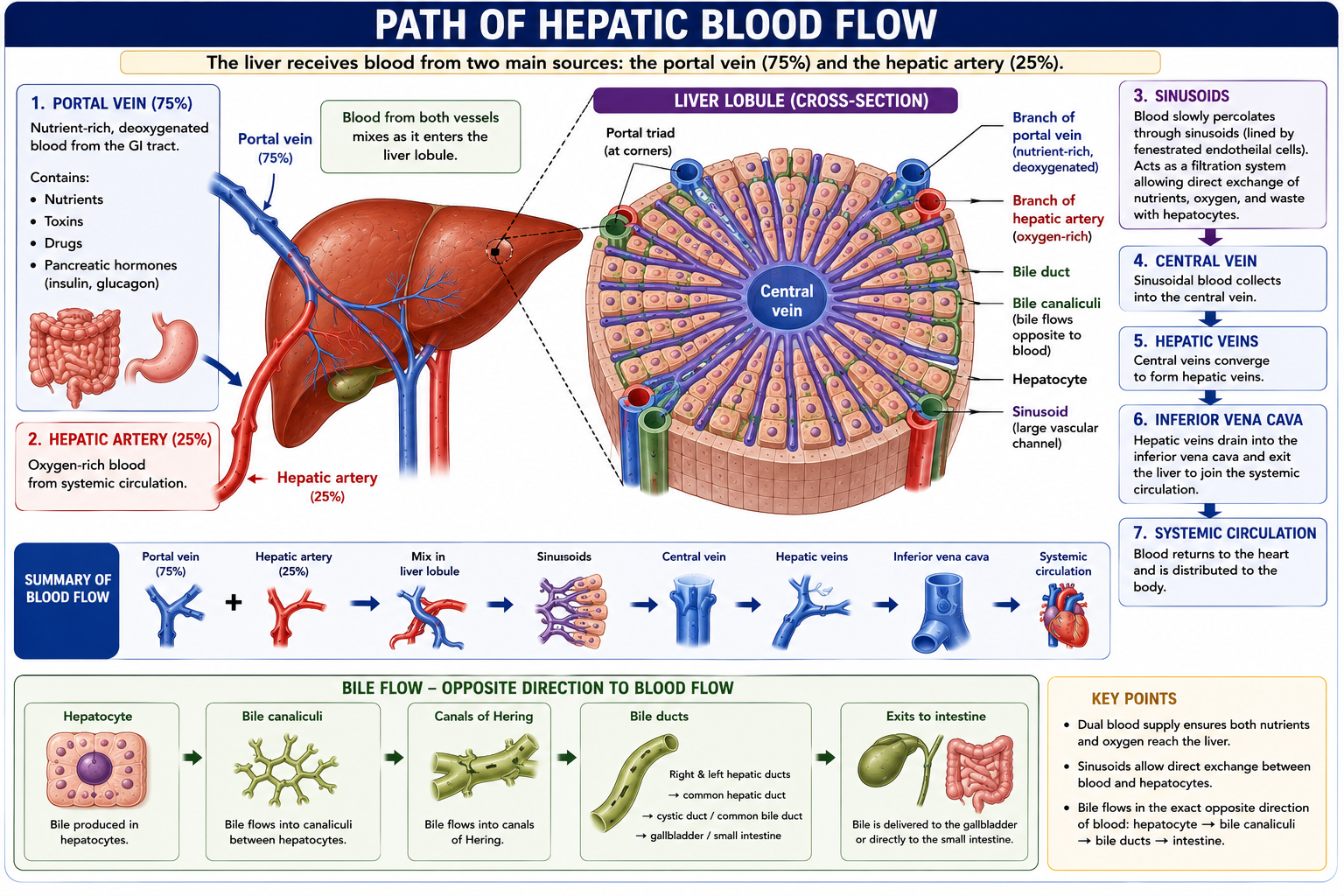
The liver receives blood from two main sources: the portal vein (75%) and the hepatic artery (25%).
The portal vein carries nutrient-rich, deoxygenated blood from the GI tract (including toxins, drugs, and pancreatic hormones like insulin and glucagon).
The hepatic artery provides oxygen-rich blood.
Blood from both vessels mixes as it enters the liver lobule and slowly percolates through sinusoids. The sinusoids are large vascular channels lined by fenestrated endothelial cells, acting as a filtration system to allow direct exchange of nutrients, oxygen, and waste with hepatocytes.
From the sinusoids, blood flows into the central vein, then converges into the hepatic veins, and finally exits the liver into the inferior vena cava to join the systemic circulation.
Note: Bile flow travels in the exact opposite direction of blood flow (from hepatocytes to bile canaliculi, and out through bile ducts).
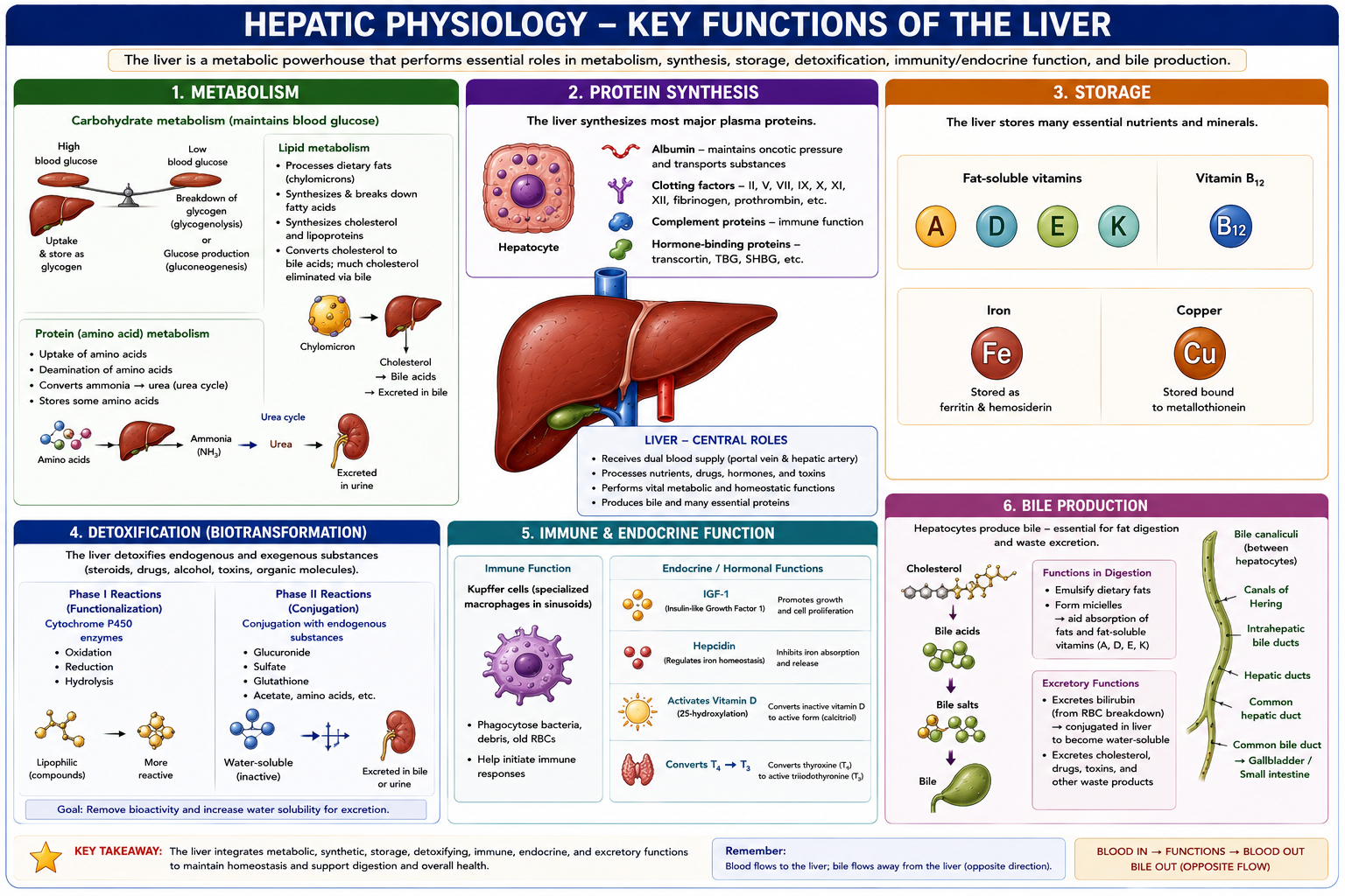
Hepatic Physiology: The liver has several critical physiological roles:
Metabolism: It maintains blood sugar by consuming, storing, or releasing glucose (gluconeogenesis/glycogenolysis). It processes dietary fats (chylomicrons) and manages cholesterol, much of which is eliminated via bile. It is also responsible for amino acid uptake and the breakdown of amino acids into urea.
Protein Synthesis: The liver synthesizes major serum proteins, including albumin, clotting factors, and hormone-binding proteins.
Storage: It is the main storage site for fat-soluble vitamins (A, D, E, K), vitamin B12, iron, and copper.
Detoxification: The liver performs Phase I (cytochrome P450 enzymes) and Phase II (conjugation) biotransformations to remove the bioactivity of steroids, organic molecules, and drugs, making them water-soluble for excretion.
Immune & Endocrine Function: Kupffer cells (specialized macrophages in the sinusoids) clear bacteria and debris. The liver secretes hormones like IGF-1 and hepcidin, activates Vitamin D, and converts T4 to T3.
Bile Production: Hepatocytes synthesize bile acids from cholesterol, which combine to form bile salts. Bile aids in the digestion and absorption of fats by forming micelles. Bile also excretes bilirubin (a breakdown product of red blood cells conjugated in the liver to become water-soluble).
Objective 2: Discuss the different types and manifestations of liver dysfunction.
a. Hepatocyte Dysfunction Dysfunction of the individual hepatocytes leads to a loss of the liver's metabolic, synthetic, and clearance abilities:
Metabolic abnormalities: Results in hypo/hyperglycemia, fat buildup in the liver, and altered lipid metabolism (inability to clear LDL).
Protein & Hormone abnormalities: Decreased synthesis of clotting factors leads to coagulopathies. The inability to clear hormones results in increased blood estrogen.
Toxin buildup: Failure to detoxify ammonia into urea leads to neurotoxic buildup, causing hepatic encephalopathy (altered mental status and confusion).
Cholestasis: Failure to secrete bile leads to malabsorption, vitamin deficiency, jaundice (elevated bilirubin), and pruritus (bile acid deposition under the skin).
b. Portal Hypertension Normal portal blood flow is a low-pressure system. When intrahepatic resistance rises, blood backs up, seeking alternate routes to the systemic circulation via portal-to-systemic shunting (bypassing the liver). Manifestations include:
Varices: Engorged blood vessels (e.g., esophageal or rectal) that can result in GI hemorrhage.
Ascites: Pathologic accumulation of fluid in the peritoneal cavity, driven primarily by elevated hydrostatic pressure forcing fluid out of capillaries, coupled with sodium/water retention.
Loss of clearance: Bypassing the liver leads to decreased Kupffer cell function (the last line of defense against bacteria/endotoxins) and malabsorption.
c. Pathophysiology of Functional Zonation The liver acinus is the smallest functional unit of the liver, divided into three zones based on blood flow and oxygen/nutrient gradients:
Zone 1 (Periportal): Closest to the arteriole, receiving the highest oxygen and nutrient delivery. It is responsible for oxidative metabolism, gluconeogenesis, ureagenesis, and is the first zone exposed to absorbed toxins.
Zone 2 (Midzonal): A transitional area with intermediate oxygen and nutrients.
Zone 3 (Centrilobular): Furthest from the arteriole, receiving the lowest oxygen and nutrient concentration. It is responsible for glycolysis and ketogenesis. Due to its location, Zone 3 has high CYP450 activity and is the most prone to hypoxia and injury.

Objective 3: Pathophysiology of Liver Disorders
a. Acute Hepatitis
Pathophysiology/Pathogenesis: An inflammatory process causing liver cell death via necrosis or apoptosis. In viral hepatitis, the virus replicates inside a hepatocyte, triggering cell death and inflammation. Toxic hepatitis disrupts cellular homeostasis and causes glycogen depletion.
Epidemiology/Risk Factors: Caused by viral infections (HAV/HEV via fecal-oral route; HBV/HCV/HDV via blood or sex). Also caused by toxic exposure (acetaminophen is the most common cause of acute liver failure in the US) or prolonged, chronic alcohol consumption.
Clinical Presentation/Manifestations: Can be asymptomatic, or present in distinct phases:
Prodrome (pre-icteric): Lasts 3-4 days. Manifests as malaise, fatigue, low fever, anorexia, altered taste/smell, nausea, and right upper quadrant (RUQ) abdominal pain.
Icteric Phase: Lasts 1-4 weeks. Characterized by jaundice, direct hyperbilirubinemia, severe pruritus, weight loss, and a large, tender liver.
Convalescent Phase: Disappearance of constitutional symptoms alongside markedly elevated liver enzymes.
b. Chronic Hepatitis
Pathophysiology/Pathogenesis: Defined as a combination of liver cell necrosis and inflammation lasting more than 6 months. Driven by either an inadequate immune response resulting in persistent viral infection or an immune-mediated attack. Repeated acute injuries lead to necrosis, fibrosis, and eventual regeneration (cirrhosis).
Epidemiology/Risk Factors: Chronic viral hepatitis is the most common cause of chronic liver disease in the US. Other causes include metabolic syndrome/Nonalcoholic Fatty Liver Disease (decreased lipid clearance), drugs/poisons, and autoimmune disorders.
Clinical Presentation/Manifestations: Ranges from asymptomatic to liver failure. Early signs include fatigue, malaise, low-grade fever, mild intermittent jaundice, and mild hepatosplenomegaly. Late disease presents with variceal bleeding, coagulopathy, encephalopathy, and ascites.
c. Cirrhosis
Pathophysiology/Pathology: It is an irreversible distortion of normal liver architecture. It is not an acute process, but rather a progressive insult characterized by hepatic injury, increased deposition of fibrous tissue/collagen (scarring), and regenerative nodules surrounded by scar tissue.
Clinical Presentation/Manifestations: Results in progressive hepatocellular dysfunction and Portal Hypertension (portal venous pressure > 6 mmHg). Key complications include:
Ascites: Excess fluid in the peritoneal cavity.
Hepatorenal Syndrome: Kidney injury caused by renal vasoconstriction, developing in response to systemic arterial vasodilation.
Splenomegaly/Hypersplenism: Engorgement of the spleen traps formed blood elements, causing thrombocytopenia and hemolytic anemia.
Coagulopathy/DIC: Loss of synthesis of clotting factors, and uncontrolled simultaneous clotting/bleeding due to the inability to remove fibrin degradation products.
Hypoalbuminemia, peripheral edema, hepatic encephalopathy, and hepatopulmonary syndrome.
d. Hepatocellular Carcinoma (HCC)
Note: The provided sources primarily detail the etiology/risk factors for HCC rather than its specific pathogenesis or clinical presentation.
Etiology/Risk Factors: It is strongly linked to underlying liver damage. Specific causes include Cirrhosis, HBV infection (which carries a 100-fold increase in risk), HCV, Nonalcoholic steatohepatitis (NASH), and Alcoholic cirrhosis
