Structure of skeletal, cardiac, and smooth muscle
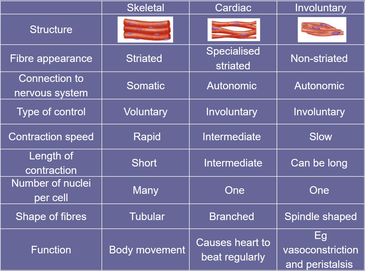
Skeletal muscle structure
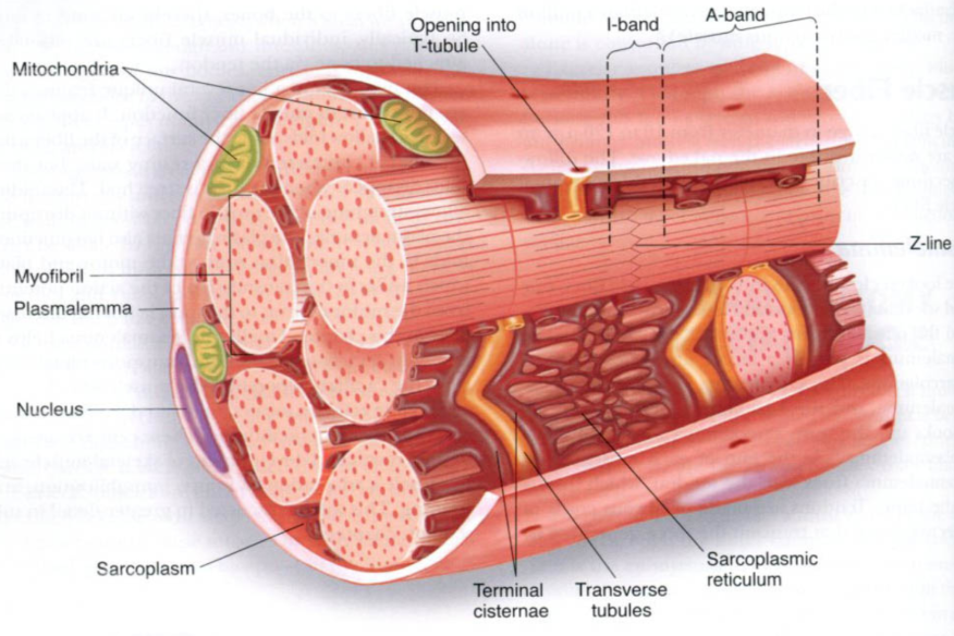
muscle fibres are highly specialised cells
multinucleate (syncytium)
many mitochondria
surrounded by a sarcolemma
sarcoplasm contains myofibrils
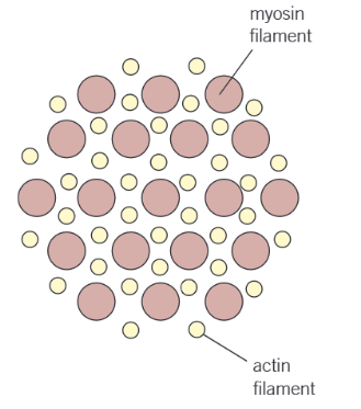
bundles of actin and myosin myofilaments
the arrangement of the actin and myosin myofilaments results in the striated appearance of skeletal muscle
the sarcolemma contains folds (invaginations) called T-tubules
link up to a specialised endoplasmic reticulum called the sarcoplasmic reticulum
calcium ions are stored here
Actin
globular protein
many actin molecules link together in a long chain
two chains twist together to form an actin filament
a fibrous protein (tropomyosin) is twisted around the actin filament
another protein (troponin) is attached at regular intervals

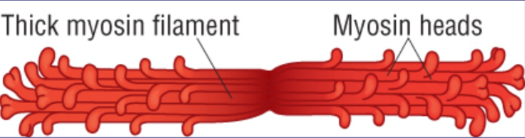
Myosin
fibrous protein
tail of the protein is attached to the M line
several molecules lie in a bundle together
the heads stick out to form cross bridges
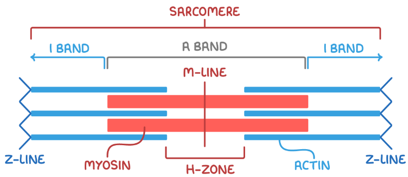
Sarcomere structure
Light bands (I band)
areas appear light as they are the region where the actin and myosin filaments do not overlap
dark bands (A band)
appear dark because of the presence of thick myosin filaments
edges are particularly dark as the myosin is overlapped with actin
Z lines
mark the boundaries of each sarcomere unit
M line
central line of sarcomere
H zone
area with only myosin
central lighter region within the A band