Week 1
Anatomy - physical structures of the body, from large, visible organs (gross anatomy) to microscopic components such as cells and tissues (microscopic anatomy). It involves techniques like dissection and modern imaging to understand how body parts are organised and connected.
Physiology - how these structures function, emphasising the chemical and physical processes that sustain life, including the body’s ability to maintain homeostasis.
Gross and Microscopic Anatomy (a) Gross anatomy considers large structures such as the brain. (b) Microscopic anatomy can deal with the same structures, though at a different scale (e.g. nerve cells)
The cell is the smallest independently functioning unit of life.
A tissue is a group of similar cells performing a specific function.
An organ is a distinct body structure composed of 2 or more tissues performing one or more physiological roles.
An organ system consists of multiple organs that collaborate to carry out major functions or meet the physiological needs of the body.
Levels of Structural Organisation of the Human Body. The organisation of the body often is discussed in terms of six distinct levels of increasing complexity.
Chemical level
Cellular level
Tissue level
Organ level
Organ system level
Organismal level
The eleven organ systems of the body:
Integumentary (encloses internal body structures)
Skeletal (Supports the body)
Muscular (Enables movement)
Nervous (Detects and processes sensory information, activates body responses)
Endocrine (Secretes hormones and regulates bodily processes)
Cardiovascular (Delivers oxygen to tissues, equalises temperature)
Digestive (Processes food, removes waste from undigested food)
Urinary (Controls water balance in body, removes waste from blood)
Respiratory (Delivers oxygen to blood, removes carbon dioxide)
Lymphatic (Returns fluid to blood, defends against pathogens)
Reproductive (Produces sex hormones, supports embryo until birth)
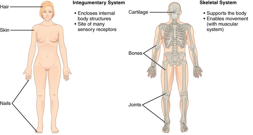
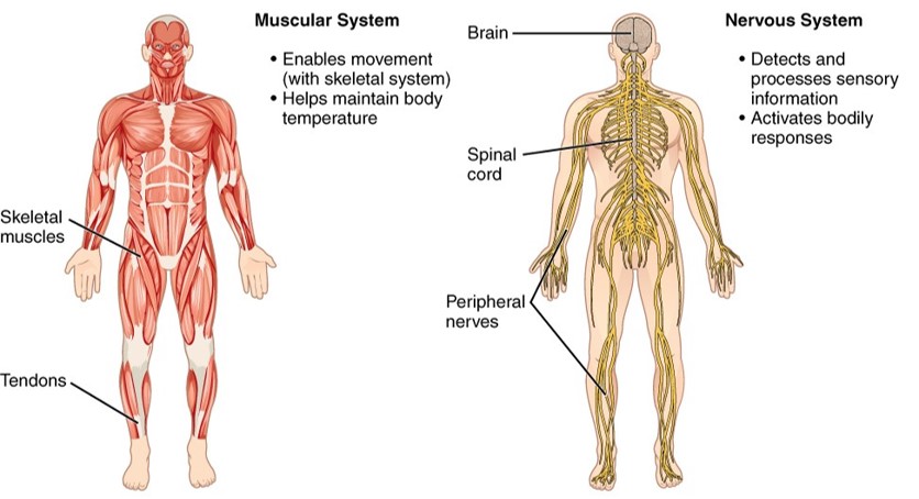
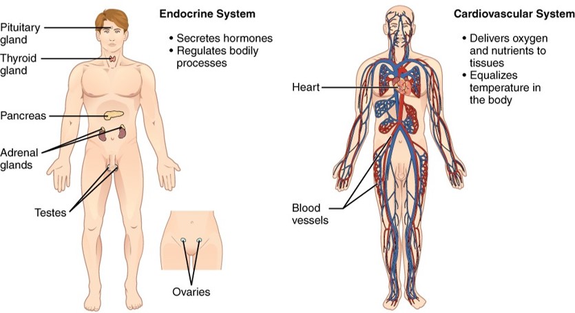
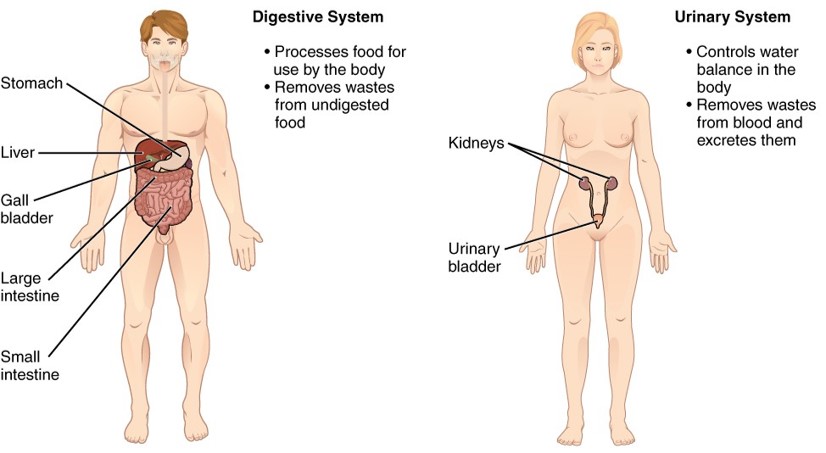
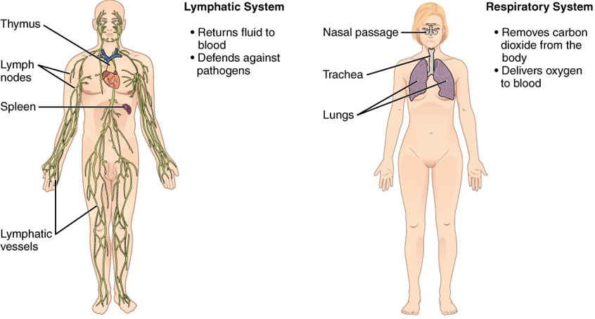
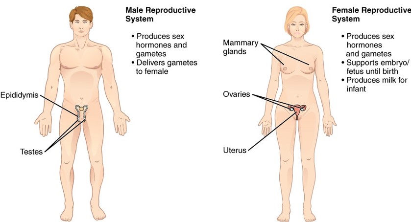
HOMEOSTASIS
Maintaining homeostasis means the body constantly monitors and adjusts its internal conditions—such as temperature, blood pressure, and nutrient levels—to stay within a normal range.
Each parameter has a set point, the ideal value around which a normal range fluctuates. For example, the set point for human body temperature is approximately 37°C
Negative feedback: Reverses deviations from set point to restore balance
Examples: If blood pressure drops, your heart beats faster and blood vessels constrict, blood pressure rises and the response diminishes.
Positive feedback: Amplifies changes
Examples: Woman goes into labour, baby’s head stimulates stretch receptors, oxytocin is released, oxytocin causes stronger uterine contractions, loop continues until baby delivered.
You cut your finger. Platelets stick to the site of injury and release chemicals. Chemicals attract more platelets forming a clot, loop continues until bleeding stops, then clotting stops.
THE HUMAN CELL
Human cells have a flexible membrane that encloses cytoplasm, a water based fluid containing organelles.
Cell Membrane (aka plasma membrane)
Phospholipid bilayer and phospholipid structure. The phospholipid bilayer consists of two adjacent sheets of phospholipids, arranged tail to tail. The non-polar (hydrophobic) lipid tails associate with one another, forming the interior of the membrane. The polar (hydrophilic) phosphate heads contact the fluid inside and outside of the cell.
Nucleus (most prominent of organelles)
The nucleus stores all genetic instructions for manufacturing proteins within DNA.
Passive transport
Passive transport moves substances down their concentration gradient (from high to low concentration) without using cellular energy (Adenosine triphosphate - ATP). It includes:
Simple diffusion: Movement of small, nonpolar molecules directly through the lipid bilayer.
Example: Oxygen diffusing into cells.
Facilitated diffusion: Movement of larger or polar molecules via membrane proteins.
Example: Glucose entering cells through a glucose transporter.
Osmosis: Diffusion of water across a semipermeable membrane.
Water moves toward areas with higher solute concentration.
Isotonic: Equal solute concentration inside and outside the cell; optimal cell function.
Hypertonic: cells lose water and shrink, higher solute concentration outside
Hypotonic: cells gain water and may burst, more solute in cell, lower solute concentration outside
Filtration: Movement of fluid and solutes driven by pressure gradients, not concentration.
Example: Kidney filtration and nutrient delivery in capillaries.
Active transport
Active transport moves substances against their concentration gradient (from low to high concentration) using ATP and often involves membrane proteins/protein pumps.
Sodium-Potassium Pump The sodium-potassium pump is found in many cell (plasma) membranes. Powered by ATP, the pump moves sodium and potassium ions in opposite directions, each against its concentration gradient. In a single cycle of the pump, three sodium ions are extruded from and two potassium ions are imported into the cell.
4 MAIN TISSUE TYPES
Epithelial tissue
Muscle tissue
Nervous tissue
Connective tissue

EPITHELIAL TISSUE
Epithelial tissues can be grouped into two major types: surface epithelia that line internal surfaces and cover external surfaces of the body; and glandular epithelia that form all glands in the body.
Surface epithelial tissues are classified according to the shape of the cells and number of the cell layers formed. Cell shapes can be squamous (flattened and thin), cuboidal (boxy, as wide as it is tall), or columnar (rectangular, taller than it is wide). Similarly, the number of cell layers in the tissue can be one—where every cell rests on the basal lamina—which is a simple epithelium, or more than one, which is a stratified epithelium and only the basal layer of cells rests on the basal lamina. Pseudostratified (pseudo- = “false”) describes tissue with a single layer of irregularly shaped cells that give the appearance of more than one layer.

Simple squamous epithelium
The cells in simple squamous epithelium have the appearance of thin scales. Squamous cell nuclei tend to be flat, horizontal, and elliptical, mirroring the form of the cell. The endothelium is the epithelial tissue that lines vessels of the lymphatic and cardiovascular system, and it is made up of a single layer of squamous cells. The mesothelium is a simple squamous epithelium that forms the surface layer of the serous membrane that lines body cavities and internal organs.
Simple cuboidal epithelium
In simple cuboidal epithelium, the nucleus of the box-like cells appears round and is generally located near the centre of the cell. These epithelia are active in the secretion and absorption of molecules. Simple cuboidal epithelia are observed in the lining of the kidney tubules and in the ducts of glands.
Simple columnar epithelium
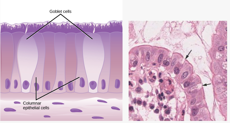
In simple columnar epithelium, the nucleus of the tall column-like cells tends to be elongated and located in the basal end of the cells. Like the cuboidal epithelia, this epithelium is active in the absorption and secretion of molecules. Simple columnar epithelium forms the lining of some sections of the digestive system and parts of the female reproductive tract. Ciliated columnar epithelium is composed of simple columnar epithelial cells with cilia on their apical surfaces. These epithelial cells are found in the lining of the fallopian tubes and parts of the respiratory system, where the beating of the cilia helps remove particulate matter.
Glandular epithelium
A gland is a structure made up of one or more cells modified to synthesise and secrete chemical substances. Most glands consist of groups of epithelial cells. A gland can be classified as an endocrine gland, a ductless gland that releases secretions directly into surrounding tissues and fluids (endo- = “inside”), or an exocrine gland whose secretions leave through a duct that opens directly, or indirectly, to the external environment (exo- = “outside”).
Pseudostratified epithelium
Pseudostratified columnar epithelium is a type of epithelium that appears to be stratified but instead consists of a single layer of irregularly shaped and differently sized columnar cells. In pseudostratified epithelium, nuclei of neighboring cells appear at different levels rather than clustered in the basal end. The arrangement gives the appearance of stratification; but in fact all the cells are in contact with the basal lamina, although some do not reach the apical surface. Pseudostratified columnar epithelium is found in the respiratory tract, where some of these cells have cilia.
Both simple and pseudostratified columnar epithelia are heterogeneous epithelia because they include additional types of cells interspersed among the epithelial cells. For example, a goblet cell is a mucous-secreting unicellular “gland” interspersed between the columnar epithelial cells of mucous membranes.
Stratified Epithelium
A stratified epithelium consists of several stacked layers of cells. This epithelium protects against physical and chemical wear and tear. The stratified epithelium is named by the shape of the most apical layer of cells, closest to the free space. Stratified squamous epithelium is the most common type of stratified epithelium in the human body. The apical cells are squamous, whereas the basal layer contains either columnar or cuboidal cells. The top layer may be covered with dead cells filled with keratin and therefore appear non-nucleated. Keratin is a protein produced to provide protection against abrasion and drying out. Keratinised epithelia are always stratified due to their locations in areas of high physical trauma and are localised only in areas of the body exposed to the external environment. The skin and the hard palate of the mouth are lined with this dry, keratinised stratified squamous epithelium. The lining of the oesophagus, vagina, most parts of the oral cavity and pharynx, and anal canal consists of non-keratinised, stratified squamous epithelium.
Muscle tissue
Muscle cells are excitable; they respond to a stimulus. They are contractile, meaning they can shorten and generate a pulling force. When attached between two movable objects, in other words, bones, contractions of the muscles cause the bones to move.
Voluntary muscle movement: under conscious control.
Involuntary muscle movement: not under conscious control, e.g. the contraction of your pupil in bright light.
Muscle tissue is classified into three types according to structure and function: skeletal, cardiac, and smooth.
All three muscle tissues have some properties in common; they all exhibit a quality called excitability as their plasma membranes can change their electrical states (from polarised to depolarised) and send an electrical wave called an action potential along the entire length of the membrane. Contractility allows muscle tissue to pull on its attachment points and shorten with force.

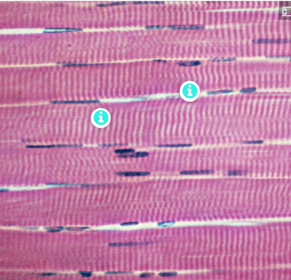
Skeletal muscle is attached to bones and its contraction makes possible locomotion, facial expressions, posture, and other voluntary movements of the body. Forty percent of your body mass is made up of skeletal muscle. Skeletal muscles generate heat as a by-product of their contraction and thus participate in thermal homeostasis. Shivering is an involuntary contraction of skeletal muscles in response to perceived lower than normal body temperature. The muscle cell, or myocyte (myo = muscle; cyte = cell), numbers remain relatively constant throughout life. Skeletal muscle tissue is arranged in bundles surrounded by connective tissue. Under the light microscope, muscle cells appear striated with many nuclei squeezed along the membranes. The striation is due to the regular alternation of the contractile proteins actin and myosin, along with the structural proteins that couple the contractile proteins to connective tissues. The cells are multinucleated as a result of the fusion of the many myocytes that fuse to form each long muscle fibre.
Smooth muscle tissue contraction is responsible for involuntary movements in the internal organs. Smooth muscle (so-named because the cells do not have striations) is present in the walls of hollow organs like the urinary bladder, uterus, stomach, intestines, and in the walls of passageways, such as the arteries and veins of the circulatory system, and the tracts of the respiratory, urinary, and reproductive systems. Each cell is spindle shaped with a single nucleus.
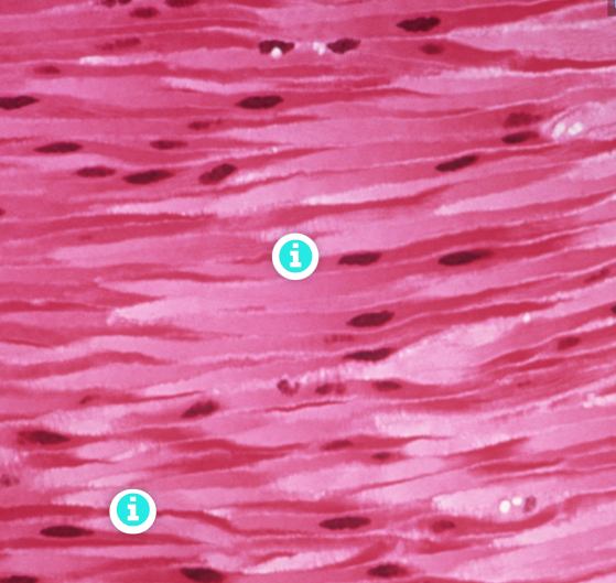
Cardiac muscle forms the contractile walls of the heart. The cells of cardiac muscle, known as cardiomyocytes, also appear striated under the microscope. Unlike skeletal muscle fibres, cardiomyocytes are single cells typically with a single centrally located nucleus. A principal characteristic of cardiomyocytes is that they contract on their own intrinsic rhythms without any external stimulation. Cardiomyocytes attach to one another tightly with specialised cell junctions called intercalated discs. Attached cells form long, branching cardiac muscle fibres that allow the cells to synchronise their actions. The cardiac muscle pumps blood through the body and is under involuntary control.

NERVOUS TISSUE
Nervous tissue is composed of two types of cells, neurons and glial cells. Neurons are responsible for the computation and communication that the nervous system provides. They are electrically active and release chemical signals to target cells. Glial cells, or glia, are known to play a supporting role for nervous tissue.
Neurons are the cells considered to be the basis of nervous tissue. They are responsible for the electrical signals that communicate information about sensations, and that produce movements in response to those stimuli, along with inducing thought processes within the brain. An important part of the function of neurons is in their structure, or shape. The three-dimensional shape of these cells makes the immense numbers of connections within the nervous system possible.
Glial cells, or neuroglia, are the other type of cell found in nervous tissue. They are considered to be supporting cells, and many functions are directed at helping neurons complete their function for communication. The name glia comes from the Greek word that means “glue". There are several types of glial cells. Some are found in the central nervous system (brain and spinal cord) and others in the peripheral nervous system (peripheral nerves).
CONNECTIVE TISSUE
Connective tissue is the most widely distributed primary tissue in the human body. As its name suggests, one of its key roles is to support and connect other tissues and organs. In contrast to epithelial tissue, where cells are tightly packed with minimal extracellular space, connective tissue cells are scattered throughout an extensive extracellular matrix. This matrix, produced by the cells embedded within it, is fundamental to the function of the tissue.
The matrix consists primarily of a ground substance, which is often interwoven with fibres. While the ground substance is typically fluid, it can also be mineralised and solid, as seen in bone tissue. Despite the diversity of connective tissues, they generally share three defining components: cells, abundant amorphous ground substance, and fibres. The composition and structure of these elements vary depending on the tissue’s specific function. For example, the rigid ground substance in bone provides structural support, while certain connective tissues house specialised cells such as phagocytes, which help eliminate pathogens and cellular debris.
Connective tissues proper:
Connective tissue proper or general connective tissues can be classified into two major types based on the density of connective tissue fibres- Loose and Dense
Loose connective tissue:
Loose connective tissue is found between organs, where it cushions, supports, and binds structures together. It facilitates the diffusion of water, salts, and nutrients to nearby cells. There are three types: areolar, adipose, and reticular.
Areolar tissue is the most common type. It contains all connective tissue cell types and fibres in a loosely arranged, web-like pattern. It fills spaces around muscles, surrounds blood and lymph vessels, supports abdominal organs, and lies beneath most epithelia, forming part of epithelial membranes.
Adipose tissue consists mostly of adipocytes, with little extracellular matrix. A large number of capillaries are also present to allow rapid storage and mobilisation of lipid molecules.
Reticular tissue is a mesh-like, supportive framework for soft organs such as lymphatic tissue, the spleen and the liver. Fibroblasts produce the reticular fibres that form the network onto which other cells attach. It derives its name from the Latin reticulus, which means “little net.”
Dense connective tissue:
Dense connective tissue has a higher concentration of collagen fibres than loose connective tissue, giving it greater tensile strength. It exists in two forms: regular and irregular.
Dense regular connective tissue features parallel collagen fibres, providing resistance to stretching in one direction. Tendons and ligaments are examples, though ligaments contain more elastic fibres, allowing greater flexibility. Collagen fibres appear wavy in photomicrographs due to their natural crimping, which straightens under tension.
Dense irregular connective tissue has collagen fibres arranged randomly or in layered orientations, offering strength in multiple directions. It is found in the dermis, organ capsules, periosteum, endosteum, and deep fascia. Photomicrographs show collagen bundles in both cross-section and longitudinal views due to the irregular fibre arrangement.

Specialised Connective Tissue
Specialised connective tissues include supportive and fluid connective tissues. Two major forms of supportive connective tissue, cartilage and bone, allow the body to maintain its posture and protect internal organs.
Cartilaginous tissue
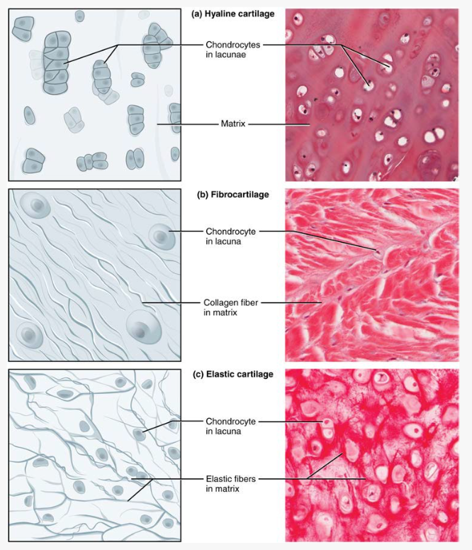
Cartilaginous tissue has a distinctive appearance due to carbohydrates bound to ground substance proteins. Within its semisolid matrix are chondrocytes (cartilage cells), each housed in a small cavity called a lacuna. Because cartilage is avascular, nutrients must diffuse through the matrix to reach the chondrocytes, contributing to its slow healing.
There are three main types of cartilage: hyaline, fibrocartilage, and elastic cartilage.
Hyaline cartilaginous tissue, the most common type of cartilage in the body, consists of short and dispersed collagenous fibres and ground substance. Under the microscope, tissue samples appear clear due to the collagneous fibres being so thin; this appearance is reflected in its name 'hyalos' meaning glass-like. The surface of hyaline cartilage is smooth. Both strong and flexible, it is found in the rib cage as costal cartilages, in the trachea and bronchial tree, in most laryngeal cartilages (thyroid and cricoid), in the nose and covers bones where they articulate to form moveable joints (articular cartilage). It makes up the template of the majority of the embryonic skeleton before bone formation commences through endochondral ossification. A plate of hyaline cartilage at the ends of bone allows continued growth until adulthood (epiphyseal growth plate).
Elastic cartilaginous tissue contains elastic fibres as well as collagenous fibres and ground substance. This tissue gives rigid support as well as elasticity. The auricle of the ear, the auditory (Eustacian) tube and the epiglottis of the larynx are composed of elastic cartilage.
Fibrocartilage is tough because it has thick bundles of collagenous fibres dispersed through its matrix. The menisci in the knee joint, pubic symphysis and the intervertebral discs are examples of fibrocartilage. Very little ground substance is located in this cartilaginous tissue; its high fibrillar content enables it to resist high compressive forces while still retaining its firm support. It is therefore located in area of high loading and stress.
Bone tissue
Bone is a highly vascular, mineralised connective tissue with a biphasic composition: one-third organic (cells, fibres, and ground substance) and two-thirds inorganic (mineral crystals). This dual structure gives bone its unique combination of strength and flexibility—without the organic phase, bone would be brittle; without minerals, it would lack rigidity.
Bone protects internal organs, supports body structure, regulates calcium homeostasis, and serves as the primary site for blood cell production (haematopoiesis) within the bone marrow. Its rigid extracellular matrix is rich in collagen fibres, which provide a scaffold for mineral deposition. The main mineral, hydroxyapatite, forms from calcium phosphate and calcium carbonate, incorporating other salts like magnesium hydroxide, fluoride, and sulfate during calcification.
Unlike cartilage, bone heals relatively quickly due to its rich blood supply.

Cortical bone
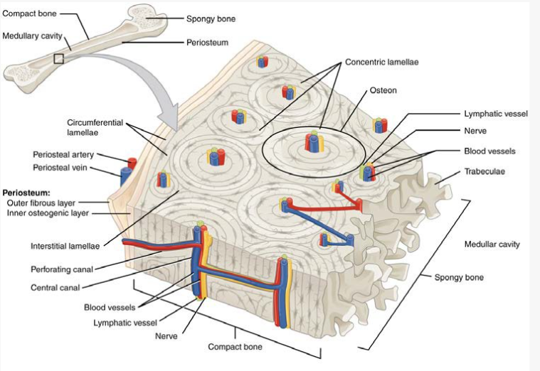
Cortical bone forms the outer layer of all bones and is especially prominent in the shafts (diaphyses) of long bones. Its structural unit is the osteon (Haversian system), composed of concentric rings of calcified matrix called lamellae. At the centre of each osteon is a central canal (Haversian canal) containing blood vessels, nerves, and lymphatics. These connect to deeper bone layers via Volkmann’s canals.
Osteocytes reside in lacunae between lamellae and communicate through tiny channels called canaliculi, which allow nutrient and waste exchange.
Trabecular bone
Trabecular bone consists of a network of trabeculae aligned along lines of stress, providing structural support while reducing bone weight. Unlike cortical bone, its osteocytes are not arranged in concentric rings but are housed in lacunae within the trabecular matrix. The spaces between trabeculae often contain red bone marrow, where haematopoiesis occurs. Trabecular bone is more abundant in the ends of long bones (epiphyses and metaphyses), where it accommodates multidirectional forces at joints.

Fluid connective tissue
Blood and lymph are fluid connective tissues. Cells circulate in a liquid extracellular matrix containing nutrients, salts and wastes. More information regarding fluid connective tissues can be found in the cardiovascular system chapter (blood) and the lymphatic system chapter (lymph).
ANATOMICAL TERMINOLOGY
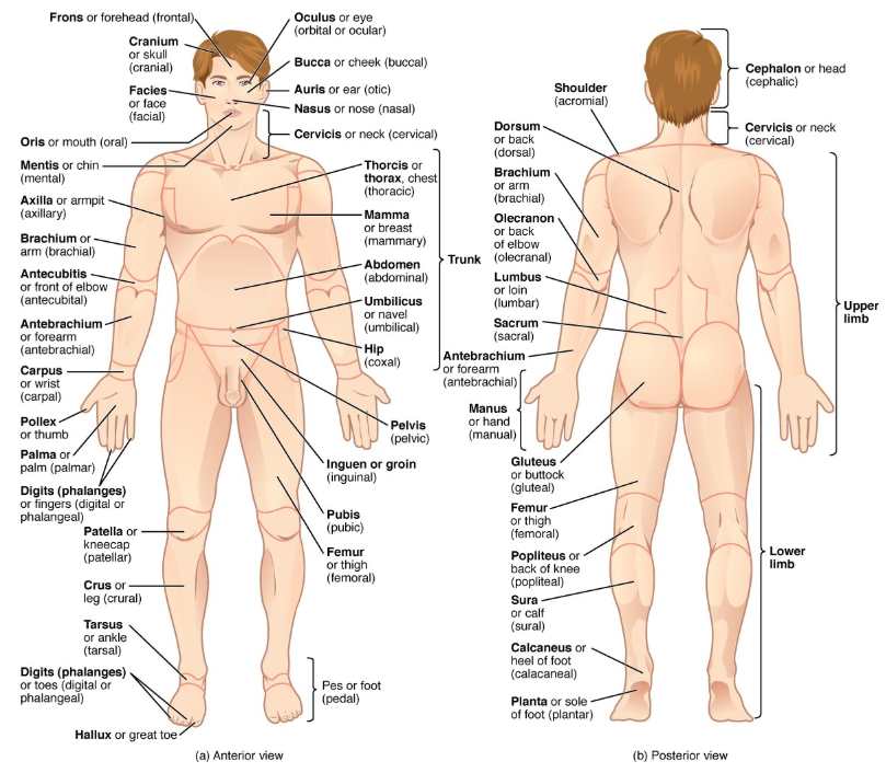
Axial Region
Head:
Regions include the frontal, orbital (eye), nasal, oral (mouth), and mental (chin) regions.
Neck:
Includes the sternocleidomastoid, cervical (neck), and supraclavicular (above the collarbone) regions.
Trunk:
Thorax: Regions include the pectoral (chest), deltoid (shoulder), axillary (armpit), and infrascapular (below the shoulder blade) regions.
Abdomen: Divided into lumbar region posteriorly (lower back) and umbilical region anteriorly (where the umbilicus is located).
Pelvis: Includes the sacral (end of the vertebral column) and anal regions.
Appendicular Region (Limbs)
Upper Limb:
Brachium: The arm.
Cubital: The elbow region, both anterior and posterior.
Antebrachium: The forearm.
Carpal: The wrist.
Manus: The hand, including the pollex (thumb) and digits (fingers).
Lower Limb:
Coxal: The hip.
Femoral: The thigh.
Patellar: The front of the knee.
Popliteal: The back of the knee.
Crural: The shin, the front of the lower leg.
Sural: The calf, the back of the lower leg.
Tarsal: The ankle.
Pedal: The foot, including the plantar (sole) and calcaneal (heel) regions.
Anatomical Planes:
Coronal
Sagittal
Transverse
Directional anatomical terms
Anterior (or ventral) Describes the front or direction toward the front of the body. The toes are anterior to the foot.
Posterior (or dorsal) Describes the back or direction toward the back of the body. The popliteus is posterior to the patella.
Superior (or cranial) describes a position above or higher than another part of the body proper. The orbits are superior to the oris.
Inferior (or caudal) describes a position below or lower than another part of the body proper; near or toward the tail (in humans, the coccyx, or lowest part of the spinal column). The pelvis is inferior to the abdomen.
Lateral describes the side or direction toward the side of the body. The thumb (pollex) is lateral to the digits.
Medial describes the middle or direction toward the middle of the body. The hallux is the medial toe.
Proximal describes a position in a limb that is nearer to the point of attachment or the trunk of the body. The brachium is proximal to the antebrachium.
Distal describes a position in a limb that is farther from the point of attachment or the trunk of the body. The crus is distal to the femur.
Superficial describes a position closer to the surface of the body. The skin is superficial to the bones.
Deep describes a position farther from the surface of the body. The brain is deep to the skull.
Ipsilateral on the same side, e.g. the spleen and descending colon are ipsilateral
Contralateral on opposite side, e.g. the spleen and gall bladder are contralateral
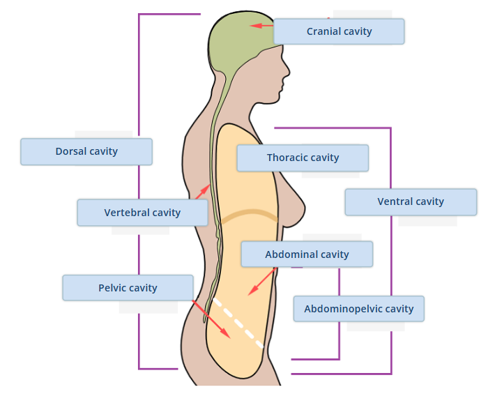
BODY CAVITIES
Posterior (Dorsal) Cavity
Cranial cavity: Contains the brain.
Spinal (vertebral) cavity: Encloses the spinal cord.
These two cavities are continuous, reflecting the uninterrupted structure of the brain and spinal cord. Both are protected by the bones of the skull and vertebral column, as well as by cerebrospinal fluid, which cushions and supports the central nervous system.
Anterior (Ventral) Cavity
Thoracic cavity: Located above the thoracic diaphragm and enclosed by the rib cage, it contains the lungs and the heart.
Abdominopelvic cavity: Situated below the thoracic diaphragm, it is the largest body cavity. Although not physically divided, it is often described in two parts:
Abdominal cavity: Contains mostly digestive organs.
Pelvic cavity: Contains mostly reproductive organs.
Anatomical variation is defined as a difference in topography (location), morphology (shape and size), developmental timing or frequency of an anatomical structure.
It presents during subadult or embryological development and results in no substantive observable interruption to physiological function.