Final Exam Review
Lecture 4: ER and Golgi
Endomembrane System:
includes the nucleus, ER, golgi, perioxisomes, lysosoymes, endosomes, cell memrbane
ER Main function:
synthesis of most lipids, proteins which are distributed
Golgi: modification, sorting, and packaginging of proteins and lipids for secretion or delivery to another organelle
Smooth ER
lipid synthesis
Rough ER
Protein Synthesis
covered in ribosomes
Soluble Protein Synthesis
following synthesis of soluble protein, the protein is released into the lumen of the ER
the signal peptide is cleaved and remains in the ER membrane
Help of translocator channel
soluble protein → ER lumen
Signal peptide → remains ER membrane
Membrane protein synthesis
signal peptide is cleaved,
protein goes into the ER lumen where it stops going in at the stop-transfer sequence
protein remains growing on the outside (cytoplasmic side)
Post translational protein modification in ER
chemical modifications of the protein that isn’t coded by DNA or RNA happening inside the ER
Glycosylation: sugar attachment
Function of Golgi
synthesis of glycoproteins, proteoglycans, polysaccharides, sphingomyelin, glycosphingolipids
site of many post-translational modifications
ex: glycosylation and phosphorylation
flippase
proteins and membranes are sorted and packed
proteins enter golgi from cis side and leave to lysosome or cell surface through trans side
Role of ER in calcium signalling
sacroplasmic reticulum = ER in muscle
mucle action potention propagated into T-tubules
Calcium is released lateral sac → ryanoid receptor → binds to troponin to remove the block by tropomyosin allowing for action potential
Lecture 5: Mitochondria and Peroxisomes
Mitochondria
produce atp
calcium signaling and release of clacium
storage and release of apoptoic molecules
source of reactive oxygen species
Mitochondrial protein synthesis and import
maternally inherited
20-30 proteins coded by mitohon. DNA
~2000 proteins make proteome
most mitochondrial proteins are made in the cytoplasm and imported into the mitochondria
mitochondria density and organization depends on the cellular needs
coupling oxidation and phosphorylation
membrane potential of mitochondria contributes to
Heat generation
ion transport
membrane leak
movement of protons into matrix with out atp being produced
this is normal and contributes to heat generation, metabolism and reactive oxygen species re
Investivation of mitochondrial function (Respirometry)
Oligomycin
decreases oxygen cellular respiration (OCR) by blocking ATP production
FCCP
increases OCR by adding more protons to the ETC to promote oxidative phosphorylation which in turn increases oxygen consumption
Rotenone
poisons complex 1
prevents NADH from reaching electron transport chain
Antimycine
blocks electron flux of complex 3 from working [reducing OCR]
Investigation of the mitochondrial function
flavanoids (E + Q) improves mitochodnrial function by increasing the space respiratory capacity of the mitochondria
Perioxisomes
break down alcohol, toxins, and Fatty acids
involved in biosynthesis of lipids
Fatty acid hydrolysis (break down) creates FADH2 and NADH which are two energy rich molecules
Lecture 6: Endocytosis and Lysosomal System
pinocytosis = small cell drinking
macropinocytosis = larger vesicles
clathrin-associated receptor-mediated endocytosis
specific
wraps things in coat/cage structure
caveolae
little cave
50-80 nm
phagocytosis
cell eating
cell receptor recognizes surface of bacteria
actin starts to push out forming the pseudopods that will wrap around the bacteria and engulf
after the psudopods wrap around the bacteria and forming the phagosome
actin is removed from phagosome since it acts like a barrier, this is to allow the lysosome.
the phagosome fuses with the endosome to get membrane parts from it while the endosome gets somethings to recycle
the primary lysosome from the golgi apparatus fuses with the phagosome to form the phagolysosome (highly acidic) → degredation of the bacteria by digestive enzymes
the secondary lysosome is where the remains of the bacteria are left, and this is where nutrients are absorbed and waste is prepared to be removed.
exocytosis adds to the membrane
endocytosis removes from the membrane
Dynamin is the “scissor” that cuts the vesicle from the membrane to separate it and allow the vesicle to travel through the cell.
Autophage: eating things that are already in the cell
1. envelope
2. seal
3. merge with lysosme
4. resulting residual body
Nucleus and Nucleolus: Lecture 7
1-5 nucleolus in a nucleus
nuclear envelope has 2 layers inner and outer layer
nuclear lamina separates the outer and inner nuclear membranes
The EJC attaches to where the introns where cut out and where the exons were glued in place. it’s like a proof of mRNA having been edited correctly.
Intermediate Filaments
tough rope like protein fibers that provide mechanical strength and structural support so that cells don’t tear under physical stress
Cytoplasmic Intermediate filaments
gives structural integrity to:
Keratins
Vimentin
Neurofilaments
Nucleolus
site of rRNA synthesis
dissolves during mitosis
made up of fibrillar centers or cores, dense fibrillar and granular components
Large 60S Subunit rRNA
has ~49 ribosomal proteins and 3 rRNA molecules
Small 40S Subunit rRNA
~33 ribosomal proteins and 1 rRNA molecule
come together to form the complete eukaryotic ribosome
Molecular weight (MW) = 4,200,000
Cytosol and Cytoskeleton 1
Microfilaments
Microtubules: part of centrosome
Intermediate filament: provides structural support
Actin
Monomer = G actin (globular)
Polymer = F actin (filamentous chain)
Polarized molecule: has a growing plus end and a disassembling minus end
Microfilmaent: F-actin is stabilized by capping at both ends
1. G active goes through polymerization to become F-actin
2.Tropomyosin like a long rope, wraps around the F-actin helix to cover the myosin-binding sites during muscle rest
Troponin sits on tropomyosin and acts like a calcium-driven switch for muscle contraction
Cap z: protein that caps the plus end of the filament to stabilize
Tropomodulin: protein that caps the minus end of the filament to stabilize and prevent it from unraveling
alpha - actinin: binds the actin filament to the Z disk
Myosin 2 Thick Filaments
Energy-Dependent Movement of Thin filaments
1. hydrolyze ATP so that ADP and Pi remain bound to myosin head
2. Pi then releases tight binding of actin filament
3. Incudes a conformation change the powerstroke
Release ADP binding of ATP , as we prepare for the new cycle (goes back to state of step one)
Cytosol and Cytoskeleton 2
Microtubules
composed of tubulin
heterodimer of alpha and beta tubulin
requires GTP to polymerize
Has a growing plus end and a disassembling minus end
Plus end: has GTP on its terminal dimer
Minus end: has microtubule organizing center
Microtubule function
transport and secretion of vesicular cargo
separation of chromoses during mitosis and mesiosis
structural stability of cilia and flagella
motion
Dynamic Instability
the constant contest between growth and degradation of microtubules
GTP bound plus end adds to it
GDP bound plus end adds at a slower rate, which makes the disassembly as the minus end faster, favoring degradation
Regulation of Microtubule Stability
Capping at + end by (microtubule associated proteins, MAPs) helps prevent disassembly
Chemical Modifications: removing the amino acid Tyrosine from the end of the alpha-tubulin subunit makes the microtubule tougher and more long-lived
Tau and High Mr MAPs bind to the side to help the microtubues bundle together and stay anchored
Stathmin and Katamin are MAPs that favor slicing and disassembly of microtubules when the cell needs to tear down its network
Mediated Transport of Cargo
mediated by motor proteins
plus end is the axon terminal (outward transport)
N-terminal motor domain
typically members of the kinesin family
Kinesin plays no role in flagellar bending or beating
Kinesin is for intraflagellar transport to deliver fresh building material for ciliary maintenance
minus end is the nerve cell body ( backward transport)
C-terminal motor domain
typically members of the dynein family
Dynein does the motions
Centrosome
composed of 2 centrioles with each composed of 9 triplets of microtubules in a circle
centrioles are perpendicular to each other
which are surrounded by a cloud of microtubule binding proteins in which microtubule centers bind their minus end to the nucleating site(the pore structures of the centrosome matrix)
Intermediate Filaments (IFs)
composed of heterogenous groups of fibrous proteins
Main function: To provide structural Stability that maintains the integrity of cells and tissues
their formation doesn’t require the breakdown or input of chemical energy like ATP
No polarity within the filaments (Both ends of the intermediate filament are structurally Identical) They lack the plus and minus end as we saw with microfilaments and microtubules
Primary functions are structural
2 dimers then line up with opposite ends to create a balance and symmetry
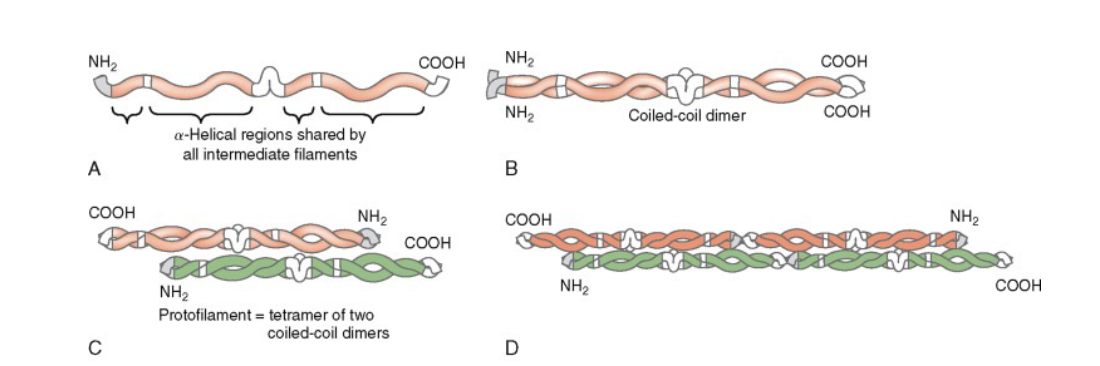
Cell Adhesion and Motility
anchoring of cells within tissues
adhesion is important for cell migration and motility
Homophilic binding
same binding
ex: cadherin and cadherin
Ig-like
Heterophilic binding
different: example: carbohydrate binding to selectin
Integrins
heterodimers of alpha and beta chains
cell adhesion to ECM and some cells
role in cell motility
Intercellular Junctions
Tight Junction
seals neighboring cells to prevent leakage
claudin of one cell binds to the claudin of another cell
occludins of one cell binds to the occludins of another cell
Adherens Junction
joins actin bundles between neighboring cells
spot and zonula
Cadherin mediated (Ca2+ dependent binding)
Desmosome
joins intermediate filaments of one cell with another
intermediate filament → intermediate filament
many together impart great tensile strength to epithelial cells
Gap junction
forms channel to allow small-water soluble molecules, ions
cell to cell channel
communication junction
homophilic binding
connexon-mediated
hemidesmosome
anchors intermediate filaments in a cell to the basal lamina
intermediate filament → basal lamina
contains keratin filaments
Junction morphology
Zonula: belt, that wraps around the entire perimeter of the cell
Tight and Adherens junctions
Macula: spot
adherens
desmosomes
hemidesmosomes
Process Protrusion
Contractile Bundle
stress fibers
parallel filaments going in opposite directions
allowing motor proteins like myosin to bind and pull
Lamellipodium
Dense, cross-linked, branched network of actin filaments
forms flat, sheet like protrusion at the leading edge to push the cell membrane forward
Filopodium
Tight, parallel, unbranched bundles of actin filaments all pointing in the same direction
chemotaxis
movement of a cell/organism in response to a chemical stimulus

The Cell Cycle and Mitosis Lecture
Cell cycle → replication and division of cell
2 daughter cells inherit same genetic info from mother
Cell division is needed
repair/regeneration
(s)exual reproduction (mesiosis)
asexual reproduction (mitosis)
growth
Labile cells
contanstly dividing, ex: skin, oral mucosa, hematopoietic cells
Stable Cells
only divide when needed, liver, replaces lost tissue
Permanent cells
never divide, neurons, cardiac myocytes
Senescence: biological process of getting old
Cyclosporine: immunosuppresent to preven organs rejecting a transplant
Phases in cell cycle
G1
preparing to divide
increasing cell size, RNA and protein synthesis
5 hours
G0 phase
cells exit G1 and enter resting state (G0)
cell performs function without actively preparing to divide
permanent state for some cells, temporary for others if they get the right signal
S
2n to 4n dna content, duplication of centrosomes (centrioles)
DNA is replicated
7 hours
G2
synthesis of proteins
tubuiln for mitotic spindles
synthesis of maturation promotiong factors
increase cell size and organelles
3 hours
M
nuclear division (mitosis) and cytokinesis (cell cytoplasm division)
1 hour
1. Prophase
chromosomes condense
centrosomes separate and spindles start to assemble
2. Prometaphase
breakdown of nuclear envelope
chromosomes attach to spindle via kinetochore
3. Metaphase
line up at equator (metaphase plate)
sister chromatids of chromosome attached to spindle on opposite poles
4. Anaphase
centromere is broken as sister chromatids go to opposite poles.
anaphase promoting complex (APC) triggers chromatid separation
degrades cyclin b through ubiquitination (tagging) to turn off Cdk1 so the cell can finish dividing
5. Telophase
chromosomes arrive at poles
nuclear envelope reassembles
chromosome de-condenses
division of cytoplasm with formation of contractile ring
6. Cytokinesis
cell is divided in half by contractile ring
Cell Cycle control system
proteins the ensure the cell cycle runs smoothly
each process has to be completed before the next one begins
regulated at critical points of the cycle by feedback from the process
G1 checkpoint
size, nutrients, growth factors, DNA damage
G2 checkpoint
is all DNA replicated
is all DNA damage repaired
Checkpoint in Mitosis
are all chromosomes properly attached to the mitotic spindle?
Cyclins
trigger kinase activity (there is not enzymatic activity on their own.
Cyclin dependent protein kinases (CDKs)
Cyclin dependent protein kinases inhibitors (CDKIs)
suppresses CDKs
bind directly to cyclin-CDK complex
regulation of the G1-S phase transition
7 types: p15, p16, p18, p19, p21, p27, p57
Cyclin D: activates CDK4 and CDK6 which function in Progression past G1 and into S phase
Cyclin E & A: CDK2 initiate DNA synthesis in S phase
Cyclin B: initiates CDK1 G2 → M phase
CDK-activating kinase (CAK)
constant throughout the cell cycle and phosphorylates the CDK as soon as the cyclin and CDK complex is formed
think of the CDK as the car, cyclin is the key to unlock, and CAK is the ignition to get the car started.
Regulation of CDK activities
Inhibitory kinases
wee1, wee2
Phosphatase
enzymes that remove phosphate groups, refersing the effects of kinases
ex: Cdc25
Regulation of Cyclin
Anaphase-promoting complex (APC): A ubiquitin ligase that degrades cyclin B to separate sister chromatids
G1/S phase control by p53 and Rb
p53 and Rb are tumor suppressor genes (anti-oncogenes)
p53 = guardian of the genome
activates p21
involved in DNA repair, cell cycle control, and cell death
prevent entry of cells into S phase
DNA damage arrest in G1
Activate p53
Transcription factor for p21
p21 inhibits cyclin-cdks
cell repairs damage (to go on to S phase) or cell dies
Rb tumor suppressor
Rb gene mutation causes malignancies
small-cell carcinoma of lung, breast cancer, bladder cancer
p16 Tumor Suppressor
CDK inhibitor that binds to the cyclin D and CDK4 combo
Mitogens
growth factors that bind to its receptor on the cell membrane ultimately leading to mitosis
Oral cancer is 3% of yearly cancer diagnosis
Increase mitogens
increase cyclin D1
decrease in p16, p53, Rb
Meiosis Lecture
Somatic cells
diploid, one set from each parent
somatic mutations impact the individual, but cannot be passed down to offspring
Germ Line Cells = gametes, sperm and eggs
sex cells
sperm or eggs (haploid)
combine during fertilization to make new individual (diploid)
mutations can be passed down to offspring
Mitosis = asexual reproduction
offspring identical
Meiosis = sexual reproduction
offspring: diversity
Reshufflying the genes to help a species sruvive in an unpredictable environment
2N → 4N → 2N → N
one doubling of DNA, followed by 2 cell divisions
Crossing over during prophase 1 leads to exchange of DNA = diversity
Chromosomes are randomly sorted due to independent segregation or independent assortment
metaphase 1
Meiosis 1: Prophase 1
bivalent (tetrad) forms when 2 homologous chromosomes come together
synapsis is when they come together and align along their lengths
Synaptonemal complex: the fibers that connect the 2 homolgous chromoomes when they long against one another
Cohesion Rings
found around the 2 sister chromatids of one homologous chromosome
generates tension around the centromere
Crossing Over: point is called chiasmata
helps position and stabilize bivalent
The farther the distance between 2 genes = greater chance of recombination
Prometaphase 1
Synaptonemal complex breaks down at late prophase 1, even though the chiasmata is still connected, even after crossing over occurred
nuclear envelope breaks down
microtubules move towards the center attaching to the kinetochores, with one homologous chromosome facing each pole
Metaphase 1
line up via independent assortment
Anaphase 1
chiasmata is broken
centromeres are still in place holding the sister chromatids to each other
the 2 homologous chromoosmes now move to opposite poles of the cell
telophase 1
chromosomes gather into nuclei, and the original cell divides
Prophase 2
chromosome condenses again, after a brief interphase (interkinesis) in which DNA is not replicated
metaphase 2 - anaphase 2
chohesions in centromere degrade, sister chromatids separate
Telophase 2
4 haploid cells
Aneuploidy
Abnormal # of chromosomes
caused by nondisjunction
Amphitelic: normal
Monotelic: only one spindle attaches to one of the sister chromatids, none of the other
Syntelic
both sister chromatids attach to the same microtubule pole
Merotelic: Most dangeorus error
a single sister kinetochore connects to one pole, but its companion kinetochoresi s pulled inboth directions at once, catching microtubules from both the left and right
This can trick and bypass the cell’s safety checkpoint
leads to stuck tug-of-war in anaphase leading to lagging chromosome that leads to severe DNA damage or cancer
Chromosomes
Chromosome = a centromere, 2 telomeres, an origin of DNA replication for every 100,000 base pairs
P arm is the short arm
Q Arm is the long arm

DNA is first orgnaized into chromatin by nucleosmes
nucleosome = beads on a string
Chromatin = DNA + nucleosomes(8 histone proteins)
histones are basic (positively charged)
Euchchromatin = 11 nm fibers
Heterochromatin = 30 nm fibers
Karyotype = display of full set of chromosomes of cell
Giemsa-banded karyotype
Red knobs: genes that econde for large rRNA
Aneuploidy: Trisomy 21
Oral and craniofacial findings
periodontal disease
low caries rate
delayed tooth eruption
hypodontia
microdontia
midflash hypoplasia
Reciprocal chromosomal translocation
large sections of chromosomes swap locations
example: philadelphia translocation causes leukemia
chromosome9 and 22
Centromeric Chromatin (center that hold the sister chromatids together) is in the form of consistutive heterochromatin which has little or no genes expressed
Telomeres
keep normal chromosome ends from arresting cell cycle
keep ends from being joined by repair mechanisms
contain high G-C content
must be replaced each cell division by telomerase in germ cells and stem cells
Telomerase uses RNA as a template for DNA synthesis (reverse transcriptase)
Telomerase is inactive in most cells of the body (cells that have a limited number of divisions, in post mitotic cells, and permanent cells (neurons that never replicate)
Hayflick Limit
number of time somatic cell population can divide before cell division stops
Genes and Non-Genes Lecture
mendelian definition: a gene is heritable trait
Modern definition: a gene encodes a functional product (either protein or RNA)
Alleles: variations in DNa sequence among copies of a gene
Genome: sequence of all the DNA
The modern gene
Coding Region: opening reading frames are the coding sequence
Exons contain ORF and any untranslated regions (UTRs)
Simple genes: have one version of their mRNA
no introns to splice out
Complex genes: have their RNA psliced into variant mRNAs by removing introns and linking different exons to make different versions = Protein Isoforms
Solitary Genes
only one copy of a gene
if there are multiple copies of the same exact gene, then it’s for the purpose of increasing the total output of that product (protein or RNA)
found in tandem array that are separated by spacer to act as a insulator preventing read thorugh

Gene Families
Have more than one member
are duplication of a gene
the genes started out identical but diverged over time to yield variants that perform different tasks
some family members don’t have functions = Psudogenes
Types of RNA
hnRNA is the precursor for m, t, r, sn, and mi RNA
98.5% of the genome is non-coding
50% of DNA is unique
50% repeated seqneces
45% transposons (mobile DNA)
genome tries to make these elements transcriptionally silent
DNA and histone methylation are installed on them
2 types
DNA transposons
move as DNA, leave one site and enter another. “cut and paste”
proteins (transposase) are needed to cut the DNA sequence out and move it to another site
Retrotransposons
move like a retrovirus with an RNA intermediate using reverse transcriptase.
RNA template converted to DNA
then integrate into the host genome
They replicate themselves, thus creating a new copy but remaining in their oringial location
“copy and paste
Endogenous retrovirus
ancient viral DNA that is embedded in humans
can make RNA and also make proteins that are not useful to the cell or could cause problems
DNA methylation keeps them “off”
Non-retrovirus-like retrotransposons
are the most abundant mobile DNA elements in mammals
34% on human DNA
LINE: Long interspersed elements (6 kb)
SINE: Short interspersed elements (100-400 bp each)
Simple Sequence Repeats
Microsatellite DNA
1-13 bp reapeated less than 150 times that are cuased by backward slippage during DNA replication
They don’t move around in the genome: they expand or contract in one place
Satellite DNA
mostly repeats of 14-500 bp
often at centromeres and telomeres
Telomeres
DNA fingerprinting
identify individuals based on their qunite pattern of Satellite or Microsatellite DNA
NIH ENCODE Project
seeks to map non-coding gene elements, the regulatory seqeunces - exons, introns, promotes, enhancers, transcription binding sites
CRISPR-Cas9
Eukaryotic Gene Regulation and Epigenetic Lecture
main function of gene is to make RNA → to make proteins
Gene expression: the production of RNAs and proteins from a DNA blueprint
RNA Polymerase 1: large precursor of the rRNAs (5.8S, 18S, 28S)
RNA Polymerase 2: heterogenous nuclear RNAs (hnRNAs) which are precursors to mRNAs, miRNA, and most snRNA
RNA Polymerase 3: precursor to 5S rRNA, tRNAs
Promoter
region of DNA upstream of a gene where proteins like RNA polymerase and transcription factors bind to initiate transcription
Class 1-3
Class 2 Promoters are recognized by RNA Polymerase 2 and are the most complex and best studied (transcription)
Enhancers and Silencers are DNA elements that stimulate or depress transcription of associated genes
Transcription factors
Euk. RNA polymerases are unable to bind to promoter by themselves like bacteria. So they rely on the transcription factor to guide them to the promoter
General Transcription Factors
weak extent, basal level of transcription
ex: skin cell nucleus
Gene-specific transcription factors (activators)
help cells exert fine control over transcription.
ex: liver cells nucleus
repressor proteins
brain cell nucleus
Chromatin
fold DNA into the small volume of a cell’s nucleus
Histones
carry a positive charge which binds tightly to the negatively charged DNA
Nucleosome: first order of chromatin folding, core of histones which DNA winds around
Epigenetics
chemical changes in DNA or histones that regulates gene expression without altering the DNA sequence
Histone Tail modifications: Reversible
Acetylation
opens up chromatin to allow transcription
provides rapid control of gene expression in response to external stimuli
Performed by Histone Acetyltransferases (HATs)
Removed by histone deacetylases (HDACs)
Methylation
Can be used for both chromatin condensation (repress transcription) and open chromatin (promoting transcription)
Performed by histone methylatransferases (HMT)
Removed by histone demethylases (HDM)
Phosphorylation
Ubiquitination
SUMOylating
DNA Methylation
the most STABLE of epigenetic modifications
strongly repress transcription by blocking transcription factors
contritubtes to the formation of Heterochromatin for long term repression of transcription
Euchromatin
11nM fibers
transcription activated
light-stained on electron micrograph
Histone tails acetylated
Histone tails methylated in ways that factor transcriptoin
DNA not methylated
Heterochromatin
30 nM strands
transcription repressed
dark-stained on electron micrograph
Histone tails deacetylated
histone tails methylated in a way that represses transcription
DNA methylated
Genetic and Obesity
1/3 adults and 1/5 children in US are obese
Obesity = BMI ≥ 30
monogenic Obesity: single gene mutation, rare, onset early childhood
caused by loss-of-function mutations in a single gene
often inherited autosomal r or Dominant
typically involves genes in the leptin-melanocortin pathway
Common Obesity: polygenic + environment, very common, childhood/adulthood
Genome Wide Association studies (GWAS) to understand genetic basis of obesity
FTO Locus is the first GWAS locus to be associated with obesity
codes for alpha-ketoglutarate dependent dioxygenase
enzyme found on chromosome 16
Non-coding intronic variants within FTO are strongly associated with BMI
where people homozygous for the top risk vairant weigh ~3kg more than non-carriers
FTO region directly interacts with the promoters of IRX3
IRX3 is a functional long-ranged target of obesity-associated variants with FTO
Epigenome-wide association study (EWAS) of DNA methylation
shows that variation in epigenetic modifications (dna methylation) is associated with BMI and metabolic diseases
Genetics 1: Genes and Alleles
Monogenetic Disorder
inherited single gene disorder
ex: hypodontia, amelogenesis imperfecta, epidermolysis bullosa (extreme canker sores), gingival fibromatosis, enlargement of gingival tissues
Hemizygous
having a single copy of a gene in an otherwise diploid cell
x-linked genes in men
Complete (classic) dominance
when heterozygous is indistinguishable from homozygous dominant
Incomplete (partial) domiance
one allele is not completely dominant over the other
results in blending or expression of both alleles in the phenotype
red plus white = pink
Co-dominance
contributiosn of both alles are visible
both black and white spots visible
Blood type
D or d display classic dominance
ONly D allele confers Rh+ blood
Genetics 2: Genes and Alleles
Pea plants are a good model
could get “true breeding plants”
reproduce quickly and sexually
gametes, make and female sex cells
self fertilizing
many traits that existed in only 2 forms
Unit Factors
basic unit of heredity
phenotype: expression of these traits
genes: unit factors
alleles: various versions of genes (unit factors)
Mendels first law: law of segregation
genes for a specific trait separate randomly into their 2 alleles, so that each gamete receives one or the other with equal chance.
Mendel’s second law: independent assortment
genes for different traits are inherited independently of each other if they are on different chromosomes
Linked Genes
are unable to assort independently
genes passed down together as a group
Linkage: when genes are located on the same chromosome, which results in their inheritance together, unless they are separated by crossing over during meiosis 1 prophase 1
The maximum rate of recombination for linked genes is 50%
Interference (I)
when a crossover event happens, it reduces the odds that another crossover will occur
Coefficient of coincidence (C) = observed double crossover/ expected double crossover
I = 1 - C
Genetics 3: Sex based genes
sex chromosomes are heteromorphic (dissimilar)
Barr body: highly condensed inactivated chromosome found by the nuclear envelope
Lyon hypothesis: a single X chr is randomly inactivated in early development
passed down to all progenitor cells
Sex-linked disease
diseases on the X chromosome
Y linked inheritance - Holandric inheritance
only males affected and passed from father to all his sons
X linked alleles always appear in males since they only have 1 X chromosome
ex: Colorblindness
X-linked Dominant disorders
m and f are affected
higher frequency in female than male
affected males pass on gene to all their daughters
no male to male transmission
ex: fragile X syndrome, rickets
X-linked recessive disorders
affected individuals usually have unaffected (carrier) parents
usually only males are affected
affected males pass on genes to all daughters
daughters of female carriers have ½ chance to be carriers (no phenotype)
sons of female carriers have ½ chance to be affected
ex: hemophilia, Duchenne muscular dystrophy, Malaria
Duchenne Muscular Dystrophy
Normal at birth
slow progression of muscle weakness
male genetic fitness is zero
1/10000
Hemophilia (royal disease)
inability to clot blood
Sex-influenced inheritance
when the sex of the individual influences the expression of a phenotype that is not limited to one sex or another
ex: pattern baldness
sex-limited
when the expression of a specific phenotype is absolutely limited to one sex
ex: plumage of domestic fowl
Monoallelic gene expression
only the maternal or paternal allele is expressed
Prader-Willi Syndrome
deletion on chromosome 15
paternal allele inherited and expressed
Angelman’s syndrome
only maternal inherited allele
Point Mutation
Transition
change of a pyrimidine to another pyrimidine. or purine to purine
Transversion
purine to pyrimidine and vice versa
Missense mutation
amino acid change does occur due to altered codon
sickle cell anemia: one nucleotide subsititon of glutamic acid is replaced by valine in the hemoeglobin gene
valine is hydrophobic
nonsense mutation
turns normal codon to a stop codon
Trinucleotie Repeats
tandem sequence of 3 nucleotides within a gene is repeated many times expanding the size of the gene
in people with TNRE disorders, the length of a trinucleotide repeat is increased above a certain ciritical size
this increase in size in future offspring is what causes earlier onset in future generations = genetic anticipation
If the repeats occur in the coding sequence of a gene
expansion of CAG = glutamine
protein aggregartes are toxic and implicated in many diseases
also depends on who you inherit from
Huntington disease
TNRE more likely to occur if inherited from father
Myotonic muscular dystrophy
TNRE are more likely to occur if inherited from mother
Sickle Cell Anemia
point mutation, glutaminc acid is replaced by valine (hydrophobic)
Chromosome 11 is affected, recessive inheritance
hemoglobin molecules are made up of 2 alpha chains and 2 beta chains linked together
point Mutation in HBB (hemoglobin Beta chain)
hemoglobin = 4 heme (4Fe) and 4 globing chains
Cancer Genetics
Solid Tumors
dividing cells that don’t die appropriately
uncontrolled growth in solid tissues like bone, muscle, organs
Liquid tumors
originate in blood or lymphatic system (leukemia, lymphoma)
Malignant tumor
metastasis: travels to other body regions to form new tumors
Carcinoma: cancers of epithelial cells of endodermal (gut epithelial cancers) or ectodermal: skin, neural epithelial
Sarcomas: cancers of mesodermal origin
Blastomas: come from immature precursor cells or embryonic tissues
Hallmarks of Cancer (same functional capabilities)
Genomic instability and inflammation are enabling characteristics that drive the acquisition of hallmarks of cancer
6 Original hallmarks
Self-sufficiency in growth signals
Insensitivity to anti-growth signals
Evading apoptosis
Limitless replicative potential
Sustained angiogenesis
Tissue invasion and metastasis
Viruses associated with human cancers
DNA Viruses
Papovavirus family
Hepatitis virus
RNA viruses
human t-cel leukemia virus
HIV, AIDS
Genes Commonly mutated in Cancer
Proto-oncogenes
promote survival and proliferation
gain of function
mutations are dominant
arise by point muatation
Tumor-suppressor genes
inhibit cell survival or proliferation
loss of function
both alleles of the gene must be lost
mutations are recessive
arise by deletion, point, methylation
Caretaker genes
repair or prevent DNA damage
loss of funtion
mutations are recessive
arise by deletion, point, methylation
Conversion of a proto-oncogene to an oncogene
1. Mutated hyperactive form is made (qualitative change)
point mutations that make the protein hyperactive
fusion of protein with oother proteins leading to hyperactivity
2. Too much of the normal protein is made (quantitative change)
translocations
gene amplifications: multiple copies created
c-myc is a transcription factor that regulates many genes including those the control proliferation, growth, and survival
Amplification of c-myc
Types of Proto-oncogenes/Oncogenes
Growth factors and receptors: PDGF AND EGFR
EGFR mutation are common in head and neck, non-small lung carcinoma (NSCLC), and gliomas
G Proteins: ras
3 RAS genes (HRAS, KRAS, NRAS) belong to most common oncogenes
All 3 genes encode for small GTPases, which are cell membrane attached cytosolic proteins, attached to GDP or GTP
Role of p53 in prevention of carcinogenesis
Inactivates at least 70% human cancers
stops replication at G1/S boundary
actiaves DNA repair
intiates apoptosis if DNA damage cannot be repaired
Rb tumor suppressor
Rb and E2F complex prevents the cell from entering the S phase
Phosphorylation of Rb by cyclin-CDK releases E2F, the cell would then transition into the S phase
p16 binds to cyclin-CDK complex to inactivate it and prevent it from separating Rb and E2F
Fanconi anemia (FA)
suppressed immune systems and rare hereditary disease
more susceptible to oral cancer
caused by mutation in FA genes (FANCA to FANCV) that serve tumor suppression roles in DNA repair
Evidence of Epigenetic alterantion
DNA methylation silces suppressor gene
smoking increaes DNA methltransferase activity, enhancing methyaltion of the p16 promoter which stops cyclin-CDK from separating Rb and E2F. Silenced p16 would lead to cell growth = increase in cancer
Demethylating previously methylated oncogenes
alcohol causes DNA hypomethylation of PI3
Histone modification
Histone Deacetylases (HDACs) removes acetyl group from histones, leading to gene suppression (heterochromatin)
Non-coding RNAs
Cell Death
Apoptosis and autophagy
cell dies quietly and contains its contents
Necrosis, necroptosis, pyroptosis
Cell spills its guts and stimulates inflammation
Nerve growth factor withdrawal triggers the mitochondrial apoptosis pathway
nervous system programs itself to respond to environment stimuli
Viruses
want to turn off apoptosis to extend the life of cells, to have more time to make new viruses
Increase or mimic the anti-apoptosis factor Bcl-2
degrade p53
stimulate anti-apoptotic kinases like AKT
Cell Death can be pathological
Oxidative stresss from Ischemia injury, where the absence of oxygen and nutrients from blood creates a condition inf which the resotration of circulation results in inflammation and oxidative damage
ex: stroke, acute kidney injury, myocardial infarction
Programmed Cell Death (PCD)
Apoptosis
primary PCD pathway
suicide
Autophagy
self consume
Necroposis
regulated necrosis with passive and active proinflammatory functions, detect pathogens and promote tissue repair
Pyroptosis
PCD as part of an inflammatory reaction, detect pathogens and promote tissue repair
Ferroptosis
iron -dependent form of programmed cell death that involves accumulation of lipid peroxides and oxidative stress leading to membrane damage
Not PCD
Necrosis
uncontrolled death → irreversible injury
killing the cells by an outside agent
loss of blood flow (ischemia)
physiacal trauma
Cell membranes swell and become permeable
lytic enzymes destroy the cellular contents, which leak out into the intercellular space leading to an inflammatory response
Apoptosis
rapid clearnace to prevent an inflammatory reaction which is caused by intercellular contents being spilled out of the cell.
phosphatidylserine (PS) on the cell surface is the “eat me” signal for macrophage to quickly clear away the cell and its contents so that there is no inflammation
Intrinsic Apoptosis
cellular stress, initiator is caspase 9
executioner is caspase 3/7
1. Cellular stress (heat, radiation, and growth factor depreivation)
2. Pro-apoptotic members of the BH3-only proteins (PUMP, BID, BIM)
3. They block the pro-survival BCL-2 like proteins ( BCL-2, XL, MCL-1)
4. This inhibition activates effectors of apoptosis (BAK, BAX), which assemble into large complex
5. holes begin to form in the mitochondrial outer membrane (MOMP) this is the point of no return
6. Mitochondria releases cytochrome C into the cytoplasm where it binds to apoptoic peptidase activator factor 1 (APAF1), which forms a apoptosome
7. Apoptosome activates a cascade of caspase which starts with the initiator caspase 9, and that activates the effectors caspases 3 and caspase 7
Extrinsic Apoptosis
1. Ligands like FASL is recognized by the receptor on the cell called FAS
1. Ligand like TNF is recognized by the receptor on the cell called TNFR1
2. Once the receptor is activated , the tail inside of the cell changes shape, which allows it to bind to an internal adaptor protein called FADD (FAS associated via death domain)
3. FADD acts like a bridge to recruit the inactive pro-caspase-8 to the site. Together the clustered receports, FADD + procaspase 8 lock together to form the DISC (death induced signaling complex)
4. The job of DISC is to bring the pro-caspase-8 close enough to each other so that they can cleave an activate one another . which becomes an initator enzyme that goes onto slice and activate executioner caspases liek caspase 3
initiator is caspase 8
executioner is caspase 3/7
Caspases
Cystein and aspartic acid -ase (enzyme)
synthesized as inactive pro-caspase precursors that are cleaved by other caspases or signals when the time for cell death is here
Cytotoxic T lymphocytes (CTL)
FASL (FAS Ligand) on Cytotoxic T Lymphocyte kills cells that have FAS receptor by the extrinsic apoptosis pathway
Autophagy
protects and kills stressed cells
non-pathological process by which a cell digests cell components (proteins, organelles) that are dysfunctional/unnecessary and recycles them.
Cytoplasmic constituents are incorporated into an autophagosome, which then fuses with a lysosome for degradation of the components: Macroautophagy
Autophagy is active after cell injury as it’s trying to save itself and during times of starvation when resources are limited
Necroptosis
regulated necrosis with passive and active pro-inflammatory functions
RIPK1 (receptor interacting serine/threonine-protein kinase 1)
key molecule in non-apoptotic cell death pathway
MLKL (mixed lineage kinase domain-like) proteins
Pyroptosis
programmed cell death with collateral damage
sensor molecules in cells recognize invasion by virus or bacteria → inflammasome protein complex
Inflammasome activates Caspase 1
Caspase 1 then activates pro-infalmmatory cytokines IL-18 and IL-1ß, which are released upon cell lysis, triggering inflammatory response
Necrotic Bone Disease
Osteomyelitis
caused by bacterial infection
Location
The Mandible is the most common affected in head and neck area since it has a limited blood supply
the only blood supply to the mandible is the inferior alveolar artery
Local factors
bone dysplasia
failed hardware
Most common
Odontogenic infection
Trauma (mandible)
Systemic factors
diabetes mellitus
collagen vascular disorders
tobacco, alcohol
psychiatric diagnosis
Medication-related osteonecrosis of the jaw (MRONJ)
Multiple myeloma, osteoporosis, metastic breast cancer
Cancer Drugs
Radiation Therapy
Photon
the most common type of radiation treatment
aka intensity-modulated radiotherapy (IMRT)
beam of energy same as xray
passes through body, through tumor, and exits out the other side of body
gives off the greatest dose near the surface of the skin
Proton
advanced form of radiation for precise radiation delivery
focused proton beam that ravels a limited and controlled distance into the tissue
gives off gretest dose just before the beams ends in the tumor/target
Brachytherapy
radioactive metallic seeds
permanent or temporary implants
placed in or near tumor
low, med, and high doses available
Osteoradionecrosis (ORN)
When bone tissue dies due to damage from radiation therapy
a disease process of bone and soft tissue
3-H Principle
tissue effects (bone and soft tissue) that cause impaired healing
Hypovascularity: lack of blood vessels
Hypoxia: lack of oxygen
Hypocellularity: lack of living, functioning cells within a tissue
Histology
Biofilms
Mangement of necrotic bone disease
antibiotic therapy
dirupt the infectious foci
bipsy to rule out other pathology
surgicaly remove any foreign bodies
reconstructurion
supplemental (hyperbaric oxygen)