Lab 17: Muscles of Upper Limb
Section 1: Muscles that Move the Shoulder
Introduction
As we start learning about muscles of the upper body, lets clarify what “muscles acting on” means and recognize the difference between movement at the shoulder and movement of the arm. Recall that the pectoral girdle, also known as the shoulder girdle, consists of the clavicle and scapula. These bones support the arm allowing for attachment of the proximal end of the humerus to the axial skeleton via the shoulder joint. Muscles that attach to the clavicle, scapula, and humerus either help stabilize ( hold it steady) and position (elevate, depress, protract, retract, or rotate) the shoulder girdle or move the arm at the shoulder joint. Therefore, we will consider these muscles and determine whether they function to stabilize and position the pectoral girdle or move the arm.
Muscles that move the shoulder overall generally have axial skeleton origin (stationary attachment site). The insertion site (moveable attachment site) is the bones of the pectoral girdle. These attachment site arrangements result in movements of the shoulders overall. Although we are discussing shoulder movement overall, the scapula of the pectoral girdle is loosely attached to the thoracic cage and is capable of considerable movement. Therefore, muscles that move or “act on” the shoulder or pectoral girdle are also referred to as muscles that move the scapula. The clavicle, in comparison, braces and helps support the movement of the scapula. We will now stay with the description of “muscles that move the scapula” from here our when describing this group of muscles.
Possible movements of the scapula include the following:
Protraction: Scapula moves laterally and anteriorly (as in a punching movement).
Retraction: Scapula moves medially and posteriorly (as in clasping hands together behind your back).
Elevation: Scapula moves superiorly (as in lifting a weight above your head).
Depression: Scapula moves inferiorly (as in pulling down a cat from a tree).
Rotation: Inferior angle of the scapula can move medially or laterally.
Anterior Muscles that Move the Shoulder
Muscles that move the scapula are located either in the anterior thorax or in the posterior thorax. The anterior muscles include the pectoralis minor and serratus anterior (Figure 17.1). The pectoralis minor is deep to the large pectoralis major. When it contracts, it pulls the scapula in an anterior direction (protraction). If the scapula remains in a fixed position, the pectoralis minor can assist in forced inhalation. The serratus anterior, named for the "saw-toothed" appearance of its attachment to ribs, also protracts the scapula. Like the pectoralis minor, it can increase the volume of the thoracic cavity and assist with forced inhalation when the scapula is fixed.
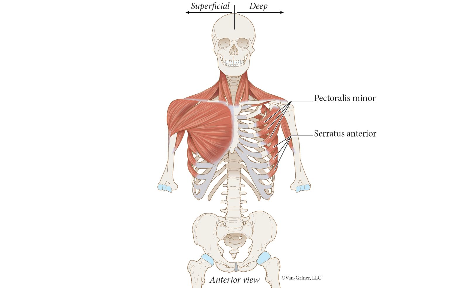
Figure 17.1Anterior Muscles that Move the Scapula
Posterior Muscles that Move the Shoulder
The posterior muscles that move the scapula include the trapezius, levator scapula, rhomboid major, and rhomboid minor (Figure 17.2).
Each trapezius muscle (Figure 17.2) is a large, superficial triangular sheet that extends from the occipital bone to the inferior thoracic vertebrae. Since the trapezius contains fibers running in multiple directions, it is able to perform multiple actions. In addition to rotational movement of the scapula, the trapezius can cause either elevation or depression, depending on which of its fibers are contracting.
The straplike levator scapula (Figure 17.2) lies deep to the trapezius. It extends from the first four cervical vertebrae to the medial border of the scapula. When it contracts, it elevates the scapula as its name suggests.
The paired rhomboid muscles, rhomboid major and rhomboid minor (Figure 17.2), also lie deep to the trapezius. They extend from the spinous processes of the upper thoracic vertebrae to the medial border of the scapula. They retract and elevate the scapula.
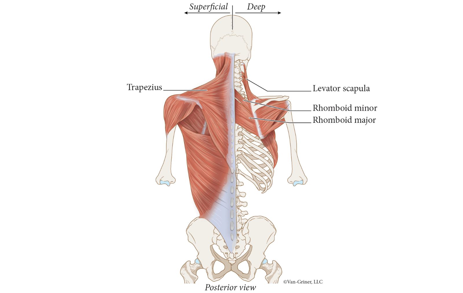
Figure 17.2Posterior Muscles that Move the Scapula
Muscles that Move the Shoulder—Actions
To study the attachment sites and actions of all the indicated anterior and posterior muscles that move the scapula, see Table 17.1. Although the muscle site attachments are listed in Table 17.1, they are used only for the purpose of understanding and mastering the muscle actions and will not be used for exam questions. However, you should be able to name and locate each indicated muscle and identify the action(s) of each.
Table 17.1
Muscles that Move the Scapula and Their Actions
Muscle | Origin | Insertion | Action |
|---|---|---|---|
Anterior Muscles | |||
Pectoralis minor | Sternal ends of ribs 3–5 | Coracoid process of scapula | Scapula protraction |
Serratus anterior | Lateral surfaces of most ribs | Medial border of scapula | Scapula protraction |
Posterior Muscles | |||
Trapezius | Occipital bone Spinous processes of C7–T12 | Spine and acromion of scapula; clavicle | Elevation, depression, rotation of scapula |
Levator scapulae | Transverse processes of C1–C4 | Medial border of scapula | Elevation of scapula |
Rhomboid major | Spinous processes of T2–T5 | Medial border of scapula | Retracts and elevates scapula |
Rhomboid minor | Spinous processes of C7–T1 | Medial border of scapula | Retracts and elevates scapula |
Section 2: Muscles that Move the Arm
In this section, we will continue our discussion of understanding the difference between muscles acting on the shoulder versus muscles acting on the arm. We noted previously that muscles that act on the shoulder originated from the axial skeleton and then inserted on the bones of the pectoral girdle. This resulted in movement or positioning of the scapula. When considering muscles that act on the arm, these muscles originate at the scapula or axial skeleton and then insert at the humerus. This results in movement of the arm.
There are nine muscles that cross the shoulder joint and insert at the humerus. Seven of these nine muscles have scapular origin and the remaining two have axial origin.
Muscles of Axial Origin that Move the Arm
Of the two muscles that are of axial skeleton origin, one is located on the anterior side of the thorax, and the other is on the posterior side of the thorax. Anteriorly, the pectoralis major (Figure 17.3) is a convergent (triangular) muscle with multiple fiber orientations. Because its extensive origins converge on a common insertion site from different angles, there are multiple actions. Use Figure 17.3 to help you visualize these actions. In addition, the human nervous system can "talk to" groupings of muscle fibers within the muscle; for example, contraction of the middle fibers can primarily cause adduction of the arm by moving the arm towards the torso.
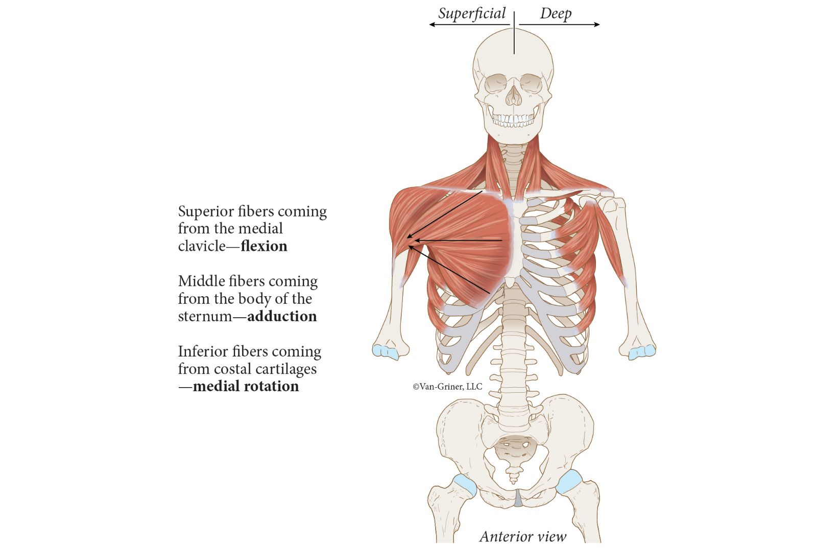
Figure 17.3Anterior Muscles that Moves the Arm (Axial Origin)
Posteriorly, in the lower half of the back (dorsum) is the large, superficial latissimus dorsi muscle (Figure 17.4). Like the pectoralis major, it has broad origins and the same insertion site. Two of its actions, adduction and medial rotation, are the same as those of pectoralis major. However, the third action—arm extension—antagonizes the action of pectoralis major.
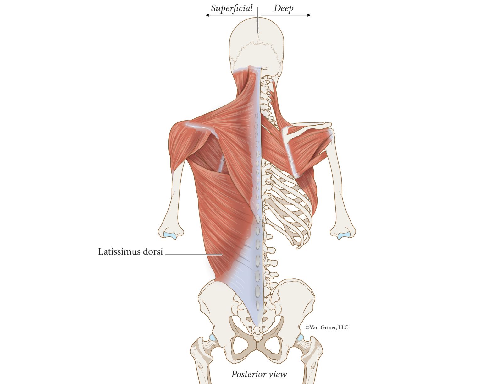
Figure 17.4Posterior Muscles that Moves the Arm (Axial Origin)
Muscles of Scapular Origin that Move the Arm
Now, let’s discuss the muscles that cross the shoulder joint to move the arm and originate at the scapula. The first muscle we will discuss is also a large superficial muscle called the deltoid muscle (Figure 17.5). This thick muscle forms the rounded curvature of the shoulder. It has multiple origins including the clavicle, the spine of the scapula, and acromion of the scapula; therefore, it can be seen on anterior, lateral, and posterior views. The middle fibers of the deltoid, seen on a lateral view, are the prime mover in arm abduction. The anterior and posterior fibers of the deltoid, since they insert on the same deltoid tuberosity from different directions, antagonize each other with flexion and extension of the arm. Rhythmic contractions of the anterior, then the posterior deltoid, give rise to the characteristic human "arm swing" when walking.
Another muscle that crosses the shoulder joint and has scapular origin is the coracobrachialis muscle (Figure 17.5). This muscle is named for its attachments as it extends from the coracoid process of the scapula to about mid-shaft on the humerus. Its action is to flex and adduct the humerus.
The next muscle up for discussion is the teres major muscle (Figure 17.5). Teres major originates from the inferior lateral border of the scapula and inserts on the lesser tubercle of the humerus. While it is certainly not a big muscle that moves the arm, it is near the superior fibers of the latissimus dorsi muscle. It has the same three actions as those of latissimus dorsi—adduction, medial rotation, and extension, and is referred to by some as the "lat's little brother."
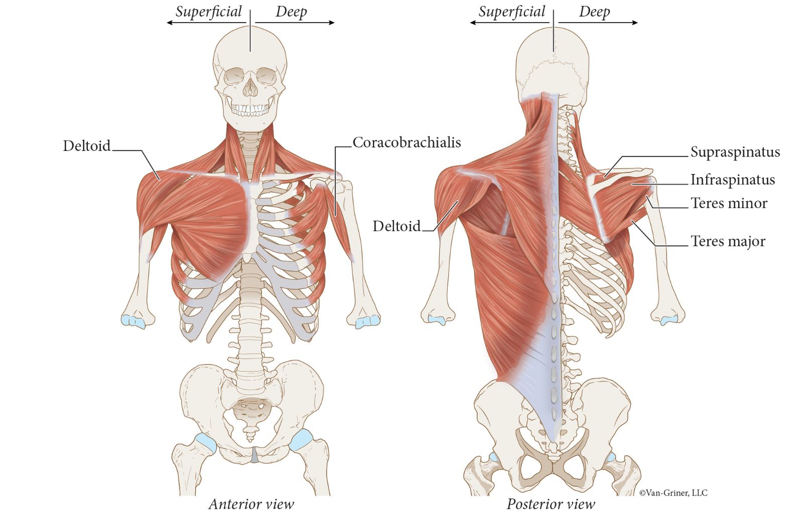
Figure 17.5Muscles of Scapular Origin that Move the Arm
Muscles of Scapular Origin that Move the Arm—Rotator Cuff Muscles
As we conclude our study of the muscle acting on the arm and are of scapular origin, let’s consider the shoulder joint for a moment. The bony articulation at the shoulder joint, between the head of the humerus and the glenoid cavity, is inherently unstable due to the shallow depression of the glenoid cavity. The tendons of the four remaining muscles are called the "rotator cuff" muscles because they function to help attach the humerus to the scapula. Unfortunately, these muscles and tendons are important clinically as they can be damaged and require surgical repair.
The rotator cuff muscles are the supraspinatus (Figure 17.5), infraspinatus (Figure 17.5), teres minor (Figure 17.5), and subscapularis (Figure 17.6) muscles. An acronym that may help you remember them is "SItS." Notice that the "t" is lower case to designate that this is teres minor rather than the teres major. Three of the muscles can easily be seen in Figure 17.5, but to view the subscapularis, we need an anterior view in which the thoracic cage has been removed as in Figure 17.6.
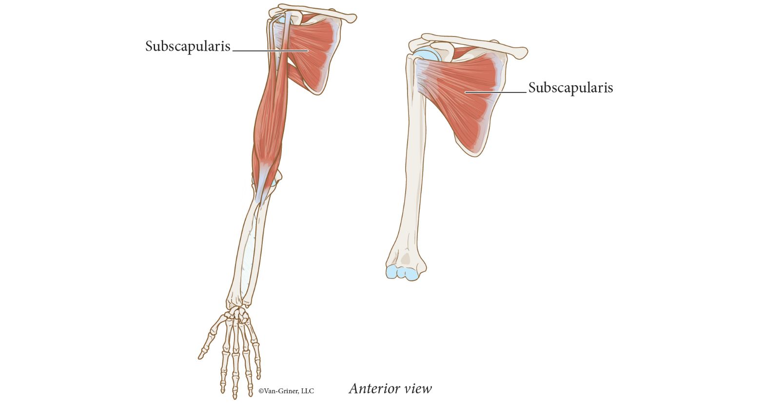
Figure 17.6Supraspinatus Muscle of the "Rotator Cuff"
Muscles that Move the Arm—Actions
To study the attachment sites and actions of all the indicated muscles that move the arm, see Table 17.2. Although the muscle site attachments are listed in Table 17.2, they are used only for the purpose of understanding and mastering the muscle actions and will not be used for exam questions. However, you should be able to name and locate each indicated muscle and identify the action(s) of each.
Table 17.2
Muscles Actings on the Arm and Their Actions
Muscle Name | Origin | Insertion | Action |
|---|---|---|---|
Muscles of Axial Origin | |||
Pectoralis major | Medial clavicle | Proximal humerus | |
Sternum | Flexes arm | ||
Adducts arm | |||
Medially rotates arm | |||
Rib cartilages | |||
Latissimus dorsi | Spinous processes of Vertebrae T5–L5 | Proximal humerus | Extends arm |
Iliac crest | Adducts arm | ||
Lower ribs | Medially rotates arm | ||
Muscles of Scapular Origin | |||
Deltoid (middle fibers) | Acromion of scapula | Deltoid tuberosity | Abducts arm |
Deltoid (anterior fibers) | Clavicle | Deltoid tuberosity | Flexes arm |
Medially rotates arm | |||
Deltoid (posterior fibers) | Spine of scapula | Deltoid tuberosity | Extends arm |
Laterally rotates arm | |||
Coracobrachialis | Coracoid process | Shaft of humerus | Flexes and adducts arm |
Teres major | Lateral border of scapula | Proximal humerus | Extends arm |
Adducts arm | |||
Medially rotates arm | |||
Muscles of Scapular Origin and Rotator Cuff Muscles | |||
Supraspinatus | Supraspinous fossa | Greater tubercle of humerus | Abducts arm |
Infraspinatus | Infraspinous fossa | Greater tubercle of humerus | Laterally rotates arm |
Teres minor | Lateral border of scapula | Greater tubercle of humerus | Laterally rotates arm |
Subscapularis | Subscapular fossa | Lesser tubercle of humerus | Medially rotates arm |
Section 3: Muscles of the trunk involved in Respiration
The last group of muscles that we will study for this lab involves the muscles of the trunk that are involved in respiration. Changes in volume of the thoracic cavity drive the cycles of inspiration and expiration. These changes in volume of the thoracic cavity are largely due to the alternate contraction and relaxation of the diaphragm (Figure 17.7).
The diaphragm, which separates the thoracic and abdominal cavities, is a dome-shaped muscle at rest. When it is stimulated to contract by the phrenic nerve, it flattens out, which increases the volume of the thoracic cavity. The increase in volume of the thoracic cavity leads to decreased air pressure compared to atmospheric air; therefore, air moves in. The diaphragm is the prime mover in the process of inspiration. In Figure 17.7, openings are shown in the diaphragm for various structures to pass between the thorax and the abdomen.
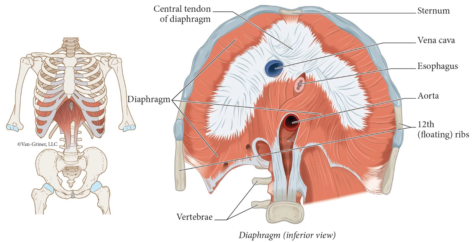
Figure 17.7Muscles of Respiration—The Diaphragm
Intercostal muscles lie between the ribs. The external intercostals (Figure 17.8) are the most superficial layer; their fibers extend from a superior rib downward to insert on the next inferior rib (see Figure 17.8). Their action synergizes the diaphragm in expansion of the thoracic cavity.
The internal intercostals (Figure 17.8) are deep to the external intercostals and are involved in forced expirations. Their fibers extend from an inferior rib to insert on the next superior rib (see Figure 17.8).
Deep to both the external and internal intercostals are the innermost intercostals (Figure 17.8). Their function is poorly understood.
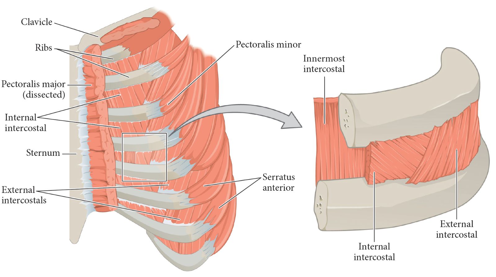
Figure 17.8Muscles of Respiration—Intercostal Muscles
Muscles of Respiration—Actions
To study the actions of the indicated muscles of the trunk involved in respiration, see Table 17.3. You should be able to name and locate each indicated muscle and identify the action(s) of each.
Table 17.3
Muscles of Respiration and Their Actions
Muscle | Action |
|---|---|
Diaphragm | Inspiration (prime mover) |
External intercostals | Inspiration (synergist) |
Internal intercostals | Expiration (forceful only) |