Protein Identification Lecture 5
Protein Identification
Overview of Techniques for Protein Analysis
Electrophoresis: A technique used to separate proteins based on their size and charge.
Determination of Molecular Weight: Techniques such as mass spectrometry are employed to ascertain the molecular weight of proteins.
Protein Sequencing: Methods like Edman degradation are used for determining the amino acid sequence of proteins.
Western Blotting: A technique for detecting specific proteins in a sample using antibody-based processes.
Localization: Techniques to determine the cellular or tissue location of proteins using specific antibodies.
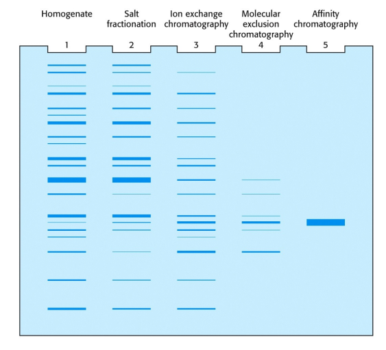
Electrophoresis: used to determine molecular weight and the purification of you protein
Native polyacrylamide gel
SDS-denatured polyacrylamide gel
2D electrophoresis
General Principles
Direction of electrophoresis is influenced by the charge of macromolecules in an electric field.
Utilizes a porous gel medium to separate proteins during the electrophoresis process.
Some proteins will be linear when migrating down the gel, others will be spherical. This has to with the electronegativity of the protein. It also deals with the amount of positive or negative charge.
Polyacrylamide Electrophoresis
Polyacrylamide: A gel matrix used in electrophoresis to separate proteins.
Components:
Acrylamide: The primary polymer used for constructing the gel.
Methylenebisacrylamide: A cross-linker in the gel formation.
Persulfate: Starts the polymerization process, generating radicals.
TEMED (N,N,N',N'-tetramethyl ethylenediamine): Stabilizes the radicals formed during polymerization.
Types of Electrophoresis
Native Polyacrylamide Gel:
Proteins maintain their biological activity in the gel.
Migration is influenced by: size, charge, shape, and oligomerization state (if the protein has subunits).
SDS-Denatured Polyacrylamide Gel: Process that allows for uniform analysis. Denatured before and after
The denaturing of the protein allows for the primary structure to be present, no matter if it is secondary, tertiary or quaternary.
Any protein will interact with SDS-gel
Sodium dodecyl sulfate (SDS) binds to proteins at an average ratio of one SDS per two amino acid residues, eliminating the native charge so that proteins can be separated primarily by size.
It is proportional to the amount of amino acids you have, for example:
If you have 100 amino acid sequence, you will have 50SDS. This will result in a negatively charged protein, dominated by the SDS molecule.
Proteins are often stained with Coomassie Blue for visualization.
2D Gel Electrophoresis:
First Dimension: Isoelectric focusing using polyampholytes to separate proteins based on their isoelectric point.
Proteins will migrate towards their respective pH where they have no net charge (pI). (x-axis)
Second Dimension: SDS-PAGE is employed after isoelectric focusing to further separate the proteins by size. (y- axis)
2D allows for two levels of analysis:
Comparison of protein expression in different cells
Identification of specific proteins by using mass spec.
Relative Mobility (RM)
Defined as:
Utilizes a marker for scale comparison during electrophoresis.
Chromatography Techniques Related to Electrophoresis
Ion Exchange Chromatography: A technique that separates proteins based on their charge.
Affinity Chromatography: Allows for the purification of proteins based on specific interactions.
Size Exclusion Chromatography: Separates proteins based on their size.
Protein Mass Determination
Mass Spectrometry Techniques
Mass Spectrometry (MS): used to determine the precise molecular weight of a protein.
Electrospray Ionization (ESI): A technique to generate ionized proteins from a solution through an electric spray.
This allows for a ratio of the mass to the charge.
The ions are analyzed to determine their mass, which is crucial for identifying proteins and their quantities.
Key Components of ESI Mass Spectrometry Setup
Ion Trap: Stores ions prior to mass analysis.
Uses nitrogen gas to assist in the focusing of ions.
A mass detector captures the data generated by the ionized proteins.
MALDI Mass Spectrometer:
Matrix-assisted laser desorption-ionization (MALDI)
Uses UV laser to activate the solid matrix to carry proteins
1) Protein sample is ionized
2) electrical field accelerates ions
3) Lightest ions arrive at the detector first
4) Laser triggers a clock
Peptide Sequencing
Edman Degradation
A method for sequencing amino acids in proteins, specifically from the N-terminus.
Can sequence up to 50 amino acid residues from N-terminal of protein. After 50, the yield is very low.
Utilizes phenyl isothiocyanate to label the amino acids for identification.
It attaches to an amino acid, and then it is released to form a new compound. (ex: PTH-alanine).
Chemical and enzyme cleavage to make small polypeptides from a big protein
Sequencing Process Steps
Labeling of the N-terminal amino acid.
Cleavage to release the labeled amino acid while leaving a shorter polypeptide.
Ability to sequence up to 50 amino acid residues.
Emphasizes the importance of chemical and enzymatic cleavage to generate manageable polypeptides for sequencing.
Polypeptide Cleavage Methods: used to cut sequence into more manageable chunks.
Cyanogen bromide (CNBr): Cleaves at the C-side of Methionine residues.
Hydroxylamine: Targets bond between Asparagine and Glycine.
Trypsin (BASIC): Cleaves at the C-side of Lysine and Arginine.
Chymotrypsin (BIGGER): Targets C-side of aromatic amino acids (Tyr, Trp, Phe, Leu, Met).
How do you understand the order of proteins after they are cleaved?
Tryptic peptides and chymotryptic peptides can overlap in order to understand the final sequence. Since different amino acid groups are targeted by specific chemicals.
Antibody Techniques for Protein Identification
Antibodies Overview
Antibody: A protein produced by an animal in response to an antigen, which is recognized by a specific
Epitope: a specific amino acid group that is recognized by antibody
Types of Antibodies:
Polyclonal Antibodies: Derived from multiple sources and recognize multiple epitopes.
Antigens have multiple surfaces, and these antibodies will recognize a specific surface protein on the antigen. These work together collectively.
Think: Collection of monoclonal antibodies
Monoclonal Antibodies: Identical antibodies from a single cellular source, recognizing a specific epitope.
Antigen Structure:
Two binding sites in order to recognize different antigens.
Heavy Chain
Light Chain
Interactions between Antibody and Antigen
This is a very strong interaction. This bond has only been seen biologically, and has not been able to be recreated.
You cannot separate antigen and antibody. Because of this, it allows for high specificity in identification.
Preparation of Monoclonal Antibodies
Give a rat a specific antigen, this will fuse in polyethylene glycol. The mice’s cells will fuse with the tumor cells. These fused cells can be grown in mass cultures to produce antibodies or can be injected into rats to induce tumor growth and isolate the antibodies that way. The final step is to purify the antibody.
Western Blotting Technique:
When the primary antibody is injected into a different species, for example, mouse antibody into rabbit antibody, this allows the secondary antibody to attach to the primary antibody.
Proteins are transferred to a polymer sheet after electrophoresis.
Primary Antibody: Specific antibody binds to the protein of interest.
Secondary Antibody: Tagged with an enzyme, it binds to the primary antibody.
Enzyme substrate is added, allowing visibility of the protein band through color development.
Use of photographic film for exposure to capture binding events, creating an autoradiogram.
SDS Gel Electrophoresis is transferred to a polymer sheet. The polymer sheet is then added to specific antibody mixture. and is washed to remove the unbound antibody. The proteins on the polymer sheet being exposed to antibody. The overlay of the photographic film is exposed and developed. The protein band remains and you now know what specific antibody detects your protein.
ELISA (Enzyme-linked Immunosorbent Assay)
Types of Elisa:
Indirect ELISA (specific antibody): Used to test for specific antibodies; involves binding antigen to a well and adding a specific antibody.
Sandwich ELISA(specific antigen): Used to detect specific antigens; utilizes two monoclonal antibodies and observes substrate conversion into a color product for quantification.
Protein Localization Techniques
Methods
Use of specific antibodies tagged with enzymes or electron-dense markers for visualizing protein location in cells or tissues.
Staining of actin filaments in a cell was possible because of what?
A DNA binding protein in Drosophila embryo
Protein Structure Determination Techniques
Circular Dichroism (CD): Remember that polarizing light rotates at different angles.
Analyzes secondary structures of proteins to determine their content based on optical properties of peptide bonds.
The peptide bonds within different secondary structures have different optical properties.
When you have a random coil, you are able to determine the percentage of alpha helices, beta sheets, etc.
For example: Myoglobin contains 80% helix content and 20% random coil
Nuclear Magnetic Resonance (NMR) Spectroscopy: This allows for analysis of tertiary and quaternary structures.
Provides atomic structure of proteins in solution, particularly for small proteins.
Requires knowledge of the amino acid sequence and multiple spectroscopic measurements to construct and validate 3D structures.
1) Amino acid sequence
2) measure the multidimensional spectra
3) interactions in space
4) Final structure
X-ray Crystallography
Procedure involves:
Screening crystallization conditions.
Collecting diffraction data from protein crystals.
Using computational means to simulate protein structure and match with diffraction data for validation.
Screen Conditions for Protein Crystals:
Hanging drop method
X-ray source comes from above and diffracts through the crystal. the diffracted beams are captured on a detector, and this information is used to further analyze the structure of the protein.
Chemical Synthesis of Polypeptides
Applications
Medical Purposes: Create peptide hormones, anti-cancer reagents, and enzyme inhibitors.
Research Purposes: Aid in specificity testing for antibodies, and functional investigations of protein motifs.
Costs and Trends in Synthesis
Historical cost trends have highlighted the substantial decrease in peptide synthesis prices—$100 per amino acid in 1995 to approximately $3 per amino acid in 2009.
Chemical Synthesis of Polypeptides Overview:
t-Boc Strategy: Utilize t-Butyloxycarbonyl (t-Boc) protected amino acids for controlled synthesis.
DCC Activation: Employ Dicyclohexylcarbodiimide (DCC) to facilitate the coupling of activated amino acids.
Deprotection (CF3COOH): Sequentially remove protective groups to progress through the peptide synthesis cycle.
Final Release: Cleavage with HF to release the synthesized polypeptide from the resin.
This process is very useful for small peptides, however there is still an issue with yield. Yield will become low when long polypeptides are synthesized
The yield of each step is about 0.99, the more steps you have (longer amino acid sequence) the lower yield you will receive along the way.