NEUROANATOMY: The Human Nervous System
DIVISIONS OF THE NERVOUS SYSTEM
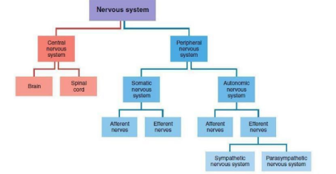
Central Nervous System (CNS) - Located within the skull and spine (brain and spinal cord)
Peripheral Nervous System (PNS) - Located outside the skull and spine
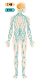
PERIPHERAL NERVOUS SYSTEM
Somatic Nervous System -Interacts with the external environment. It is composed of afferent nerves that carry sensory signals from the skin, skeletal muscles, joints, eyes, ears and so on, to the CNS, and efferent nerves that carry motor signals from the CNS to the skeletal muscles.
Autonomic Nervous System - Regulates the body’s internal environment. It is composed of afferent nerves that carry sensory signals from internal organs to the CNS, and efferent nerves that carry motor signals from the CNS to the internal organs.
2 Kinds of Efferent Nerves:
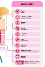
Sympathetic Nerves - The division of the autonomic nervous system that coordinates arousal.
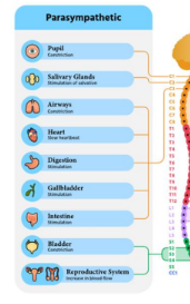
Parasympathetic Nerves - The division of the autonomic nervous system responsible for rest and energy storage.
BLOOD-BRAIN BARRIER - impedes the passage of many toxic substances. In the rest of the body, the cells that compose the walls of blood vessels are loosely packed; as a result, most molecules pass readily through them into surrounding tissues.
In the brain, however, the cells of the blood vessel walls are tightly packed, thus, forming a barrier to the passage of many molecules, particularly proteins and other large molecules. The degree to which therapeutic or recreational drugs can influence brain activity depends on the ease with which they penetrate the blood-brain barrier.
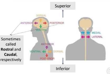
ANATOMICAL DIRECTIONS & PLANES OF SECTION
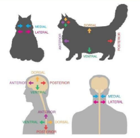
Sometimes called Rostral and Caudal, respectively
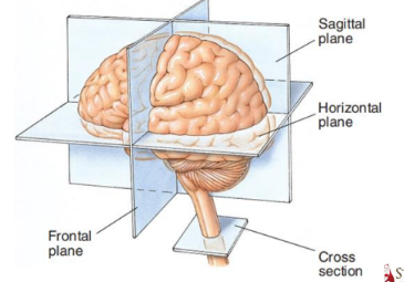
PLANES OF SECTION
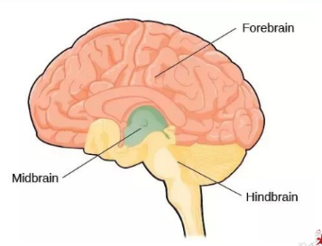
MAJOR DIVISIONS OF THE BRAIN
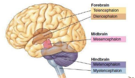
The first indications of the developing brain are three swellings that eventually develop into the adult forebrain, midbrain, and hindbrain.
Before birth, the initial three swellings in the neural tube become five: telencephalon, diencephalon, mesencephalon, metencephalon, and the myelencephalonand
MAJOR STRUCTURES OF THE BRAIN
MYELENCEPHALON -Sometimes called the medulla Composed largely of tracts carrying signals between the rest of the brain and the body
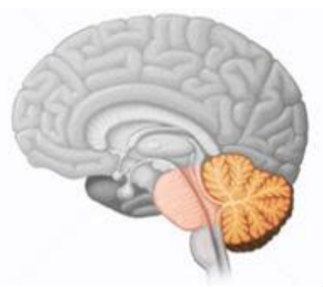
Reticular Formation - a complex network of about 100 tiny nuclei that occupies the central core of the brain stem from the posterior boundary of the myelencephalon to the anterior boundary of the midbrain
The various nuclei of the reticular formation are involved in a variety of functions— including sleep, attention, movement, the maintenance of muscle tone, and various cardiac, circulatory, and respiratory reflexes
Pons - participates in the management of states of consciousness.
Cerebellum - participates in balance, muscle tone, muscle coordination, some types of learning, and possibly higher cognitive functions in humans.
MESENCEPHALON
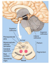
MESENCEPHALON 2 Divisions:
Tectum (roof) is the dorsal surface of the midbrain; composed of two pairs of bumps, the colliculi (little hills).
Inferior Colliculi - have an auditory function (posterior).
Superior Colliculi - have a visual-motor function
2 Divisions:
Tegmentum is ventral to the tectum
Contains three colorful structures of particular interest to biopsychologists:
Periaqueductal Gray - plays a role in mediating the analgesic (pain-reducing) effects of opiate drugs
Substantia Nigra - involved in sensorimotor functions
Red Nucleus - involved in sensorimotor functions
DIENCEPHALON
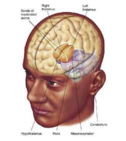
Thalamus - Large, two-lobed structure that constitutes the top of the brain stem ○ Information relay station
Hypothalamus - Located just below the anterior thalamus Plays an important role in the regulation of several motivated behaviors (e.g., eating, sleep, and sexual behavior).
TELENCEPHALON - The largest division of the human brain. Mediates the brain’s most complex functions. It initiates voluntary movement, interprets sensory input, and mediates complex cognitive processes such as learning, speaking, and problem solving.
Cerebral Cortex - layer of tissue covering the cerebral hemispheres
Gray Matter - composed of small, unmyelinated neurons
White Matter - the layer beneath the cortex; mainly composed of large myelinated axons
Fissure - large furrows in a convoluted cortex
Sulcus - small furrows
Gyrus - ridge between fissure and sulcus
4 Lobes of the Brain
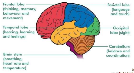
Neocortex (New Cortex) - About 90% of human cerebral cortex is neocortex It is six-layered cortex of relatively recent evolution. By convention, the layers of neocortex are numbered I through VI, starting at the surface.
LIMBIC SYSTEM - A circuit of midline structures that circle the thalamus Involved in the regulation of motivated behaviors—including the four Fs of motivation: fleeing, feeding, fighting, and sexual behavior Include the amygdala, the fornix, the cingulate cortex, and the septum
Amygdala - major processing center for emotions
Fornix - acts as the primary outgoing pathway from the hippocampus, and thus its most recognized function is its involvement in memory
Cingulate Cortex - involved in emotional processing and inhibitory control, in attentional processing, and in sleep staging
Septum - indicates a subcortical structure in the forebrain that is found near the midline of the brain
SPINAL CORD - Comprises two different areas: an inner H-shaped core of gray matter and a surrounding area of white matter Pairs of spinal nerves are attached to the spinal cord— one on the left and one on the right—at 31 different levels of the spine. Each of these 62 spinal nerves divides as it nears the cord, and its axons are joined to the cord via one of two roots: the dorsal root or the ventral root. all dorsal root axons, whether somatic or autonomic, are sensory (afferent) unipolar neurons. In contrast, the neurons of the ventral root are motor (efferent) multipolar neurons with their cell bodies in the ventral horns