Biochemistry term 4 week 7
Proteins: Relation of Structure and Function
Four levels of protein structure:
Primary Structure: The sequence of amino acids.
Secondary Structure: Localized folding, such as alpha-helices and beta-sheets.
Tertiary Structure: The overall three-dimensional structure, including the arrangement of secondary structures.
Quaternary Structure: The arrangement of multiple polypeptide chains in multi-subunit proteins.
Examples of Proteins
Fibrous proteins:
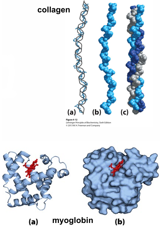
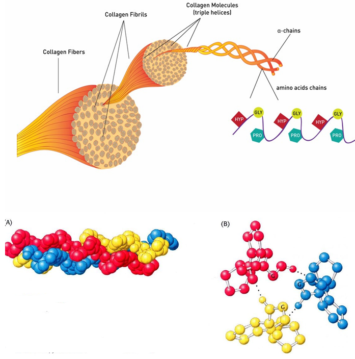
Collagen
Keratin
Globular proteins:
Hemoglobin
Myoglobin
Fibrous Proteins
Collagen and Keratin
Filaments or sheets
Water insoluble (many hydrophobic amino acids)
Structural proteins, form bones, cartilage, hair
Globular Proteins
Hemoglobin and Myoglobin
Spherical, globular shape
Water-soluble
Enzymes, hormones, plasma proteins
Fibrous Proteins: Collagen
Three separate polypeptides are supertwisted about each other, forming a right-handed superhelical twist.
Many triple-helices assemble into a collagen fibril.
Covalent bonds between triple helices (by modified lysine side chains).
Amino acid sequence is generally a repeating tripeptide unit: Gly-X-Y (X often Proline, Y often Hydroxyproline).
Only Gly residues can be accommodated at the very tight junctions between the individual chains.
Within a strand: no H-bonds. Instead, the helix is stabilized by steric repulsion of the pyrrolidine rings of Pro and Hyp.
Between strands: H-bonds.
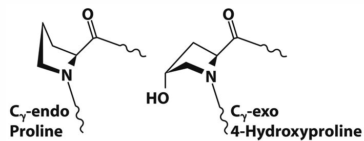
Fibrous Proteins: Collagen - Hydroxyproline
Forces the proline ring into a favorable pucker.
Offers more hydrogen bonds between the three strands of collagen.
The post-translational processing is catalyzed by prolyl 4-hydroxylase and requires α-ketoglutarate, molecular oxygen, and ascorbate (vitamin C).
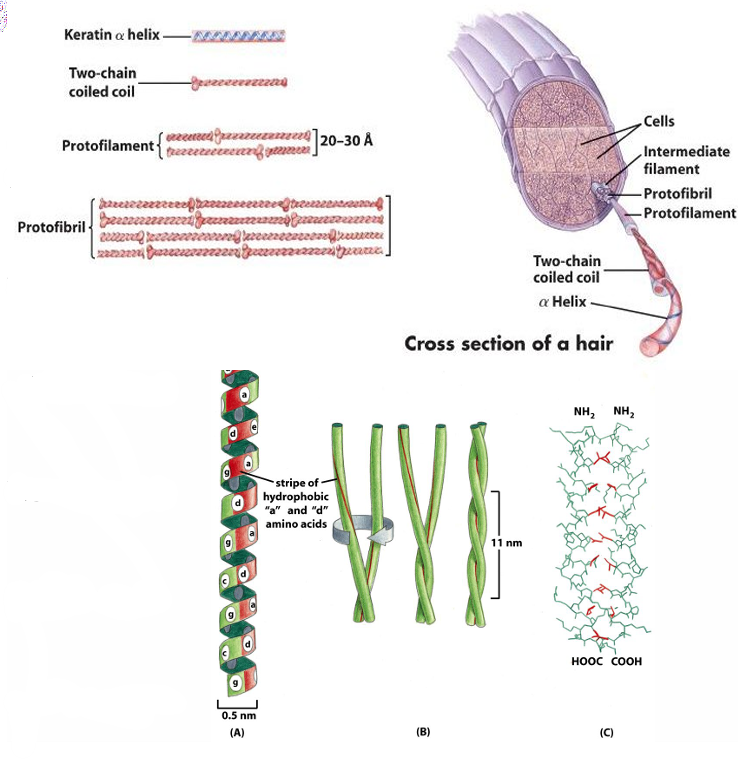
Fibrous Proteins: α-Keratin
Two α-helices intertwine with each other to form a supertwisted coiled coil (rich in hydrophobic amino acids).
The α-keratin helix is right-handed, while the helical path of the supertwists is left-handed.
The tertiary structure of keratin is quite simple (left-handed super helix; coiled coils).
The quaternary structure can be the result of the assembly of coiled coils into supramolecular complexes.
Many amino acids with hydrophobic residues allow close packaging of the two polypeptide chains.
Many cysteines for the disulfide bonds between the chains.
The strength of fibrous proteins is enhanced by covalent cross-links between polypeptide chains.
In α-keratins, these are disulfide bonds.
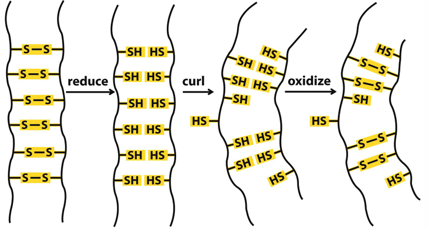
Fibrous Proteins: α-Keratin - Permanent Waving / Straightening
Disulfide bridges (cysteine) between the different coiled-coils.
Process involves reduction and oxidation of disulfide bonds to change hair shape.
Globular Proteins
Spherical, globular shape, with hydrophobic amino acid residues oriented towards the interior.
Hydrophilic side chains on the surface → water-soluble.
NOT the same as intrinsically disorganized proteins (proteins / protein segments that lack definable structure; see pt4).
Examples: enzymes, hormones, plasma proteins, hemoglobin, myoglobin.
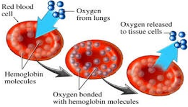
Globular Proteins: Hemoglobin
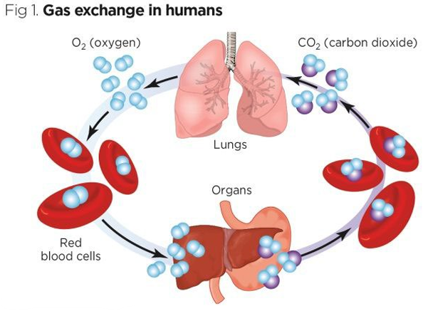
Oxygen needs to go from lungs to tissue.
Oxygen is poorly soluble in aqueous solutions → cannot be carried to tissues if simply dissolved in blood serum.
Transport mechanisms necessary for multicellular organisms.
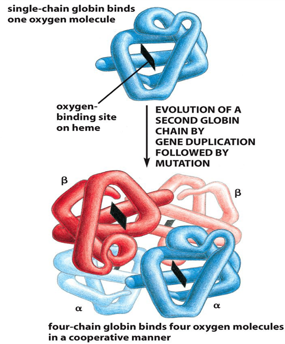
Hemoglobin consists of 4 globin subunits (myoglobin is a single chain globin).
2 α-globin subunits
2 β-globin subunits
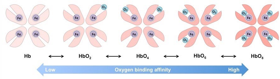
4 globin subunits → can bind 4 oxygen molecules.
Each subunit has 1 heme molecule: prosthetic group.
4 subunits work cooperatively → the binding of oxygen to a site in one chain increases the likelihood that the remaining chains will bind oxygen.
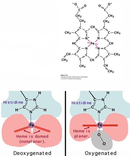
Globular Proteins: Hemoglobin - Heme
Heme group is a prosthetic group (non-protein part of protein contributing to its function).
None of the amino acid side chains present in proteins is suitable to bind oxygen.
Transition metals (such as iron) have a strong tendency to bind oxygen.
Iron interacts with a non-peptide group that is bound to protein: the heme group.
Organic ring structure porphyrin bound to .
can make 6 coordination bonds:
4 to N in the porphyrin ring.
1 to His-side chain.
1 is free to bind oxygen.
Globular Proteins: Hemoglobin – Transport
Required properties are:
Efficient uptake of oxygen at high oxygen pressure (lungs).
Efficient release of oxygen at low oxygen pressure (tissue).
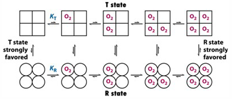
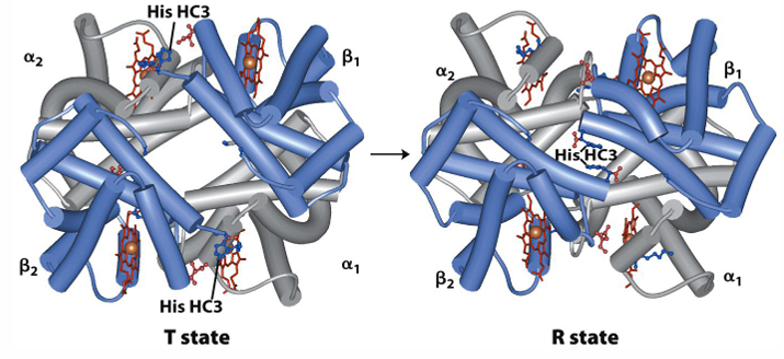
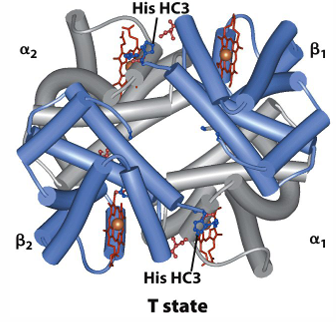
Hemoglobin exists in two conformations: T (tense) and R (relaxed).
T (tense) state: less affinity for , deoxy conformation.
R (relaxed) state: more affinity for , oxy conformation.
T state is stabilized by ion pairs at the and interface.
Binding of to a hemoglobin subunit triggers a change in conformation to the R state.
Some of the ion pairs that stabilize the T state are broken, and some new ones are formed.
High affinity in lungs.
Low affinity in tissue.
Globular Proteins: Hemoglobin: Cooperative Binding - Concerted Model
All subunits exist either in the T state or in the R state (undergo transition simultaneously).
At each level of oxygen loading, an equilibrium exists between the T and the R states.
The equilibrium shifts from T -> R when the molecule is loaded with .
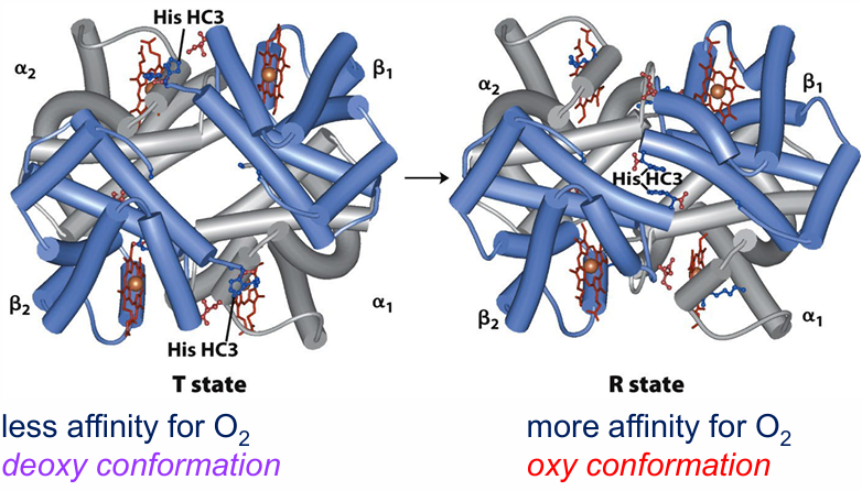
T state is stabilized by ion pairs at the α1β2 and α2β1 interface.
Binding of O2 to a hemoglobin subunit triggers a change in conformation to the R state.
→ some of the ion pairs that stabilize the T state are broken and some new are formed
Globular Proteins: Hemoglobin: Cooperative Binding of O2
Binding of oxygen to one subunit influences the conformation (shape) of the other subunits and therefore affinity for the ligand
subunits cooperate
Globular Proteins: Hemoglobin: Cooperative Binding, concerted model
all subunits exist either in the T state or in the R state (undergo transition simultaneously)
at each level of oxygen loading, an equilibrium exists between the T and the R states
the equilibrium shifts from T -> R when the molecule is loaded with O
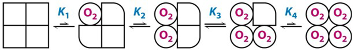
Globular Proteins: Hemoglobin: Cooperative Binding, sequential model
The binding of a molecule of in one subunit changes the conformation of this subunit.
The first change induces changes in neighboring subunits that increase their affinity for the ligand.
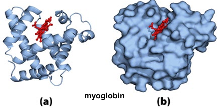
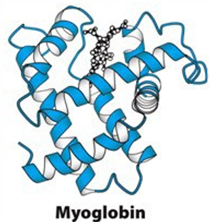
Globular Proteins: Myoglobin
Located in the muscles.
Facilitates the diffusion of through the cell for the generation of cellular energy.
Myoglobin is a single polypeptide (153 amino acid residues) with one molecule of heme
→ no cooperative binding possible.
High affinity for oxygen => Releasing only in low .
Globular Proteins: Hemoglobin - Allosteric Protein
An allosteric protein is one in which the binding of a ligand to one site affects the binding properties of another site on the same protein.
The proteins adapt other conformations induced by the binding of ligands referred to as modulators.
Modulators can be inhibitors or activators.
Normal ligand = modulator => homotropic interaction.
Normal ligand ≠ modulator => heterotropic interaction.
Globular Proteins: Hemoglobin – Allosteric Protein - Modulators
Modulators: , , and 2,3-bisphosphoglycerate (BPG).
Globular Proteins: Hemoglobin – Allosteric Regulation by and
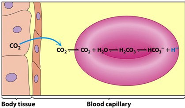
produced by the oxidation of fuels in mitochondria is hydrated to form .
dissociates to and which leads to a drop in pH in the tissue.
Hemoglobin transports about 40% of the total and 15-20% of the .
The rest of the is absorbed by the bicarbonate buffer, and the rest of as .
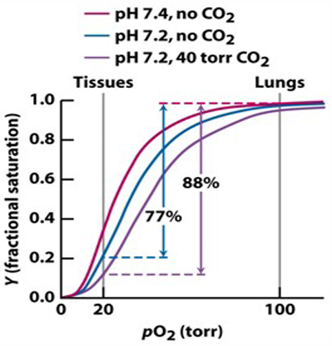
Globular Proteins: Hemoglobin – Allosteric Regulation: Low pH and
The lower the pH, the less the binding affinity of hemoglobin to oxygen.
pH of blood in lungs = 7.6.
pH of blood in tissues = 7.2.
Mechanism for the influence of : binds to residues in hemoglobin → Stabilization of T-state.
Mechanism for the influence of : binds to hemoglobin → Further stabilization of T-state.
In tissues, and are higher → pH is lower (7.2) → oxygen is released.
In lungs, is higher, and are released from hemoglobin, pH of blood is higher (7.6), and oxygen is bound.
and are allosteric modulators (inhibitors) of binding.
Globular Proteins: Hemoglobin – Allosteric Regulation: 2,3-Bisphosphoglycerate
2,3 BPG binds in the central cavity of the tetramer but only to the T state.
Stabilizes T-state → heterotropic allosteric inhibition of binding to hemoglobin by 2,3-BPG.
The more BPG present, the less binding affinity for oxygen (little effect in lungs with high oxygen pressure, higher effect in tissues).
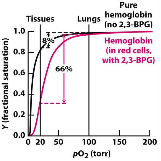
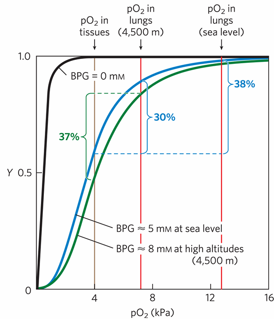
Adaptation to High Altitudes Through 2,3-Bisphosphoglycerate
At high altitude, is lower.
Upregulation of BPG concentration in blood.
Lower affinity for oxygen.
Little effect on binding of oxygen in lungs (see curve).
Considerable effect on release in tissues (see curve).
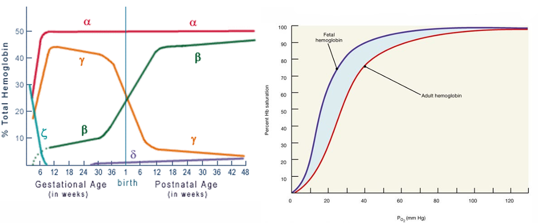
Globular Proteins: Hemoglobin – Fetal Oxygen Delivery
Fetal hemoglobin needs to have a higher affinity for oxygen than maternal hemoglobin.
Fetus synthesizes γ-subunits instead of β-subunits of hemoglobin: hemoglobin.
hemoglobin has a lower affinity for BPG than hemoglobin → higher affinity for oxygen.
The difference in oxygen affinity allows oxygen to be effectively transferred from maternal to fetal red blood cells.
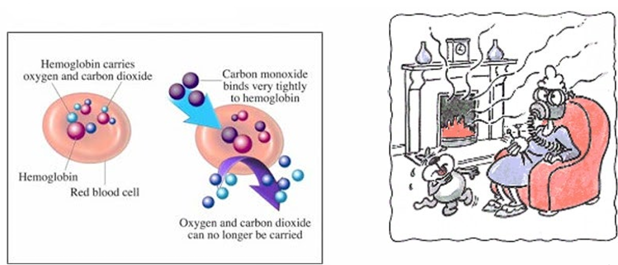
Globular Proteins: Hemoglobin – CO Poisoning
Each year in the US around 2500 people die from carbon monoxide poisoning
CO binding: competitive inhibition.
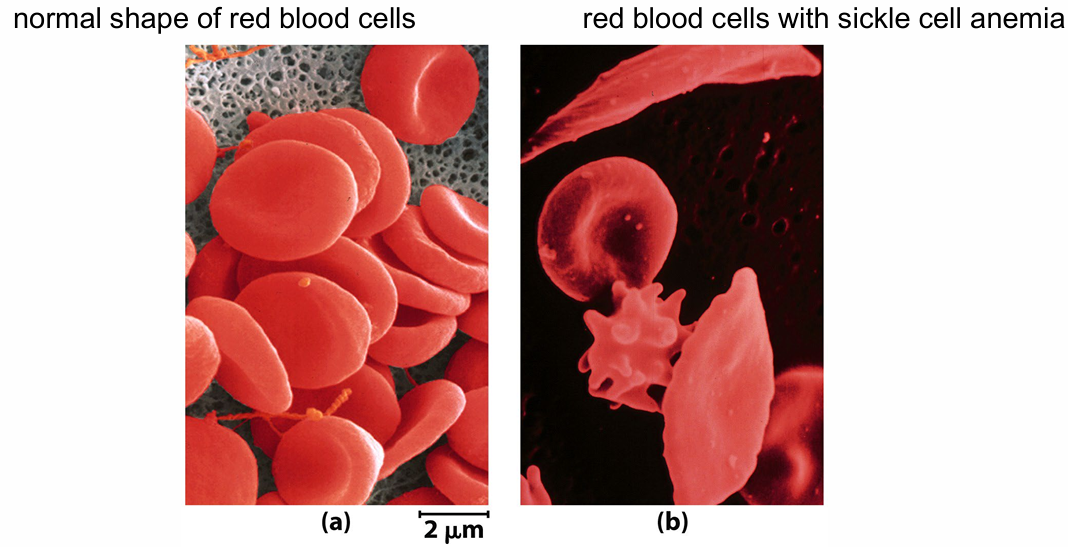
Globular Proteins: Hemoglobin – Sickle Cell Anemia
Single mutation in the β-globin chains of hemoglobin: Glutamate (Glu) to Valine (Val).
Mutated form: Hemoglobin S (normal: hemoglobin A).
In the oxy hemoglobin S (R-state): hydrophobic residues are largely buried.
In the deoxy hemoglobin S (T-state): extra hydrophobic interactions → formation of aggregates.
When hemoglobin S is deoxygenated, it becomes insoluble and forms polymers that aggregate into tubular fibers.
Red blood cells from sickle cell patients are more adherent to the walls of blood vessels.
Results: painful swelling of the extremities, a higher risk of stroke or bacterial infection, and anemia.
Revision Questions
Draw a sigmoidal and a hyperbolic oxygen-binding curve. Which of the two binding curves belongs to which oxygen-binding molecules?
- Sigmoidal curves represent cooperative binding, such as that seen in hemoglobin. Hyperbolic curves represent non-cooperative binding, like that seen in myoglobin.

What are allosteric inhibitors?
- Allosteric inhibitors are modulators that bind to a protein and reduce its activity or binding affinity at another site.
a. What is the Bohr effect? - The Bohr effect is the phenomenon where a decrease in pH (increase in H+H+ concentration) or an increase in CO2 partial pressure results in a lower affinity of hemoglobin for oxygen.
b. What is the benefit of the Bohr effect? - The benefit of the Bohr effect is that it enhances oxygen delivery to tissues with high metabolic activity, where pH is lower and CO2CO2 levels are higher.
What shows us that hemoglobin works cooperatively?
- The sigmoidal shape of the oxygen-binding curve for hemoglobin indicates cooperativity. Also, the fact that hemoglobin has different forms, T (tense) and R (relaxed).
Where do we find BPG? What is the effect of BPG? What is the influence of high altitude on BPG levels.
- BPG is found in red blood cells. It reduces the binding affinity of hemoglobin for oxygen by stabilizing the T-state. At high altitudes, BPG levels increase, leading to a lower binding affinity for oxygen, which enhances oxygen release in tissues.
Topics Discussed / Knowledge Test
Amino acids: structure, types of rest groups, possible interactions, charges.
Proteins: protein structure, motifs, domains, fibrous and globular proteins, protein families, conserved proteins.
Oxygen-binding proteins: binding curves, mechanisms, regulation, related disease.
Enzymes: characteristics, types, enzyme kinetics, inhibitors.
Protein interactions.
Structure and function of proteins.
and .
Post-translational modification and protein targeting.
20 multiple-choice questions and 10 points open questions in Biochemistry.